
Gout *** (clinc) Leschnyhan***(clinc) Purine denovo*** Purine salvage** Vit D Imp antioxidant vitamins B12 B9
Reading File
Finding Sources
Finding Sources
Good results. Now let me read the most relevant content in parallel — Gout (clinical), Lesch-Nyhan, purine metabolism from Harper's Biochemistry, Vitamin D, and B12/B9/antioxidants.
Reading File
Reading File
Reading File
Now let me read the key biochemistry content — purine de novo and salvage from Harper's, Gout clinical from Robbins, and Vitamin D + B12/B9/antioxidants.
Reading File
Reading File
Reading File
I need more content from Harper's on purine de novo synthesis and salvage, plus Vitamin D, B12, B9 sections. Let me read those now.
Reading File
Reading File
I have rich content now. Let me also get Vitamin D and purine salvage details, plus the antioxidant vitamins (C, E).
Finding Sources
Finding Sources
Reading File
Reading File
I now have comprehensive content from the textbooks. Let me compose the complete answer.
Gout, Lesch-Nyhan, Purine Metabolism, Vitamin D, B12, B9 & Antioxidant Vitamins
1. GOUT ⭐⭐⭐ (Clinical)
Definition & Pathogenesis
Gout is marked by transient attacks of acute arthritis initiated by monosodium urate (MSU) crystals deposited within and around joints. It is characterized by hyperuricemia (plasma urate >6.8 mg/dL), which is necessary but not sufficient.
Uric acid accumulates by:
- Overproduction — enzymatic defects in purine synthesis (e.g., partial HGPRT deficiency), tumor lysis syndrome
- Underexcretion (~90% of primary gout) — reduced renal excretion; urate is filtered, almost completely reabsorbed by the proximal tubule, then a fraction is secreted distally
Types
| Primary Gout | Secondary Gout | |
|---|---|---|
| Cause | Reduced excretion (idiopathic) or enzymatic defects | Drugs (diuretics, cyclosporine), renal disease, myeloproliferative disorders, HGPRT deficiency |
| Proportion | ~90% | ~10% |
Mechanism of Acute Arthritis
- MSU crystals precipitate in joints
- Resident macrophages phagocytose crystals → activate the NLRP3 inflammasome → caspase-1 activation → IL-1β release
- Massive neutrophil recruitment → cytokines, free radicals, proteases, lysosomal enzyme release
- Results in acute arthritis → spontaneously remits in days to weeks
- Repeated attacks → tophi (aggregates of urate + inflammatory tissue) in synovial membrane and periarticular tissue → cartilage damage
Clinical Features
- Podagra — classic first attack: hot, red, swollen 1st metatarsophalangeal joint (MTP joint)
- Attacks often nocturnal, precipitated by: high-purine meals, alcohol, dehydration, surgery, trauma
- Tophi — in pinna of ear, Achilles tendon, periarticular tissue (after 20–30 years of hyperuricemia)
- Uric acid nephrolithiasis — radiolucent stones
- Gouty nephropathy — urate deposition in renal interstitium
Stages of Gout
- Asymptomatic hyperuricemia
- Acute gouty arthritis (intermittent attacks)
- Intercritical gout (asymptomatic between attacks)
- Chronic tophaceous gout
Risk Factors
- Male sex, postmenopausal women
- Obesity, hypertension, metabolic syndrome
- Alcohol (especially beer — high in guanosine)
- Medications: diuretics (thiazides, furosemide), cyclosporine, low-dose aspirin
- Chronic renal disease
Investigations
- Serum uric acid >6.8 mg/dL
- Joint aspiration (gold standard): needle-shaped, negatively birefringent MSU crystals (yellow when parallel to compensator)
- X-ray: "punched-out" erosions with overhanging edges ("rat-bite erosions"), soft-tissue tophi
- 24h urinary uric acid to distinguish overproducers vs. underexcreters
Management
Acute attack:
- Colchicine (first-line; inhibits microtubule polymerization → impairs neutrophil chemotaxis and inflammasome activation)
- NSAIDs (indomethacin preferred) — avoid in renal impairment
- Corticosteroids — if NSAIDs/colchicine contraindicated
Chronic/preventive (urate-lowering therapy — ULT):
- Target serum urate <6 mg/dL
- Allopurinol — xanthine oxidase inhibitor; start low (100 mg/day), titrate; avoid in azathioprine patients (or reduce AZA dose by 75%) because xanthine oxidase metabolizes azathioprine
- Febuxostat — non-purine selective xanthine oxidase inhibitor; 40–80 mg/day; no renal dose adjustment; ⚠️ higher CV mortality vs. allopurinol in high-risk patients
- Probenecid — uricosuric (blocks URAT1 renal transporter); avoid in nephrolithiasis
- Pegloticase — recombinant PEGylated uricase; converts uric acid → allantoin (soluble); for refractory gout; risk of antibody formation and infusion reactions
- Start ULT not during acute flare (can prolong attack); cover with colchicine prophylaxis when initiating
2. LESCH-NYHAN SYNDROME ⭐⭐⭐ (Clinical)
Genetics & Enzyme Defect
- X-linked recessive (affects males; females are carriers)
- Gene: HPRT1 on chromosome Xq26–q27
- Enzyme deficiency: Hypoxanthine-Guanine Phosphoribosyltransferase (HGPRT/HPRT)
- Complete (or near-complete) absence of HGPRT
Pathophysiology
- HGPRT catalyzes the salvage of hypoxanthine → IMP and guanine → GMP using PRPP
- Without HGPRT:
- Hypoxanthine is not recycled; instead degraded → xanthine → uric acid (overproduction hyperuricemia)
- PRPP accumulates (no longer consumed by salvage) → drives de novo purine synthesis further → more uric acid
- IMP and GMP levels fall → loss of feedback inhibition → amplified de novo synthesis
- CNS mechanism unclear but likely involves dopaminergic pathway dysfunction in basal ganglia
Clinical Features (onset ~3–6 months)
| Feature | Details |
|---|---|
| Self-mutilation | Compulsive biting of lips, fingers, tongue (pathognomonic) — begins year 2–3 |
| Neurological | Hypotonia → hypertonia, choreoathetosis, spasticity, tremor, dysarthria |
| Cognitive | Moderate intellectual disability |
| Hyperuricemia | Serum uric acid 7–10 mg/dL |
| Uric acid lithiasis | Orange-sand crystals in diapers (early sign) |
| Gouty tophi | Appear in ears after age 10 |
| Behavioral | Aggressiveness, compulsive actions |
Diagnosis
- Clinical features + elevated uric acid
- HGPRT enzyme assay (in RBCs)
- HPRT1 gene mutation analysis — accurate for carriers and affected males
Management
- Allopurinol — controls hyperuricemia and prevents uric acid nephropathy, but does NOT improve neurological/behavioral symptoms
- Behavioral management (physical restraints to prevent self-harm)
- Baclofen, benzodiazepines for spasticity
- Fluphenazine — reported to suppress self-mutilation when haloperidol failed
- No cure for neurological manifestations
Key Distinction: Partial vs. Complete HGPRT Deficiency
- Partial deficiency (Kelley-Seegmiller syndrome): hyperuricemia + gout, but no neurological features, no self-mutilation
- Complete deficiency: full Lesch-Nyhan syndrome
3. PURINE DE NOVO SYNTHESIS ⭐⭐⭐
Overview
- Synthesizes purine ring from scratch using small molecule precursors
- End product: IMP (inosine monophosphate)
- 11 steps, energetically expensive (multiple ATPs, glutamine, glycine, aspartate, folate derivatives consumed)
- Occurs primarily in the liver
Precursors of the Purine Ring
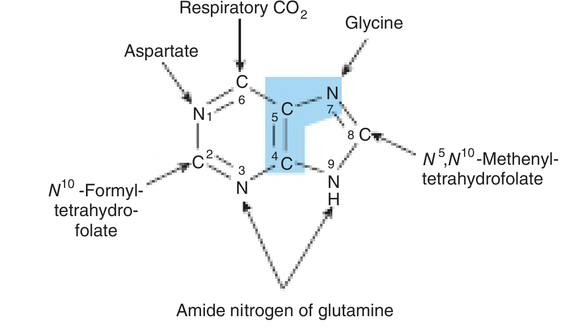
Key Steps
- PRPP synthesis (ribose-5-phosphate + 2 ATP → PRPP) by PRPP synthetase
- Glutamine donates amide N → 5-phosphoribosylamine (committed step, catalyzed by PRPP glutamyl amidotransferase)
- Sequential additions of glycine, formyl-THF (C8), glutamine (N3), CO₂ (C6), aspartate (N1), formyl-THF (C2) → closes the ring → IMP
- From IMP:
- IMP → AMP: adenylosuccinate synthetase (GTP-dependent) → adenylosuccinate lyase
- IMP → GMP: IMP dehydrogenase → GMP synthetase (ATP-dependent)
- Cross-regulation: AMP synthesis requires GTP; GMP synthesis requires ATP (mutual balance)
Regulation
- Rate-limiting factor: concentration of PRPP
- PRPP synthetase inhibited by AMP, ADP, GMP, GDP (feedback)
- PRPP glutamyl amidotransferase inhibited by AMP and GMP (feedback)
- Multifunctional enzymes in eukaryotes facilitate channeling of intermediates
Pharmacological Inhibition (anticancer/antifolate drugs)
- Methotrexate, aminopterin — inhibit dihydrofolate reductase → deplete THF → block steps 4 (C8 addition) and 10 (C2 addition)
- Azaserine, DON (6-diazo-5-oxo-L-norleucine) — glutamine analogs → block steps 2, 5, 8
- Mycophenolate mofetil (MMF) — inhibits IMP dehydrogenase → blocks de novo GMP synthesis → selectively affects T and B lymphocytes (which lack efficient salvage pathway)
4. PURINE SALVAGE PATHWAY ⭐⭐
Overview
- Recycles free purine bases (from diet or nucleic acid degradation) back into nucleotides
- Far less energy-intensive than de novo synthesis
- Particularly important in tissues with low de novo capacity: brain, RBCs, PMNs
Key Enzymes
| Enzyme | Reaction | Clinical Relevance |
|---|---|---|
| HGPRT (hypoxanthine-guanine phosphoribosyl transferase) | Hypoxanthine + PRPP → IMP; Guanine + PRPP → GMP | Deficient in Lesch-Nyhan |
| APRT (adenine phosphoribosyl transferase) | Adenine + PRPP → AMP | Deficiency → 2,8-dihydroxyadenine stones |
| Adenosine kinase | Adenosine + ATP → AMP + ADP | — |
| Deoxycytidine kinase | Phosphorylates dCyd, dGuo → dCMP, dGMP | — |
Mechanism
Purine base + PRPP → Purine-5'-monophosphate + PPi
(phosphoribosylation)
Regulation Feedback
- Salvage-generated AMP, GMP, IMP feedback inhibit PRPP synthetase and PRPP glutamyl amidotransferase, suppressing de novo synthesis
- This is why HGPRT deficiency → ↑ free PRPP → ↑ de novo synthesis → ↑ uric acid
Clinical Relevance
- Lesch-Nyhan syndrome — HGPRT deficiency (see above)
- ADA deficiency (adenosine deaminase) — accumulation of dATP inhibits ribonucleotide reductase → severe combined immunodeficiency (SCID)
- PNP deficiency (purine nucleoside phosphorylase) — dGTP accumulates → T-cell deficiency, normal B cells
- Azathioprine — prodrug metabolized to 6-mercaptopurine → incorporated via salvage → inhibits de novo purine synthesis → immunosuppression
5. VITAMIN D
Synthesis & Activation (Two-Step Hydroxylation)
7-Dehydrocholesterol (skin)
        ↓ UV light (290–320 nm)
Previtamin D3 → Vitamin D3 (cholecalciferol)  [enters dermal capillaries]
        ↓ Liver — 25-hydroxylase
25-hydroxyvitamin D [25(OH)D3] — storage form, measured in serum
        ↓ Kidney — 1α-hydroxylase (regulated by PTH, hypophosphatemia)
1,25-dihydroxyvitamin D3 = Calcitriol (active form)
- Dietary sources: fortified milk, fatty fish (salmon, sardines, tuna, cod), fish oil
- Ergocalciferol (D2) from plant sources joins the same pathway at the liver step
Actions of Calcitriol (1,25(OH)₂D₃)
- Intestine: ↑ calcium and phosphate absorption (nuclear receptor → transcription of calbindin)
- Bone: stimulates osteoclast-mediated bone resorption → releases Ca²⁺ and PO₄
- Kidney: ↑ tubular reabsorption of Ca²⁺ and phosphate
- Immune: activates macrophages via Toll-like receptor-triggered VDR expression; protective against TB
Deficiency — Rickets/Osteomalacia
- Children: Rickets — failure of bone mineralization, bowing of long bones, rachitic rosary, craniotabes
- Adults: Osteomalacia — bone pain, proximal myopathy, insufficiency fractures
High-risk groups: exclusively breastfed dark-skinned infants, elderly housebound, malabsorption (celiac, Crohn, post-gastrectomy), anticonvulsant use (induces hepatic catabolism), chronic renal failure, dark-skinned individuals in low-sun areas
Genetic Disorders of Vitamin D Metabolism
| Type | Defect | Treatment |
|---|---|---|
| Vitamin D-Dependent Rickets Type I (VDDR-I) | Autosomal recessive; deficient renal 1α-hydroxylase | Calcitriol supplementation |
| Vitamin D-Dependent Rickets Type II (VDDR-II) | Autosomal recessive; end-organ resistance (VDR mutation) | High-dose calcitriol + calcium |
Emerging Associations
- ↓ vitamin D linked to: hypertension, ↑ fasting glucose/insulin, cardiovascular disease, hip fractures, colon cancer mortality, ↑ all-cause mortality
6. IMPORTANT ANTIOXIDANT VITAMINS
Vitamin C (Ascorbic Acid)
- Water-soluble antioxidant
- Directly scavenges ROS (superoxide, hydroxyl radicals, singlet oxygen)
- Regenerates vitamin E (reduces tocopheroxyl radical back to tocopherol)
- Co-factor for prolyl and lysyl hydroxylase (collagen synthesis) → deficiency = scurvy
- Scurvy: perifollicular hemorrhages, corkscrew hairs, gingival bleeding/swelling, impaired wound healing
Vitamin E (Tocopherols/Tocotrienols)
- Fat-soluble antioxidant — major lipid-phase antioxidant
- α-tocopherol = most biologically active form
- Protects polyunsaturated fatty acids (PUFAs) in cell membranes from lipid peroxidation
- Mechanism: donates H· to lipid peroxy radical → chain-breaking antioxidant
- Deficiency: hemolytic anemia, spinocerebellar ataxia (in children with fat malabsorption), peripheral neuropathy
- Found in vegetable oils, nuts, seeds, green leafy vegetables
7. VITAMIN B12 (COBALAMIN) ⭐⭐
Structure & Sources
- Cobalt-containing vitamin; animal products only (meat, liver, dairy, eggs)
- Plant-based diets → risk of deficiency without supplementation
Absorption
- Requires Intrinsic Factor (IF) — secreted by gastric parietal cells (Castle's intrinsic factor)
- IF-B12 complex → absorbed at terminal ileum
- Transported by transcobalamin II in plasma
Causes of Deficiency
- Pernicious anemia — autoimmune destruction of parietal cells → ↓ IF → B12 malabsorption
- Gastrectomy, ileal resection, Crohn's disease (terminal ileum)
- Strict veganism
- Drugs: metformin (↓ IF-B12 absorption), prolonged PPI use, nitrous oxide (oxidizes cobalamin)
Functions
- Methylmalonyl-CoA mutase — converts methylmalonyl-CoA → succinyl-CoA (requires adenosylcobalamin)
- Deficiency → ↑ methylmalonic acid (MMA) — marker of B12 deficiency
- Methionine synthase — converts homocysteine → methionine (requires methylcobalamin); regenerates THF from methyl-THF ("methyl-folate trap")
- Deficiency → ↑ homocysteine; impaired myelin synthesis
Clinical Features of Deficiency
- Hematologic: macrocytic megaloblastic anemia (impaired DNA synthesis in RBCs), hypersegmented neutrophils
- Neurological (subacute combined degeneration of spinal cord): demyelination of posterior columns (vibration/position sense loss) + lateral corticospinal tracts (UMN signs); peripheral neuropathy
- NB: Folate deficiency also causes megaloblastic anemia but NO neurological features
Diagnosis
- ↑ MCV; megaloblastic changes on blood smear
- ↓ Serum B12
- ↑ Serum methylmalonic acid (most sensitive) + ↑ homocysteine
Treatment
- IM cyanocobalamin or hydroxocobalamin (if absorption defective)
- Oral high-dose B12 (1000 µg/day) if absorption intact
8. VITAMIN B9 (FOLATE) ⭐⭐
Sources & Absorption
- Liver, leafy green vegetables, legumes, wheat bran, fortified cereals
- Absorbed in jejunum (proximal small bowel)
- Body stores last only 3–4 months (unlike B12 which lasts years)
Active Form
- Tetrahydrofolate (THF) — carrier of single-carbon units
- Used in: purine synthesis (C2, C8 of purine ring), thymidylate synthesis (dUMP → dTMP), amino acid metabolism (serine ↔ glycine interconversion, homocysteine → methionine)
Causes of Deficiency
- Poor diet (alcoholism, poverty)
- Malabsorption: celiac disease, chronic diarrhea, total gastrectomy
- Antifolate drugs:
- Methotrexate, trimethoprim, pyrimethamine (inhibit dihydrofolate reductase)
- Oral contraceptives
- Antiepileptics (phenobarbital, phenytoin — induce hepatic enzymes, deplete folate)
- Increased demand: pregnancy, hemolytic anemia, malignancy
Clinical Features
- Megaloblastic anemia + hypersegmented neutrophils (same as B12)
- Glossitis (smooth tongue), angular cheilitis, mucosal ulcers
- Brown hyperpigmentation in palmar creases and flexures
- No neurological symptoms (key distinction from B12 deficiency)
- Neural tube defects (spina bifida, anencephaly) — with periconceptional deficiency
Treatment
- Folic acid 1–5 mg/day orally
- ⚠️ Must rule out B12 deficiency first — folate supplementation corrects anemia but allows neurological damage from B12 deficiency to progress undetected ("masking")
Supplementation
- All women of childbearing age: 400 µg/day folic acid to prevent neural tube defects
- High-risk women (prior NTD, antiepileptic use): 4–5 mg/day
Quick Summary Table
| Topic | Key Enzyme/Mechanism | Clinical Consequence |
|---|---|---|
| Gout | ↑ Uric acid → MSU crystals → NLRP3 inflammasome → IL-1β | Acute arthritis, tophi, nephropathy |
| Lesch-Nyhan | HGPRT absent → ↑ PRPP → ↑ uric acid | Self-mutilation, choreoathetosis, hyperuricemia |
| De novo purine | PRPP → IMP (11 steps); needs THF, glycine, glutamine, aspartate | Blocked by methotrexate, MMF |
| Salvage purine | HGPRT/APRT recycle free purines + PRPP → nucleotides | HGPRT deficiency = Lesch-Nyhan; ADA deficiency = SCID |
| Vitamin D | 7-DHC → D3 → 25(OH)D (liver) → 1,25(OH)₂D (kidney) | Rickets (children), osteomalacia (adults) |
| Vitamin E | Lipid-phase antioxidant; breaks lipid peroxidation chains | Hemolytic anemia, spinocerebellar ataxia |
| Vitamin C | Water-phase antioxidant; regenerates Vit E; collagen | Scurvy |
| Vitamin B12 | Methylmalonyl-CoA mutase + methionine synthase | Megaloblastic anemia + subacute combined degeneration |
| Vitamin B9 | THF → single-carbon transfers for purine/dTMP synthesis | Megaloblastic anemia (no neuro), neural tube defects |
Sources: Robbins & Kumar Basic Pathology; Harper's Illustrated Biochemistry 32nd Ed; Adams & Victor's Principles of Neurology 12th Ed; Fitzpatrick's Dermatology; Comprehensive Clinical Nephrology 7th Ed
This is a shared conversation. Sign in to Orris to start your own chat.