
Allergic conjunctivitis treatment
"allergic conjunctivitis"[MeSH Terms] AND treatment
allergic conjunctivitis eye clinical appearance
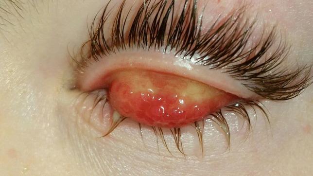
This clinical photograph displays a close-up view of a human eye with the upper eyelid everted to reveal the palpebral (tarsal) conjunctiva. The primary clinical finding is the presence of giant tarsal papillae, which are large, elevated, and closely packed, creating a distinctive 'cobblestone' appearance. The conjunctiva is highly hyperemic, exhibiting intense erythema, and shows interspersed areas of pale yellowish-white inflammatory exudate. The surrounding eyelids and eyelashes appear moist, suggestive of excessive lacrimation or mucoid discharge. This presentation is highly characteristic of the palpebral form of Vernal Keratoconjunctivitis (VKC), a chronic allergic inflammatory disease of the ocular surface. The image is a quintessential educational example of severe allergic conjunctivitis findings used in ophthalmology to differentiate inflammatory ocular surface disorders.
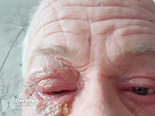
Clinical photograph of the anterior ocular adnexa showing severe blepharoconjunctivitis. Modality indicates ophthalmic photography; a close‑up frontal view of the eyelids and bulbar conjunctiva. The image depicts marked edema of the eyelid margins with diffuse hyperemia of the palpebral and bulbar conjunctiva, lash line crusting, and mucopurulent debris along the lash bases. There is conspicuous erythema, lid edema, and tearing. The anterior segment appears otherwise intact, with no obvious corneal involvement visible in this field. The appearance is consistent with acute inflammatory eyelid disease, blepharitis with conjunctivitis, and potential Dupilumab-associated ocular surface disease (OAD) given proximal timing to dupilumab initiation and rapid onset within eight weeks. Differential considerations include allergic blepharoconjunctivitis, seborrheic blepharitis, bacterial conjunctivitis, and Meibomian gland dysfunction–driven eye surface inflammation. Clinically, this image supports recognizing with high suspicion an adverse drug reaction to dupilumab presenting as ocular surface inflammation; management would focus on topical therapies, lubrication, eyelid hygiene, possible topical antibiotics if crusting/mucopurulent discharge, and coordinated care with dermatology and ophthalmology. The image is valuable for medical education, case-based review, pharmacovigilance, and research into dupilumab’s ocular adverse events and response to therapies. This documentation supports correlation with clinical history and treatment response.
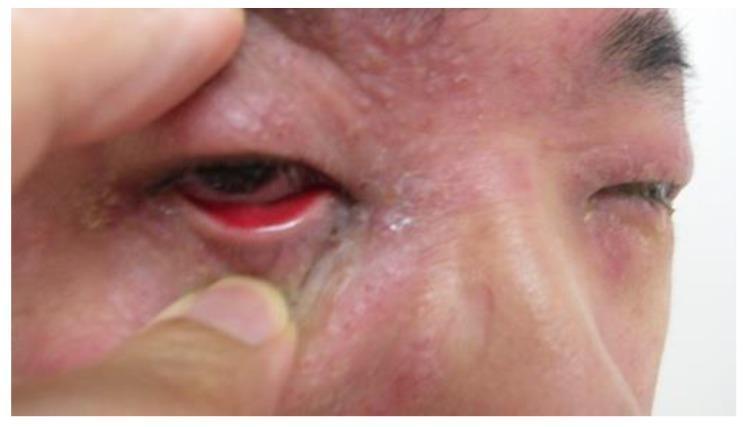
This clinical photograph shows a close-up view of a patient presenting with binocular conjunctivitis. The primary visual finding is severe, diffuse conjunctival injection (redness) and significant swelling of the eyelids (blepharedema) in both eyes. In the patient's right eye (viewer's left), the lower eyelid is manually retracted, revealing deep erythema of the palpebral conjunctiva and possible chemosis. Mucopurulent or watery discharge is visible along the lid margins and inner canthus. The surrounding periocular skin exhibits erythema and a textured, papular appearance, suggesting associated dermatitis or an allergic component such as atopic keratoconjunctivitis (AKC). The educational focus of this image is the clinical presentation of bilateral ocular inflammation, highlighting the key diagnostic signs of conjunctival hyperemia, lid edema, and ocular discharge essential for distinguishing between viral, bacterial, and allergic conjunctivitis.
Allergic Conjunctivitis: Treatment
Types at a Glance
| Type | Timing | Key Features |
|---|---|---|
| Acute allergic | Acute (pollen, grass) | Sudden chemosis, itching, watering |
| Seasonal (SAC) | Spring/summer | Pollen-driven, papillary reaction |
| Perennial (PAC) | Year-round | Dust mites, dander, fungi |
| Vernal (VKC) | Warm seasons | Boys, cobblestone papillae, corneal risk |
| Atopic (AKC) | Year-round | Adults, atopic dermatitis, corneal scarring |
Step-by-Step Treatment Ladder
1. Non-Pharmacological (First Line - All Types)
- Allergen avoidance - the most effective long-term strategy
- Cool compresses - provide immediate symptomatic relief for itching and chemosis
- Artificial tears / lubricant eye drops - dilute allergens and wash out mediators; suitable for mild symptoms
2. Topical Pharmacotherapy
- Examples: sodium cromoglicate, nedocromil sodium, lodoxamide, pemirolast (4x/day during allergic season)
- Must be started a few days before exposure for maximal effect; suitable for prophylaxis and long-term use
- Note: lodoxamide is not recommended for long-term use
- Examples: emedastine, epinastine, levocabastine, bepotastine
- Fast-acting; effective for symptomatic exacerbations; comparable efficacy to mast cell stabilizers
- Examples: olopatadine (1-2 drops once or twice daily), ketotifen (1 drop every 8-12 hours), azelastine
- Act rapidly AND prevent mast cell degranulation - particularly effective for exacerbations
- Olopatadine is considered among the most effective agents for both acute and chronic symptoms
- Example: diclofenac
- Provide adjunctive symptomatic relief; rarely used as primary agents
- Provide short-term symptomatic relief; not recommended for prolonged use
3. Topical Corticosteroids
- Reserved for moderate-to-severe cases not responding to the above
- Effective but associated with risks: raised IOP, cataract, secondary infection
- Short courses for acute flares; need ophthalmologic supervision
- In VKC/AKC: often required during severe exacerbations (e.g., prednisolone drops)
4. Systemic Therapy
- Oral antihistamines (e.g., loratadine, cetirizine 5-10 mg/day, diphenhydramine): useful for severe symptoms; note that some (especially diphenhydramine) cause significant drowsiness; oral antihistamines can also cause ocular dryness, potentially worsening symptoms
- Sedating antihistamines (diphenhydramine) may be useful specifically for nighttime relief/sleep
Vernal Keratoconjunctivitis (VKC) - Specific Treatment
- Mast cell stabilizers as long-term background therapy (cromoglicate, lodoxamide)
- Dual-action topical agents (olopatadine, ketotifen) for ongoing control
- Topical corticosteroids during severe flares - short courses essential; monitor IOP
- Topical ciclosporin (cyclosporine) 1-2% - effective steroid-sparing agent for moderate-to-severe VKC; reduces papillary hypertrophy and corneal disease
- Supratarsal injection of corticosteroid - may be used for refractory giant papillae
- Surgical removal of giant papillae or keratectomy for corneal plaques if vision is affected
- Cold compresses and cold environment (AC) help due to the condition's heat sensitivity
Atopic Keratoconjunctivitis (AKC) - Specific Considerations
- More common in adults; associated with atopic dermatitis
- Ciclosporin drops are an important steroid-sparing agent
- Risk of corneal scarring, vascularization, and perforation - requires close ophthalmologic follow-up
- Treat associated lid disease (blepharitis, staphylococcal colonization) with lid hygiene and topical antibiotics
Drug Reference Summary
| Drug Class | Examples | Use |
|---|---|---|
| Mast cell stabilizers | Sodium cromoglicate, lodoxamide, nedocromil, pemirolast | Prophylaxis, long-term |
| Topical antihistamines | Emedastine, epinastine, levocabastine, bepotastine | Acute exacerbations |
| Dual-action | Olopatadine, ketotifen, azelastine | Acute + prophylaxis |
| Topical steroids | Prednisolone, fluorometholone | Severe/acute flares only |
| Topical NSAIDs | Diclofenac | Adjunctive relief |
| Topical ciclosporin | Ciclosporin 1-2% | VKC/AKC, steroid-sparing |
| Oral antihistamines | Loratadine, cetirizine, diphenhydramine | Severe systemic symptoms |
Clinical Image
Key Points
- Olopatadine (dual-action) is one of the most effective and commonly used agents for both treatment and prophylaxis
- Mast cell stabilizers should be started before allergen season begins, not after symptoms appear
- Topical steroids are effective but should be used only short-term under ophthalmologic supervision
- Oral antihistamines can worsen dry eye, so topical therapy is preferred
- VKC and AKC are more serious forms requiring specialist input to prevent corneal scarring and vision loss