
Anatomical variations of frontal sinus in detail and simplified manner from standard ENT textbook
I'll read the medical library skill first, then search for frontal sinus anatomy and variations.
Reading File
Now let me search the library in parallel for frontal sinus anatomy and variations:
Finding Sources
Finding Sources
Finding Sources
Excellent! I have clear leads. Now let me read the key sections in parallel from Cummings and Scott-Brown's (the top ENT textbooks):
Reading File
Reading File
Reading File
Good content loading. Let me continue reading the Cummings section on frontal cells (Kuhn classification) and the Scott-Brown section on the FRONTAL SINUS heading that had the detailed classification table:
Reading File
Reading File
Reading File
Excellent data. Let me now read a bit more of the KJ Lee section (IFAC classification details), the Cummings aplasia/hypoplasia section, and get the Scott-Brown classification table:
Reading File
Reading File
Reading File
Excellent! I have very comprehensive data now. Let me also fetch the key anatomy diagram from Cummings and search for a relevant image:
Loading Image
Loading Image
Searching Images
frontal sinus cells variations anatomy Kuhn classification
I have outstanding material with excellent diagrams from Cummings. Now I have everything I need to write a comprehensive, well-structured answer. Let me compile it.
Anatomical Variations of the Frontal Sinus
Sources: Cummings Otolaryngology Head and Neck Surgery; Scott-Brown's Otorhinolaryngology Head & Neck Surgery Vol. 1; K.J. Lee's Essential Otolaryngology
1. Overview and Embryological Basis
The frontal sinus originates embryologically from an anterior ethmoidal cell - it is NOT a separate bony cavity from the start. Pneumatization of the frontal bone begins around the 16th week of gestation. At birth it exists as only a small blind pocket that is difficult to distinguish from the anterior ethmoid air cells on imaging. Radiologically visible frontal sinuses appear by age 8; significant pneumatization occurs in early adolescence and continues until about 18 years of age.
Because the sinus develops from anterior ethmoid cells, it does not have a true two-dimensional opening ("ostium") but instead connects to the anterior ethmoid complex through an hourglass-shaped space called the frontal sinus recess (FSR). The narrowest point of this hourglass is conventionally called the "frontal sinus ostium (FSO)" - though more accurately it is the waist of a continuous 3-D space, not a real ostium.
2. Basic Anatomy of the Frontal Recess (The Foundation for Understanding Variations)
The frontal recess boundaries:
| Direction | Boundary |
|---|---|
| Anterior | Frontal beak / Agger nasi cell |
| Medial | Lateral lamella / Middle turbinate |
| Lateral | Lamina papyracea |
| Posterior | Bulla ethmoidalis / Bulla lamella / Fovea ethmoidalis |
| Posterosuperior | Anterior skull base |
The frontal beak is the thick bone of the frontal process of the maxilla that projects posteriorly into the frontal recess, largely determining its anteroposterior dimension. The degree of pneumatization of the agger nasi cell determines the thickness of this beak and the position of the superior uncinate process.
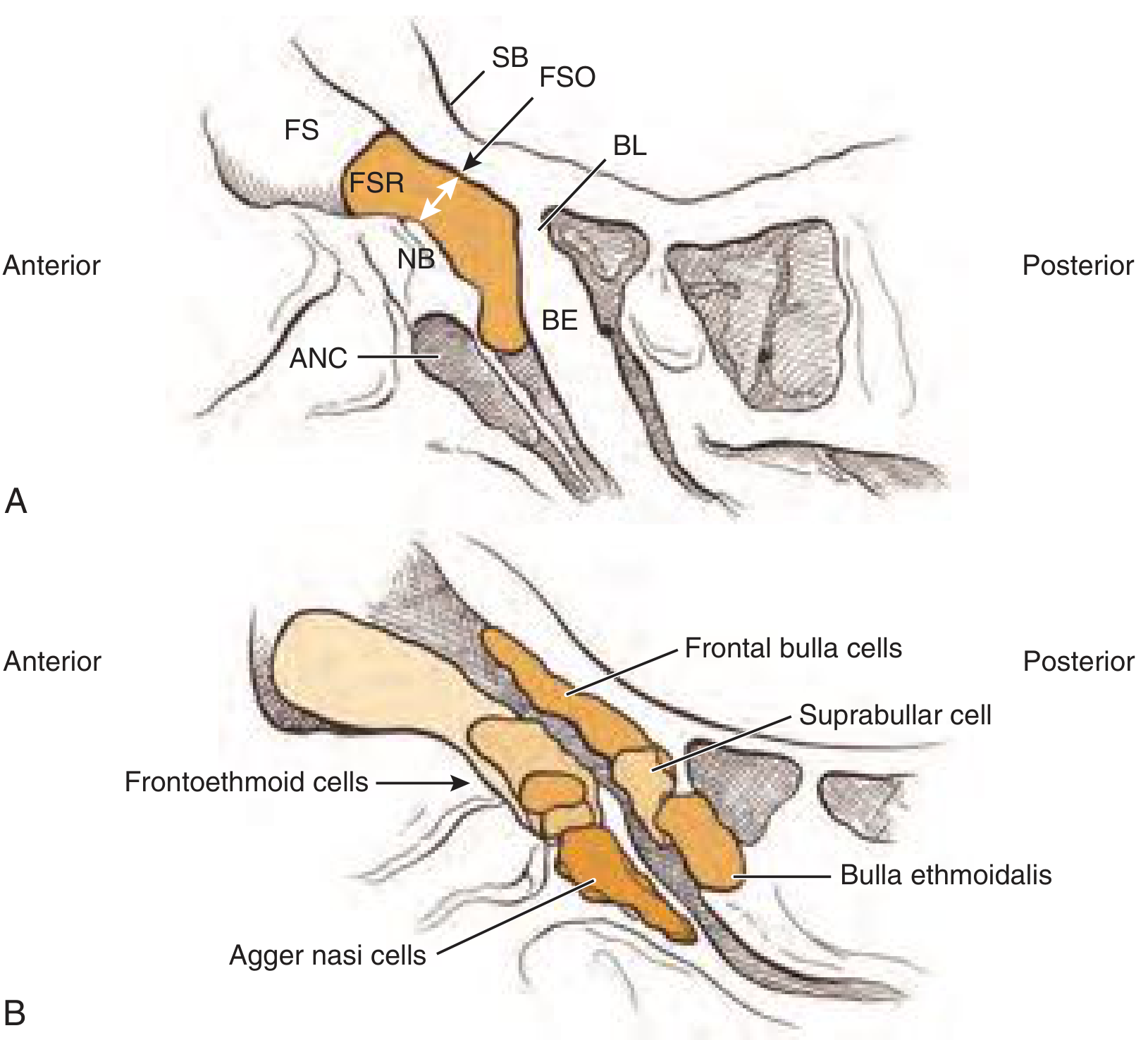
The frontal sinus recess (FSR, shaded orange) is an hourglass-shaped space with its narrowest waist at the frontal sinus ostium (FSO). The agger nasi cell (ANC) and nasal beak (NB) lie anteriorly; the bulla ethmoidalis (BE) and bulla lamella (BL) lie posteriorly.
3. Major Anatomical Variations
A. Variations in Pneumatization / Size
This is the most variable of all paranasal sinuses. Three broad patterns exist:
| Pattern | Description | Frequency |
|---|---|---|
| Aplasia | Complete absence of frontal sinus development (unilateral or bilateral) | 10-52% (varies by ethnicity) |
| Hypoplasia | Underdeveloped, very small sinus | Common |
| Asymmetry | Two frontal sinuses of markedly different sizes, separated by a deviated intersinus septum | Very common (normal finding) |
"The greatest variation in pneumatization is shown by the frontal sinus, which can be aplastic in 10% to 52% of cases based on ethnicity with either unilateral or bilateral aplasia." - Cummings Otolaryngology, p.790
B. Uncinate Process Attachment (Key Variation for Drainage Pathway)
The superior attachment of the uncinate process determines where the frontal sinus drains:
| Attachment | Drainage pathway | Frequency |
|---|---|---|
| Medial orbital wall (lamina papyracea) | Frontal recess drains medial to uncinate | 85% (most common) |
| Skull base or middle turbinate (isolated) | Frontal recess drains lateral to uncinate | 15% |
The uncinate process has multiple attachments in more than 50% of cases rather than a single attachment. Classic teaching describing only three distinct attachment patterns is neither surgically accurate nor relevant. - Scott-Brown's Vol.1, p. (block10)
Surgical rule: The frontal recess is medial to the remnant uncinate process ("vertical bar") in 85% of cases.
C. Frontoethmoidal Cells (Most Clinically Important Variation)
These are anterior ethmoidal cells that pneumatize around the frontal recess. They can obstruct the frontal sinus drainage pathway. Three classification systems exist:
Classification System 1: Kuhn Classification (Original)
| Type | Description |
|---|---|
| Type 1 | Single cell superior to the agger nasi cell (does NOT enter the frontal sinus) |
| Type 2 | A tier (2 or more cells) above the agger nasi cell (does NOT enter the frontal sinus) |
| Type 3 | Single cell that extends from the agger nasi into the frontal sinus |
| Type 4 | An isolated single cell entirely within the frontal sinus (no apparent connection to recess) |
Classification System 2: Modified Kuhn / Wormald Classification
| Type | Description | Change from Kuhn |
|---|---|---|
| Type 1 | Same as Kuhn | No change |
| Type 2 | Same as Kuhn | No change |
| Type 3 | Cell extending into the frontal sinus <50% of vertical sinus height | Modified |
| Type 4 | Cell extending into the frontal sinus >50% of vertical sinus height | Modified |
Reason for modification: The original Kuhn Type 4 (truly isolated cell) is extremely rare. Modern parasagittal CT imaging shows that most "Type 4" cells actually drain into the frontal recess. Wormald therefore re-defined Types 3 and 4 based on the percentage of frontal sinus height occupied.
Classification System 3: International Frontal Sinus Anatomy Classification (IFAC) - Current Standard
This system replaced eponymous types with descriptive anatomic names:
| IFAC Term | Equivalent Kuhn Type | Location | Description |
|---|---|---|---|
| Supra agger cell | Type 1/2 | Anterior | Anterior-lateral cell above the agger nasi; does NOT enter frontal sinus |
| Supra agger frontal cell | Type 3/4 | Anterior | Anterior-lateral cell that extends INTO the frontal sinus |
| Supra bulla cell | Suprabullar | Posterior | Cell above the bulla ethmoidalis; does NOT enter frontal sinus |
| Supra bulla frontal cell | Frontal bulla cell | Posterior | Cell originating above the bulla that pneumatizes along the skull base INTO the frontal sinus (pushes drainage pathway anteriorly) |
| Frontal septal cell | Intersinus septal cell | Medial | Pneumatizes the intersinus septum; drains into one frontal sinus; displaces the frontal drainage pathway laterally |
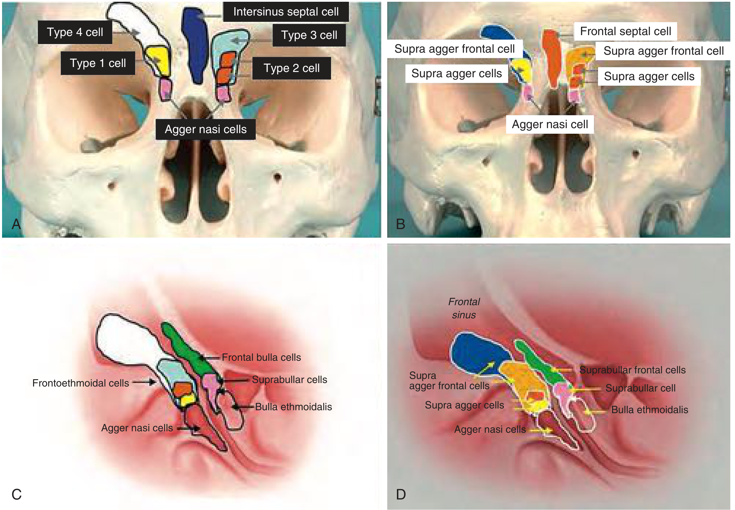
(A, C) Kuhn nomenclature; (B, D) Corresponding IFAC nomenclature. Note the colour-coded cell types around the agger nasi and their relationship to the frontal sinus.
D. Agger Nasi Cell
- The anteriormost ethmoid air cell; present in virtually all individuals
- Its degree of pneumatization determines the anteroposterior dimension of the frontal recess and the thickness of the frontal beak
- A well-pneumatized agger nasi narrows the frontal recess from the anterior side
E. Supraorbital Ethmoid Cells (SOEC)
- Posterior to the frontal sinus, the orbital plate of the frontal bone can be extensively pneumatized, producing supraorbital ethmoid cells
- Present in approximately 65% of Caucasian patients; much lower incidence in East Asian populations (ethnic variation)
- These cells extend across the roof of the orbit, posterior to the frontal recess
Surgical hazards of SOEC:
- The anterior ethmoid artery is more likely to be present in a mesentery (unsupported by bone) and is highly vulnerable to injury
- A larger area of orbit and skull base is exposed within the surgical field
- The opening of SOEC can be mistaken for the frontal sinus opening - SOEC opens more posteriorly and laterally than the true frontal sinus
F. Intersinus Septal Cell (Frontal Septal Cell)
- Pneumatization of the interfrontal sinus septum
- Drains into one frontal sinus, medial to the frontal ostium
- Displaces the frontal drainage pathway laterally
- May extend into the crista galli
G. Suprabullar and Frontal Bulla Cells
| Cell | Origin | Effect |
|---|---|---|
| Suprabullar cell | Superior to ethmoidal bulla | May narrow posterior frontal recess |
| Frontal bulla cell | Bulla itself, pneumatizing into posterior frontal table/anterior skull base | Pushes drainage pathway anteriorly; can mimic a Type 4 Kuhn cell on coronal CT |
Large frontal bulla cells can appear as isolated cells within the frontal sinus and be mistaken for a type 4 cell on coronal CT - they are readily distinguished on axial images.
4. Simplified Summary Table
| Variation | Where? | Effect | Key Feature |
|---|---|---|---|
| Aplasia/Absent sinus | No frontal sinus at all | No drainage issue (nothing to drain!) | 10-52% by ethnicity |
| Hypoplasia | Tiny sinus | Prone to obstruction | Common |
| Asymmetry | Two unequal sinuses | Normal variant | Very common |
| Uncinate attachment to LP (85%) | Frontal recess medial to uncinate | Normal drainage | Most common |
| Agger nasi hypertrophy | Narrows frontal recess from front | Obstruction risk | Anteriormost ethmoid cell |
| Type 1 frontal cell | Above agger nasi, single | May narrow recess | Kuhn/Wormald |
| Type 2 frontal cells | Above agger nasi, multiple tiers | Greater narrowing | Kuhn/Wormald |
| Type 3 frontal cell | Extends <50% into frontal sinus | Obstructs outflow | Can be removed endoscopically |
| Type 4 frontal cell | Extends >50% into frontal sinus | Major obstruction | May need extended Lothrop |
| Supraorbital ethmoid cell | Posterior, over orbital roof | Danger zone for AEA | 65% Caucasians |
| Intersinus septal cell | Medial, in septum | Lateralizes drainage | Displaces outflow laterally |
| Frontal bulla cell | Posterior, from bulla | Pushes drainage anteriorly | Can mimic Type 4 |
5. Clinical Relevance (Why These Variations Matter)
-
Surgical planning: CT scan must be carefully reviewed preoperatively to identify agger nasi cell, uncinate attachment, lamina papyracea, height of lateral lamella, and position of the anterior ethmoid artery.
-
Risk of obstruction: The frontal recess is the narrowest and most complex of all sinus drainage pathways. Minor mucosal swelling in the setting of anatomic crowding (multiple frontal cells) can precipitate chronic frontal sinusitis.
-
However, anatomic variations are NOT significantly more common in CRS patients than in controls - meaning anatomy alone rarely causes disease. In diffuse bilateral disease, mucosal pathology and host immunity are primary.
-
Anterior ethmoid artery (AEA) on mesentery: This dangerous variation - where the AEA is not protected by a bony canal but hangs freely in the ethmoid space - is significantly more common when SOEC are present. Retraction or injury causes severe bleeding and possible orbital hematoma.
-
The "building block" approach (Wormald): Mentally visualize the frontal recess in 3D as individual cell "building blocks" around the surgical box - helps surgeons plan dissection safely.
References:
- Cummings Otolaryngology Head and Neck Surgery, Frontal Sinus chapter, pp. 871-872 (block9) and Surgical Anatomy, pp. (block10)
- Scott-Brown's Otorhinolaryngology Head & Neck Surgery Vol. 1, Ch. 87 and Ch. 99 (block9, block10)
- K.J. Lee's Essential Otolaryngology, Keros Classification and Frontal Sinus Cells section (block3)
This is a shared conversation. Sign in to Orris to start your own chat.