
Explain the mechanism of acute respiratory distress syndrome
Reading File
ARDS involves diffuse alveolar damage driven by an overwhelming inflammatory cascade that disrupts the alveolar-capillary barrier, leading to non-cardiogenic pulmonary edema, refractory hypoxemia, and reduced lung compliance.
Searching sources...
Finding Sources
Searching PubMed
"acute respiratory distress syndrome" AND pathogenesis
Good. I have strong textbook hits. Let me read the most comprehensive sections in parallel - the Goldman-Cecil and Murray & Nadel chapters, plus the Sabiston block.
Reading File
Reading File
Reading File
Now let me get the dedicated ARDS chapter from Murray & Nadel (block 32) and the Goldman-Cecil ARDS chapter directly.
Reading File
Finding Sources
Let me search specifically for the ARDS pathophysiology chapter in Murray & Nadel and Goldman-Cecil.
Finding Sources
Finding Sources
Finding Sources
Finding Sources
Excellent - I have Harrison's 22E (2025) content on ARDS. Let me now read the full ARDS section from Harrison's and Robbins.
Reading File
Reading File
I have excellent detail from Harrison's. Let me get the full mechanism section from Harrison's that covers the phases of ARDS.
Reading File
Let me find the pathophysiology and phases section from Harrison's ARDS chapter.
Reading File
I now have comprehensive material from Harrison's 22E (2025) covering the full ARDS mechanism. I also have data from Robbins, Murray & Nadel, Sabiston, and Goldman-Cecil. I have enough to write a thorough, well-organized answer.
Loading Image
I now have all the material I need. Here is the complete, well-organized answer.
Acute Respiratory Distress Syndrome (ARDS): Mechanism
Definition and Overview
ARDS is a clinical syndrome of severe, rapid-onset dyspnea, refractory hypoxemia, and diffuse bilateral pulmonary infiltrates leading to respiratory failure. It results from diffuse alveolar damage (DAD) caused by an overwhelming inflammatory response to a direct or indirect lung insult. By the Berlin (2012) definition, it is classified as:
- Mild: PaO2/FiO2 200-300 mmHg
- Moderate: PaO2/FiO2 100-200 mmHg
- Severe: PaO2/FiO2 <100 mmHg
(A 2024 global definition no longer requires arterial blood gas, accepting SpO2/FiO2 ratios and HFNO.)
Annual incidence is approximately 60 cases per 100,000 population; ARDS accounts for ~10% of all ICU admissions, with mortality commonly exceeding 30%.
- Harrison's Principles of Internal Medicine 22E (2025), Chapter 312
Causes: Direct vs. Indirect Lung Injury
| Direct Lung Injury | Indirect Lung Injury |
|---|---|
| Pneumonia | Sepsis |
| Aspiration of gastric contents | Severe trauma / multiple fractures |
| Pulmonary contusion | Head trauma |
| Near-drowning | Burns |
| Toxic inhalation | Multiple transfusions |
| Drug overdose | |
| Pancreatitis | |
| Post-cardiopulmonary bypass |
Over 80% of cases are caused by pneumonia, sepsis, aspiration, trauma, and multiple transfusions. - Harrison's 22E, Table 312-1
The Three Phases of ARDS
The natural history is divided into three pathologically distinct phases:
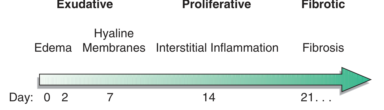
Time course of ARDS development and resolution - Harrison's 22E, Fig. 312-1
Phase 1 - Exudative Phase (Days 0-7)
This is the acute inflammatory phase and the core mechanism of lung injury.
1. Triggering Insult
Either a direct insult (e.g., pneumonia, aspiration) or an indirect, systemic insult (e.g., sepsis, pancreatitis, trauma) activates the innate immune response.
2. Neutrophil Recruitment and Activation
Neutrophils are the central effector cells of ARDS. They are recruited to the alveolar and interstitial spaces by chemoattractants, particularly IL-8. Once activated, neutrophils release:
- Proteases (elastase, matrix metalloproteinases) - directly digest structural proteins of the alveolar-capillary membrane
- Reactive oxygen species (ROS) - oxidize lipids and proteins, causing membrane peroxidation
- Platelet-activating factor (PAF) and leukotrienes - amplify inflammation and increase vascular permeability
"Neutrophils and their products have a central role in endothelial and epithelial injury." - Robbins Basic Pathology
3. Cytokine Storm
A coordinated release of proinflammatory mediators drives the cascade:
- TNF-α, IL-1β, IL-2, IL-6, IL-8 - amplify neutrophil activation and endothelial damage
- IL-6 levels correlate with mortality in ARDS and contribute to lung, liver, and gut injury
- Counter-regulatory anti-inflammatory cytokines (IL-4, IL-10, IL-13, TGF-β, PGE2) are also released but are overwhelmed in severe disease
Schwartz's Principles of Surgery; Harrison's 22E
4. Breakdown of the Alveolar-Capillary Barrier
The alveolar-capillary membrane has two critical layers:
- Capillary endothelium - damaged by cytokines and oxidants, becomes hyperpermeable
- Alveolar epithelium (type I and II pneumocytes) - type I cells are preferentially destroyed; type II cells (surfactant-producing, involved in ion transport and alveolar repair) are also injured
Disruption of both layers leads to:
- Protein-rich fluid flooding the alveolar space (non-cardiogenic pulmonary edema)
- Inactivation and dilution of surfactant, increasing alveolar surface tension
- Alveolar collapse (atelectasis), particularly in dependent lung zones
5. Hyaline Membrane Formation
Leaked plasma proteins (fibrin, fibrinogen, complement) precipitate along the denuded alveolar walls to form eosinophilic hyaline membranes - the histologic hallmark of ARDS. These membranes further impair gas exchange.
Physiological consequences of the exudative phase:
- Decreased functional residual capacity (FRC)
- Severely reduced lung compliance (stiff lungs) - the lung behaves like a wet sponge with gravity-dependent consolidation
- Intrapulmonary shunting (perfused but unventilated alveoli) causing refractory hypoxemia (does not correct well with supplemental O2 alone)
- Increased dead space ventilation
In pancreatitis-associated ARDS, there is an additional mechanism: pancreatic phospholipase A2 degrades surfactant directly, while elastase and lipase increase vascular permeability. - Murray & Nadel's Textbook of Respiratory Medicine
Phase 2 - Proliferative Phase (Days 7-14)
Most patients begin recovery during this phase, but some progress to fibrosis.
- Type II pneumocytes proliferate to line denuded alveolar walls (replacing destroyed type I cells)
- Interstitial infiltration by lymphocytes and macrophages replaces the neutrophilic exudate
- Organized fibrosis begins in some patients
- Clinically: some improvement in oxygenation, but persistent tachypnea and reduced lung compliance continue
Phase 3 - Fibrotic Phase (Day 21+)
Occurs in a minority of patients who fail to resolve the proliferative phase:
- Extensive collagen deposition and fibrosis obliterates normal alveolar architecture
- Bullae and cysts may form
- Results in chronic lung disease, pulmonary hypertension, and increased risk of barotrauma
- Mechanical ventilation can further perpetuate injury in this phase
Ventilator-Induced Lung Injury (VILI): A Critical Complication of Mechanism
The edematous, consolidated ARDS lung is extremely heterogeneous: dependent zones are atelectatic or fluid-filled; non-dependent zones remain relatively aerated. When a normal tidal volume (10-12 mL/kg) is applied, all ventilation enters this small "baby lung," causing:
- Volutrauma - overdistension of patent alveoli
- Barotrauma - excessive pressure injury
- Atelectrauma - repeated opening and collapse of alveoli at end-expiration
- Biotrauma - mechanical stretch releases further cytokines (IL-6, IL-8, TNF) into the systemic circulation, contributing to multi-organ dysfunction
This is why lung-protective ventilation (6 mL/kg predicted body weight, plateau pressure ≤30 cmH2O) is the cornerstone of management. - Goldman-Cecil Medicine; Harrison's 22E
Summary of Core Mechanism
Triggering insult (sepsis, pneumonia, aspiration, trauma)
        ↓
Activation of alveolar macrophages
        ↓
Release of TNF-α, IL-1β, IL-8, PAF
        ↓
Neutrophil recruitment and sequestration in alveoli
        ↓
Neutrophil degranulation: proteases + ROS + PAF + leukotrienes
        ↓
Endothelial + epithelial (type I pneumocyte) destruction
        ↓
Alveolar-capillary barrier breakdown
        ↓
Protein-rich edema floods alveoli → surfactant inactivation → hyaline membranes
        ↓
Atelectasis, shunting, decreased compliance, refractory hypoxemia
        ↓
Exudative (0-7d) → Proliferative (7-14d) → Fibrotic (21d+)
Key Cellular Players
| Cell / Mediator | Role in ARDS |
|---|---|
| Neutrophils | Primary effectors; release proteases, ROS, and PAF to destroy the alveolar-capillary barrier |
| Alveolar macrophages | Initiate the cytokine cascade; release TNF-α, IL-1, IL-8 |
| Type I pneumocytes | Preferentially destroyed; cover ~95% of alveolar surface |
| Type II pneumocytes | Surfactant production; attempt repair; susceptible to apoptosis |
| TNF-α / IL-1β | Master proinflammatory cytokines driving the cascade |
| IL-8 | Key neutrophil chemoattractant |
| IL-6 | Amplifies acute phase response; correlates with mortality |
| Phospholipase A2 | Degrades surfactant (especially in pancreatitis-associated ARDS) |
| PAF | Increases vascular permeability; activates platelets; amplifies inflammation |
| ROS | Lipid peroxidation of cell membranes |
Sources:
- Harrison's Principles of Internal Medicine 22E (2025), Chapter 312
- Robbins & Kumar Basic Pathology
- Murray & Nadel's Textbook of Respiratory Medicine, Chapter 134
- Goldman-Cecil Medicine, Chapter 90/91
- Schwartz's Principles of Surgery; Sabiston Textbook of Surgery
date?
Today is Wednesday, May 27, 2026.
This is a shared conversation. Sign in to Orris to start your own chat.