
Modular bipolar prosthesis vs regular bipolar prosthesis
modular bipolar prosthesis hip hemiarthroplasty
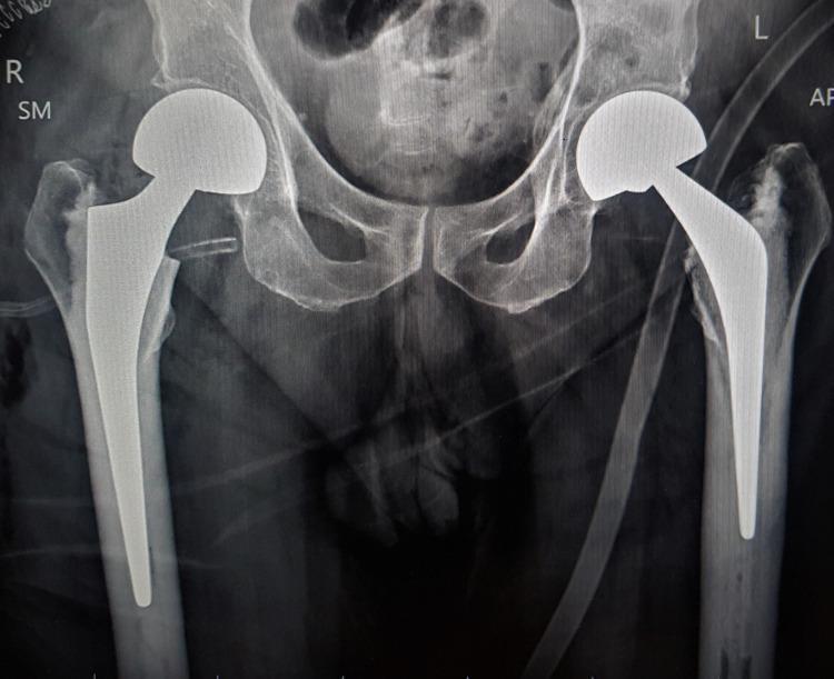
This anterior-posterior (AP) pelvic radiograph demonstrates bilateral hip hemiarthroplasty with bipolar prostheses in situ. Each prosthesis consists of a high-density, radiopaque femoral stem inserted into the medullary canal, a femoral neck, and a dual-bearing spherical head component articulating with the native acetabulum. The right hip features a recently implanted modular bipolar hemiarthroplasty, appearing well-aligned without signs of loosening. The left hip shows a long-standing cemented bipolar prosthesis, with visible cement mantle density around the femoral stem. The pelvic bony architecture, including the iliac wings, sacrum, and pubic symphysis, appears intact with age-related coarsening of the trabecular pattern. There is no evidence of periprosthetic fracture, osteolysis, or component migration. Soft tissue shadows show medical tubes and monitors consistent with an immediate postoperative setting. This image serves as a clinical example of surgical management for femoral neck fractures or advanced hip pathology using bilateral arthroplasty techniques.
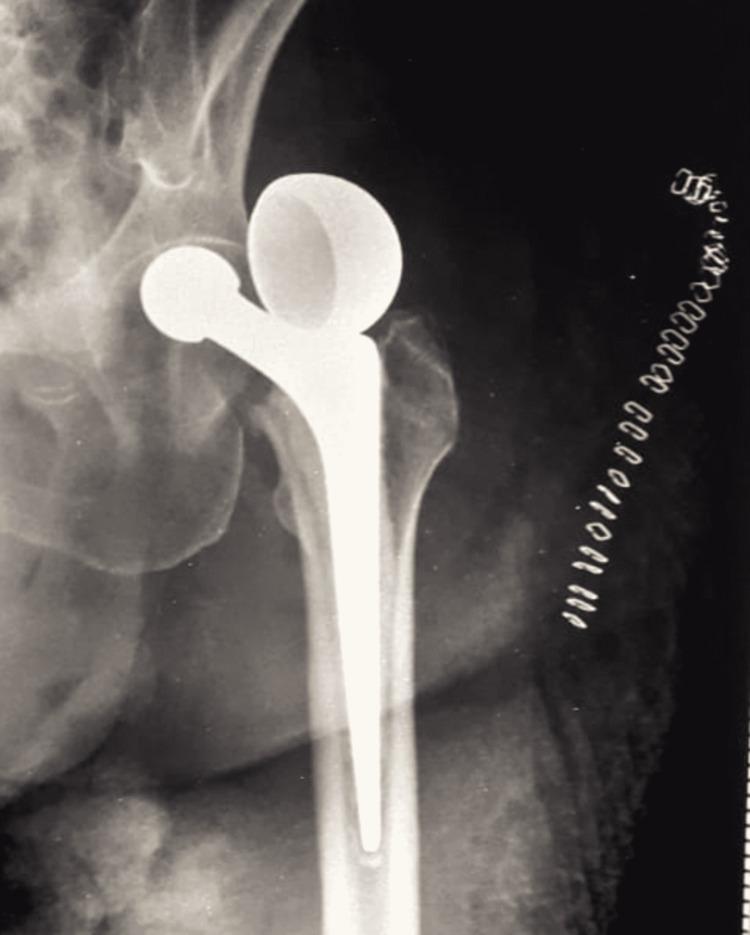
This diagnostic image is an anteroposterior plain X-ray of the left hip joint demonstrating a serious orthopedic complication following surgery. The image shows a bipolar hemiarthroplasty prosthesis with two distinct abnormalities: dislocation and uncoupling. The femoral stem remains situated within the femoral shaft; however, the bipolar prosthetic head has completely detached (uncoupled) from the femoral neck component. This dissociated prosthetic head is seen lying superior and lateral to the native acetabulum in the supracetabular soft tissues. Visible anatomical structures include the pelvis, the acetabular rim, and the proximal femur. Surgical skin staples are visible along the lateral soft tissue margin, indicating recent operative intervention. This case illustrates a rare failure mode of bipolar hip arthroplasty, where the modular components separate, leading to prosthetic dislocation and loss of joint articulation. It is a critical finding for medical students and residents in orthopedic surgery and emergency medicine.
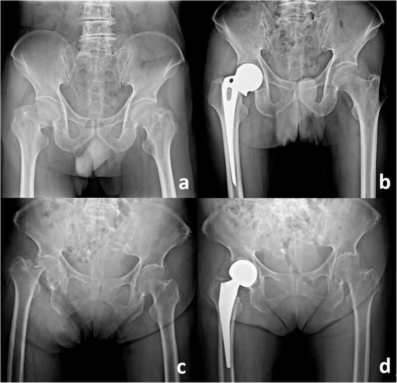
This composite of four anteroposterior (AP) pelvic radiographs illustrates acute femoral neck fractures and their subsequent management with hip hemiarthroplasty. Panels 'a' and 'c' demonstrate displaced fractures of the right femoral neck. Key features include cortical discontinuity, superior migration and external rotation of the femoral shaft, and disruption of Shenton’s line, indicating a total loss of alignment between the femoral head and the acetabulum. Panels 'b' and 'd' show the postoperative results of different hemiarthroplasty techniques for managing these fractures. Panel 'b' depicts a unipolar monoblock prosthesis (Austin-Moore type) characterized by a solid, single-component femoral stem and head fixed within the femur. Panel 'd' shows a bipolar hemiarthroplasty, featuring a modular femoral stem and a dual-mobility head designed to articulate both with the acetabulum and at a secondary internal joint. These images serve as educational examples for orthopedic radiology, highlighting the classification of intracapsular hip fractures and the selection of prosthetic implants based on patient needs and surgical protocol.
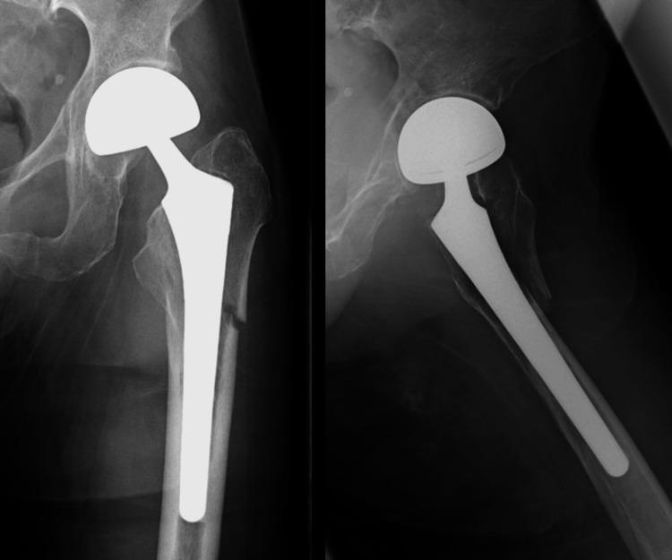
Anteroposterior and lateral radiographs of the left hip and proximal femur demonstrate a periprosthetic femoral fracture (PFF) around an existing bipolar hemiarthroplasty (BHA). The prosthesis consists of a metallic femoral stem and a modular, large-diameter femoral head articulating within the native acetabulum. A transverse, minimally displaced fracture is located mid-stem at the femoral diaphysis, consistent with a Vancouver Type B1 classification. This classification implies that the prosthetic stem remains well-fixed despite the surrounding fracture. The femoral cortex shows signs of thinning, suggesting underlying osteopenia or osteoporosis. No significant comminution, periosteal reaction, or implant subsidence is visible. This imaging is characteristic of geriatric orthopedic trauma involving existing joint replacements, requiring surgical stabilization while preserving the stable implant.
Bipolar Hemiarthroplasty: Modular vs. Non-Modular (Monoblock) Design
Background
Anatomy of a Bipolar Prosthesis
| Component | Function |
|---|---|
| Femoral stem | Fixed into the medullary canal (cemented or cementless) |
| Inner head (small) | Sits on the femoral neck taper; articulates with the outer head |
| Outer head (large) | Metal shell with polyethylene liner; articulates with acetabulum |
| Inner bearing | Between inner and outer head |
| Outer bearing | Between outer head and native acetabulum |
Modular Bipolar Prosthesis
- Variety of inner head sizes and neck lengths available as separate components
- Trial heads and neck additions allow intraoperative adjustment of leg length and soft tissue tension before final implantation
- Stem, neck offset, and head can each be selected and optimized independently
- Neck length can be adjusted (standard, +4mm, +8mm, etc.) to fine-tune leg length equality
- Head size matched to the anatomic femoral head diameter (typically 44-54 mm range)
Non-Modular (Monoblock/Fixed) Bipolar Prosthesis
- Head-neck unit comes as a single pre-assembled piece
- No intraoperative adjustment of neck length or head size beyond implant selection
- Simpler assembly - no coupling mechanism to fail
- Less flexibility if leg length discrepancy or soft tissue tension is suboptimal intraoperatively
- Limited ability to optimize offset independently
Comparison Table
| Feature | Modular Bipolar | Non-Modular (Monoblock) Bipolar |
|---|---|---|
| Head-neck assembly | Separate components, assembled intraoperatively | Pre-assembled, fixed unit |
| Intraoperative adjustability | High - neck length, offset, head size all adjustable | Low - limited to pre-set implant dimensions |
| Trial reduction | Possible with trial components | Generally not possible |
| Leg length optimization | Easier, fine-tuned intraoperatively | Less precise |
| Coupling failure risk | Present (requires secure locking) | None (no coupling mechanism) |
| Conversion to THA | Easier - stem can be retained, only head exchanged | More complex - may require full revision |
| Inventory requirement | Larger (multiple head/neck options needed) | Smaller |
| Cost | Generally higher | Generally lower |
| Dual articulation | Yes (inner + outer bearing) | Yes (inner + outer bearing) |
Unipolar vs. Bipolar - Clinical Context
- Acetabular wear at 12 months: 20% in unipolar vs. 5% in bipolar - but this difference disappears at 4-year follow-up (Inngul et al., RCT of 120 patients)
- Meta-analysis (Zhou et al., n=1100): No marked difference in acetabular erosion between unipolar and bipolar long-term
- Revision rates: Campbell's Operative Orthopaedics (15th ed., 2026) evidence table: "Bipolar may slightly lower revision rates" (Moderate strength recommendation)
- Functional outcomes: Miller's Review of Orthopaedics (9th ed.): "Functional results of unipolar and bipolar prostheses are similar"
- Conversion to THA: Acetabular erosion conversion rate is 5.3% for patients under 75 years vs. 1.4% for those over 75
Indications & Selection Summary
| Scenario | Preferred Choice |
|---|---|
| Elderly, low-demand patient, short life expectancy | Unipolar or standard bipolar (either modular or monoblock) |
| Patient requiring precise leg length restoration | Modular bipolar (intraoperative adjustment) |
| Younger, more active patient needing hemiarthroplasty | Bipolar preferred over unipolar (lower acetabular wear short-term); THA should be strongly considered |
| Avoid in younger, more active patients | Bipolar alone may not be adequate - THA preferred |
| Concern about coupling failure/implant dissociation | Non-modular (fixed) bipolar avoids this risk |
| Anticipated future conversion to THA | Modular bipolar - stem retention possible |
Key Points (Sources)
- Modular bipolar design is now the contemporary standard - virtually all modern bipolar prostheses are modular
- The main advantage of modularity is intraoperative flexibility in restoring leg length and offset
- The main risk unique to modular designs is coupling mechanism failure - rare but catastrophic if it occurs
- Both unipolar and bipolar prostheses have similar long-term functional outcomes
- Bipolar prostheses are not recommended for younger, more active patients due to eventual acetabular erosion - THA is preferred in that group
- Cemented stems are recommended regardless of head design, per NICE guidelines