

Diagnosis Question number 1
megaloblastic anemia peripheral blood smear hypersegmented neutrophils macrocytes
Q.1 - Detailed Answer: Megaloblastic Anemia (Vitamin B12 Deficiency)
a) Probable Diagnosis
b) Reasons for the Diagnosis
| Finding | Significance |
|---|---|
| Pure vegetarian diet | B12 is found only in animal products (meat, fish, milk, eggs); strict vegetarians are at high risk |
| Pallor (pale skin) | Reflects anemia |
| Hemoglobin = 9 g/dL | Confirms anemia |
| MCV = 108 fL (normal: 80-100 fL) | Macrocytosis - hallmark of megaloblastic anemia |
| MCH = 48 pg (normal: 27-32 pg) | Elevated, consistent with macrocytic cells |
| Large RBCs on smear | Macro-ovalocytes - characteristic of megaloblastic anemia |
| Hypersegmented neutrophils | Classic finding - neutrophils with ≥5 lobes; highly specific for megaloblastic anemia |
| Tingling & numbness in lower limbs | Neurological involvement (subacute combined degeneration of spinal cord) - unique to B12 deficiency, NOT seen in folate deficiency |
| Sensory loss in distal extremities | Peripheral neuropathy from B12 deficiency |
| Recurrent mouth ulcers | Due to impaired mucosal cell turnover from DNA synthesis defect |
| Fatigue | Due to anemia and tissue hypoxia |
c) Mechanism of Development of the Disease
-
B12 deficiency (due to insufficient dietary intake in this pure vegetarian)
-
B12 normally acts as a cofactor for methionine synthase, which converts homocysteine → methionine. In this reaction, N5-methyl tetrahydrofolate (N5-methyl FH4) donates its methyl group and is converted to tetrahydrofolate (FH4).
-
Without B12, this reaction fails → folate trap: folate gets "trapped" as N5-methyl FH4 and cannot be converted to FH4.
-
FH4 (and its derivative N5,10-methylene FH4) is required by thymidylate synthetase to convert dUMP → dTMP (deoxythymidine monophosphate), a building block of DNA.
-
Without adequate dTMP, DNA synthesis is impaired → cells cannot divide normally.
-
Precursor cells (especially rapidly dividing hematopoietic cells) continue to grow in size (cytoplasm matures normally) but cannot divide → megaloblasts form.
-
These abnormal large cells undergo intramedullary destruction (ineffective hematopoiesis).
- B12 is also required for methylmalonyl-CoA mutase, which converts methylmalonyl-CoA → succinyl-CoA.
- In deficiency, methylmalonyl-CoA and propionate accumulate → abnormal fatty acid synthesis → incorporation into neuronal lipids → myelin breakdown.
- This causes subacute combined degeneration (SCD) of the spinal cord - affecting the dorsal (posterior) and lateral columns.
d) Physiological Basis of Signs and Symptoms
| Sign/Symptom | Physiological Basis |
|---|---|
| Pallor | Low Hb (9 g/dL) - reduced O2-carrying capacity, compensatory cutaneous vasoconstriction |
| Fatigue | Tissue hypoxia due to reduced hemoglobin and fewer functional RBCs |
| Large RBCs / High MCV (108 fL) / High MCH (48 pg) | DNA synthesis impaired → nuclear maturation lags behind cytoplasmic maturation → cells grow abnormally large; ample hemoglobin in large cells gives high MCH |
| Hypersegmented neutrophils | Same DNA synthesis defect affects WBC precursors → neutrophil nuclei over-segment (≥5 lobes) due to delayed nuclear maturation |
| Recurrent mouth ulcers | Oral mucosal cells also rapidly dividing → impaired DNA synthesis → mucosal breakdown |
| Tingling/Numbness/Sensory loss | Subacute combined degeneration of spinal cord: dorsal column (posterior funiculus) damage → loss of proprioception and vibration sense; lateral column damage → upper motor neuron signs. Caused by abnormal fatty acid incorporation into myelin (methylmalonate pathway defect) |
e) Function, Absorption, and Metabolism of Vitamin B12
Functions of Vitamin B12
- DNA synthesis - as a cofactor for methionine synthase, it enables FH4 recycling, which is needed for dTMP synthesis (the key building block of DNA).
- Myelin synthesis / neurological integrity - via the methylmalonyl-CoA mutase reaction, enabling normal fatty acid metabolism in neurons.
- Methionine synthesis - converts homocysteine to methionine (important for protein synthesis and one-carbon metabolism).
- Red cell maturation - supports normal erythropoiesis and WBC maturation.
Absorption of Vitamin B12
-
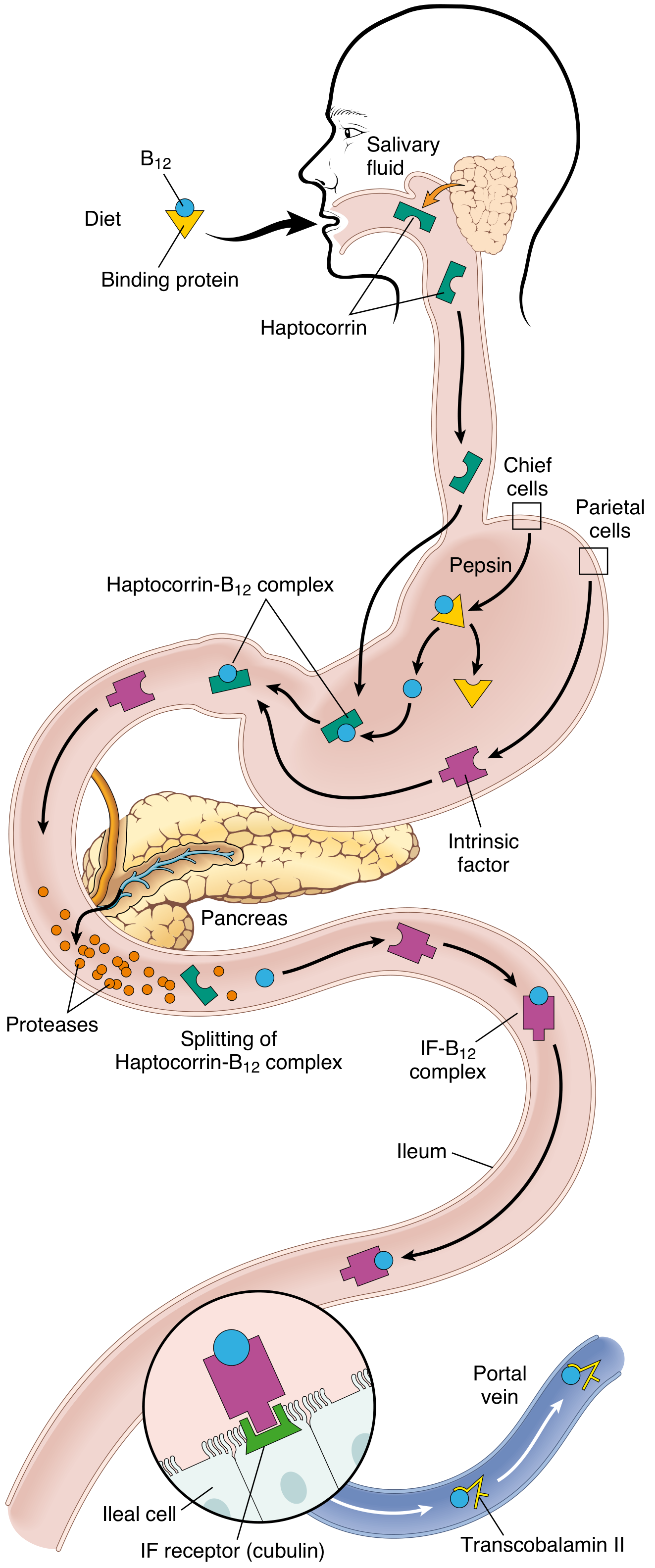
Mouth/Stomach: B12 is released from food proteins by pepsin in the stomach. It first binds to haptocorrin (R-protein), a salivary glycoprotein.
-
Duodenum: Pancreatic proteases cleave the haptocorrin-B12 complex. Free B12 then binds to Intrinsic Factor (IF), a glycoprotein secreted by parietal cells of the gastric fundic mucosa.
-
Terminal Ileum: The IF-B12 complex binds to a specific receptor called cubilin on ileal epithelial cells and is endocytosed.
-
Plasma: Within ileal cells, B12 binds to Transcobalamin II (its major carrier protein) and is secreted into the portal circulation. Transcobalamin II delivers B12 to the liver, bone marrow, and other tissues.
-
Storage: The liver stores large reserves (2-5 mg) - enough for 3-5 years, which is why dietary deficiency takes years to manifest clinically.
- Daily requirement: 2-3 μg/day
- Sources: exclusively animal products (meat, fish, eggs, dairy)
Metabolism of Vitamin B12
-
Methionine synthase reaction (uses methylcobalamin):
- Homocysteine + N5-methyl-FH4 → Methionine + FH4
- Regenerates active folate (FH4) needed for DNA synthesis
-
Methylmalonyl-CoA mutase reaction (uses adenosylcobalamin):
- Methylmalonyl-CoA → Succinyl-CoA (enters TCA cycle)
- Deficiency leads to accumulation of methylmalonic acid (useful diagnostic marker)
- B12 deficiency = macrocytic anemia WITH neurological features
- Folate deficiency = macrocytic anemia WITHOUT neurological features (folate does NOT participate in the methylmalonyl-CoA pathway)
- Elevated serum methylmalonic acid and homocysteine = B12 deficiency
- Elevated homocysteine alone (normal methylmalonic acid) = folate deficiency

Question 1 diagnose
Q.1 (Part II) - Detailed Answer: Orthostatic (Postural) Hypotension
a) Most Probable Cause of Her Light-Headedness and Fainting Sensation
- Abrupt decrease in venous return to the heart
- Reduced stroke volume and cardiac output (Frank-Starling mechanism)
- Sudden fall in blood pressure → decreased cerebral perfusion
- Result: Light-headedness, near-fainting sensation, and racing heart (reflex tachycardia)
b) Name of the Condition
- In her case, this is likely Initial Orthostatic Hypotension - an exaggerated, abrupt decrease in BP (> 40 mmHg SBP) occurring within 15-30 seconds of standing, due to a transient mismatch between cardiac output and systemic vascular resistance. This variant is common in young-to-middle-aged individuals and is benign and self-limiting.
c) Mechanism of Recovery
Phase 1 - Immediate Baroreceptor (Baroreflex) Compensation
-
Blood pools in lower limbs/abdomen → venous return falls → cardiac output falls → arterial BP drops
-
Baroreceptors (stretch receptors in the carotid sinus and aortic arch) detect the fall in arterial wall stretch (decrease in BP)
-
Baroreceptors send fewer inhibitory signals to the cardiovascular center in the medulla oblongata (vasomotor center)
-
The vasomotor center is now disinhibited, triggering:
- Increased sympathetic outflow to heart and blood vessels
- Decreased parasympathetic (vagal) outflow to heart
-
The sympathetic responses include:
- Heart: Increased heart rate (tachycardia) - she felt her heart "racing" - and increased force of contraction → increased cardiac output
- Arterioles: Vasoconstriction (increased peripheral vascular resistance, SVR)
- Veins: Venoconstriction → increased venous return to the heart (Starling effect)
-
Together, these effects restore mean arterial pressure and cerebral perfusion → light-headedness resolves
Phase 2 - As She Walked
Summary Flow Diagram
Sudden standing
↓
Blood pools in lower limbs (500-700 mL)
↓
↓ Venous return → ↓ Cardiac output → ↓ Arterial BP
↓
Baroreceptors in carotid sinus & aortic arch detect ↓ stretch
↓
↑ Sympathetic tone + ↓ Vagal tone (via medullary vasomotor center)
↓
↑ Heart rate + ↑ Contractility + Arteriolar vasoconstriction + Venoconstriction
↓
↑ Cardiac output + ↑ Peripheral resistance
↓
BP restored → Cerebral perfusion normalizes → Recovery
| Feature | Detail |
|---|---|
| Condition | Orthostatic (Postural) Hypotension |
| Trigger | Sudden standing after lying down (especially morning) |
| Mechanism | Gravity-dependent blood pooling → ↓ venous return → ↓ CO → ↓ BP → ↓ cerebral flow |
| Symptoms | Lightheadedness, near-syncope, palpitations (racing heart), visual dimming |
| Recovery mechanism | Baroreceptor reflex (sympathetic activation + vagal withdrawal) + muscle pump |
| Why transient? | Baroreflex rapidly compensates; muscle activity on walking accelerates recovery |
| Definition | SBP drop ≥ 20 mmHg OR DBP drop ≥ 10 mmHg within 3 min of standing |

Ques 1
coronary artery atherosclerosis plaque rupture thrombosis myocardial infarction pathophysiology
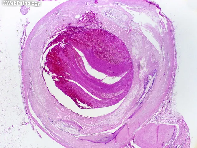
This is a light microscopic histopathology image of a human left anterior descending (LAD) coronary artery segment from autopsy. The specimen shows an atherosclerotic plaque with rupture of the fibrous cap and overlying luminal thrombosis, resulting in near-complete occlusion of the arterial lumen over approximately 1.0 cm. The arterial wall reveals intimal thickening with a lipid-rich necrotic core, cholesterol clefts, and inflammatory cells. The ruptured plaque exposes subendothelial thrombogenic material, provoking platelet aggregation and formation of an occlusive red thrombus that markedly narrows the lumen. The thrombus appears organized with layered eosinophilic material and fibrin, consistent with thrombotic components seen in acute coronary syndromes. The surrounding media may show early degenerative changes typical of atherosclerosis. This lesion corresponds to a fatal acute myocardial infarction in a patient with a history of ischemic heart disease and prior CAD. Clinically, the finding supports sudden cardiac death due to acute coronary occlusion from plaque rupture. The image emphasizes the pathophysiology of myocardial infarction: plaque instability, thrombosis, rapid flow limitation, and myocardial ischemia. It provides a classic autopsy correlate for education, teaching, and research on coronary atherosclerosis, plaque rupture, thrombosis, and sudden death. This histology image is ideal for autopsy teaching and cardiac pathology research.
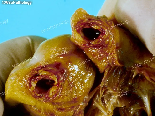
Gross pathology photograph from an autopsy showing the heart with acute coronary occlusion. Anatomical site: right coronary artery (RCA) with heavy atherosclerotic disease and a mural thrombus occluding the lumen. Plaque morphology is lipid-rich, irregular, and yellow-brown with possible calcification. The overlying thrombus is dark reddish-brown, adherent to the plaque, and partially fills the arterial lumen, producing abrupt cessation of distal flow. Associated myocardial tissue may show early signs of infarction, such as pallor or edema in the distribution supplied by the RCA. Imaging modality and technique: macroscopic gross pathology image, en-face view of the epicardial artery from autopsy; not a radiologic study and not stained. Pathophysiology: sudden plaque rupture with superimposed mural thrombosis causing acute myocardial infarction (AMI) and death within 72 hours. Clinical correlation: this lesion represents a classic mechanism of fatal coronary artery disease, leading to hemodynamic compromise and ventricular dysfunction. Diagnostic significance: demonstrates the chain of events from atherosclerotic plaque disruption to occlusive thrombosis and myocardial necrosis, illustrating the basis for sudden cardiac death in CAD. Differential considerations include plaque rupture with thrombosis, coronary vasospasm, embolic occlusion, and multivessel atherosclerosis. Educational value: aids understanding of MI pathogenesis, coronary thrombosis, and autopsy-based cardiovascular pathology.
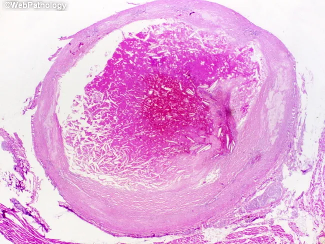
Imaging modality: Histopathology - light microscopy of an intact coronary arterial cross-section, stained with H&E. Specimen location: Left anterior descending coronary artery (LAD), proximal to mid-segment, from an adult male with known ischemic heart disease who died suddenly. Observed features: atherosclerotic plaque rupture with overlying luminal thrombosis; the lumen is nearly occluded over a 1.0 cm segment. The plaque shows a lipid-rich necrotic core and a thin, disrupted fibrous cap; the intima is thickened with yellowish plaque. The thrombus is adherent to the plaque rupture site and extends into the lumen, composed of platelets, fibrin, erythrocytes; the arterial wall demonstrates media degeneration and intimal atherosclerosis. The overall morphology is consistent with acute coronary thrombosis leading to myocardial ischemia and sudden death; histology may reveal early myocardial changes if present elsewhere. Diagnostic significance: this pattern explains sudden cardiac death due to acute myocardial infarction from an occlusive coronary thrombus following plaque rupture; differential considerations include spontaneous coronary dissection, vasospastic occlusion, or embolic events; clinical correlation with ischemic heart disease history supports the infarction mechanism. This image is valuable for education on pathophysiology of plaque rupture, thrombus formation, and fatal acute coronary syndrome; relevant to cardiology, pathology, and medical education.
Q.1 - Detailed Answer: Acute Myocardial Infarction (STEMI - Inferior Wall)
a) Likely Diagnosis (2 Marks)
| Feature | Significance |
|---|---|
| 64-year-old male | High-risk age and sex for CAD |
| Sudden crushing chest pain at 4 AM | Classic anginal chest pain; early morning is peak time for STEMI (high sympathetic tone, platelet aggregability) |
| Pallor, sweating profusely | Sympathetic activation due to pain and shock |
| Vomiting | Vagal response, common in inferior STEMI (RCA supplies AV node and also causes nausea/vomiting via vagal activation) |
| ST elevation in leads II, III, aVF | These are the inferior leads - ST elevation here = inferior wall MI |
| Heavy smoker + diabetes + hypertension | Major risk factors for atherosclerosis and coronary artery disease |
b) Pathophysiology (5 Marks)
Step-by-Step Pathophysiology:
- Smoking, hypertension, and diabetes chronically damage the vascular endothelium of coronary arteries
- Damaged endothelium loses its antithrombotic properties and becomes permeable to lipids
- LDL cholesterol enters the intima and gets oxidized → foam cells form from macrophages
- Over years, a fibro-lipid (vulnerable) plaque develops with:
- A lipid-rich necrotic core (high in tissue factor)
- A thin fibromuscular cap overlying it
- The thin fibrous cap ruptures (triggered by: sympathetic surge in early morning, shear stress, inflammation)
- The lipid core is exposed to circulating blood - this is intensely thrombogenic
- Exposed collagen and tissue factor → platelet adhesion and aggregation (via GP IIb/IIIa receptors)
- Coagulation cascade activates → fibrin mesh forms
- Result: a platelet-rich, then red occlusive thrombus completely blocks the lumen
- Complete occlusion of the coronary artery (RCA in this case) → zero oxygen delivery to the supplied territory (inferior wall of left ventricle + posterior wall + right ventricle)
- Within 20-30 min: irreversible myocyte injury begins (ATP depletes, Ca²⁺ floods in, cell membrane ruptures)
- Within 6 hours: transmural necrosis (full-thickness death of myocardium) = STEMI
- Anaerobic glycolysis produces lactic acid → acidosis → chest pain
- Pain: lactic acid + bradykinin stimulate cardiac pain fibers (referred to chest, jaw, left arm)
- Sweating + pallor: massive sympathetic activation (catecholamine surge)
- Vomiting: vagal reflex (Bezold-Jarisch reflex) - especially in inferior MI
- ST elevation: injured myocardium has altered ionic gradients → current of injury → ST elevation in overlying leads
c) Special Features of Coronary Circulation (5 Marks)
1. Highest Oxygen Extraction in the Body
- The heart extracts ~70% of oxygen from coronary arterial blood at rest (most organs extract only 25-30%)
- Since there is almost no oxygen reserve, any increase in cardiac demand must be met by increasing coronary blood flow (not by extracting more O₂ from existing flow)
- This makes the heart extremely vulnerable to ischemia when flow is reduced
2. Flow Occurs Mainly During Diastole
- During systole, ventricular contraction compresses the intramuscular coronary vessels, especially the subendocardial vessels → flow is impeded
- During diastole, myocardium relaxes → coronary vessels fill freely
- Tachycardia is dangerous: it shortens diastole disproportionately → reduces coronary filling time → ischemia risk
3. Metabolic (Local) Autoregulation is the Primary Controller
- Coronary flow is regulated primarily by local metabolic needs (oxygen demand)
- When cardiac activity increases: ATP → ADP → AMP → Adenosine is released
- Adenosine is a potent vasodilator that opens coronary arterioles in proportion to metabolic demand
- Other local vasodilators: NO (nitric oxide), CO₂, H⁺ ions, K⁺ ions, prostaglandins
4. Sympathetic and Parasympathetic Dual Innervation
- Sympathetic stimulation:
- Direct effect: alpha receptors (predominantly on epicardial vessels) → vasoconstriction
- Beta receptors (intramuscular arteries) → vasodilation
- Indirect effect: increased heart rate and contractility → increased metabolic demand → metabolic vasodilation overrides the direct vasoconstriction
- Parasympathetic (vagal): direct dilation of coronary vessels + indirect constriction (slows heart → less O₂ demand)
- Metabolic control always overrides neural control within seconds
5. End-Artery Anatomy (Limited Collaterals)
- Coronary arteries are functionally end-arteries with minimal collateral circulation in healthy individuals
- Sudden occlusion causes immediate infarction of the territory supplied
- (Chronic gradual stenosis allows some collateral development - explaining why some patients with 90% blockage have less damage)
6. Subendocardium is Most Vulnerable
- The subendocardial layer has the highest oxygen demand and faces the most compression during systole
- Blood supply to subendocardium depends entirely on diastolic flow
- In ischemia, subendocardial necrosis occurs first (before transmural infarction)
d) Cardiac Biomarkers (3 Marks)
Key Cardiac Biomarkers:
| Biomarker | Rises | Peaks | Returns to Normal | Notes |
|---|---|---|---|---|
| Myoglobin | 1-2 hrs | 6-9 hrs | 24 hrs | Earliest marker; low specificity (also from skeletal muscle) |
| CK-MB (Creatine Kinase-MB isoform) | 4-6 hrs | 12-24 hrs | 48-72 hrs | Highly specific for myocardium; used to detect re-infarction |
| Troponin I / Troponin T | 4-6 hrs | 12-24 hrs | 7-14 days | Gold standard - highest specificity and sensitivity for myocardial necrosis |
| LDH (Lactate Dehydrogenase) | 24-48 hrs | 3-6 days | 8-14 days | Used when patient presents late ("late marker") |
| AST (Aspartate Transaminase) | 6-8 hrs | 24-48 hrs | 3-4 days | Non-specific; rarely used now |
Troponin - the Gold Standard:
- Troponin I and T are regulatory proteins of the cardiac contractile apparatus (specific to cardiac muscle)
- Released only when myocyte membrane is irreversibly damaged (necrosis)
- Even small rises in high-sensitivity troponin (hs-cTn) above the 99th percentile confirm myocardial injury
- Serial troponin measurements (0, 3, 6 hours) are used: a rise and fall pattern confirms acute MI
- Troponin remains elevated for 7-14 days - useful for late presenters but cannot detect re-infarction (use CK-MB for re-infarction)
| Part | Key Answer |
|---|---|
| Diagnosis | Inferior Wall STEMI (RCA occlusion) |
| Pathophysiology | Plaque rupture → thrombosis → complete coronary occlusion → transmural myocardial necrosis |
| Special features of coronary circulation | High O₂ extraction, diastolic flow, metabolic autoregulation (adenosine), end-arteries, subendocardial vulnerability |
| Cardiac Biomarkers | Troponin I/T (gold standard), CK-MB, Myoglobin, LDH - each with specific rise-peak-fall timeline |