
Write a answer on short term regulatory mechanisms
Reading File
The question "short term regulatory mechanisms" is broad and could apply to several physiological contexts (blood pressure, blood glucose, enzyme activity, gene regulation, etc.). Let me clarify before diving in.
Asking for Preferences
Which subject/context does 'short term regulatory mechanisms' refer to?
Short-term regulatory mechanisms of blood pressure work via neural and humoral pathways acting within seconds to minutes. The main ones are: the baroreceptor reflex, chemoreceptor reflex, CNS ischemic response, and several vasoactive hormones (adrenaline, angiotensin II, ADH acting rapidly). The kidneys handle long-term regulation.
Searching sources...
Finding Sources
Finding Sources
Finding Sources
Reading File
Excellent content found. Now let me get more details on the baroreceptors, chemoreceptors, CNS ischemic response, and humoral mechanisms:
Reading File
Reading File
Reading File
Finding Sources
Reading File
Finding Sources
Reading File
Reading File
I now have rich content from both Medical Physiology (Boron & Boulpaep) and Guyton & Hall. Let me also get the humoral/hormonal short-term mechanisms and the Costanzo overview:
Reading File
Finding Sources
I now have comprehensive material from three authoritative physiology textbooks. Let me compile the complete answer.
Loading Image
I now have everything needed for a thorough, well-cited answer. Here it is:
Short-Term Regulatory Mechanisms of Blood Pressure
Short-term regulation of arterial pressure operates on a time scale of seconds to minutes, primarily through neural reflexes and a few rapidly acting humoral agents. The goal is to keep mean arterial pressure (MAP) near its set point of ~100 mm Hg.
MAP = Cardiac Output × Total Peripheral Resistance
Any mechanism that adjusts MAP does so by changing cardiac output (heart rate + stroke volume), total peripheral resistance (arteriolar tone), or both.
General Framework of Neural Reflex Loops
All neural reflex systems that regulate MAP share five components (Medical Physiology, p. 780):
- Detector (Sensor) - quantifies the controlled variable and converts it to a neural signal
- Afferent pathway - carries signal to the CNS
- Coordinating center - in the medulla; compares signal to set-point, generates error signal
- Efferent pathway - carries corrective signal back to periphery
- Effectors - heart, vascular smooth muscle, adrenal medulla
The primary sensors are baroreceptors (stretch receptors detecting vessel wall distension). Secondary sensors are chemoreceptors (detecting PO2, PCO2, and pH changes). The vasomotor center in the medulla coordinates both.
1. Baroreceptor Reflex (Primary Mechanism)
This is the most important short-term mechanism. It functions as a negative-feedback, pressure-buffer system.
Location of Baroreceptors
- Carotid sinus - wall of the internal carotid artery, just above the carotid bifurcation; afferents travel via Hering's nerve (branch of CN IX / glossopharyngeal)
- Aortic arch - afferents travel via the aortic nerve (branch of CN X / vagus)
Both are high-pressure mechanoreceptors that respond to wall stretch caused by increased intraluminal pressure.
How It Works
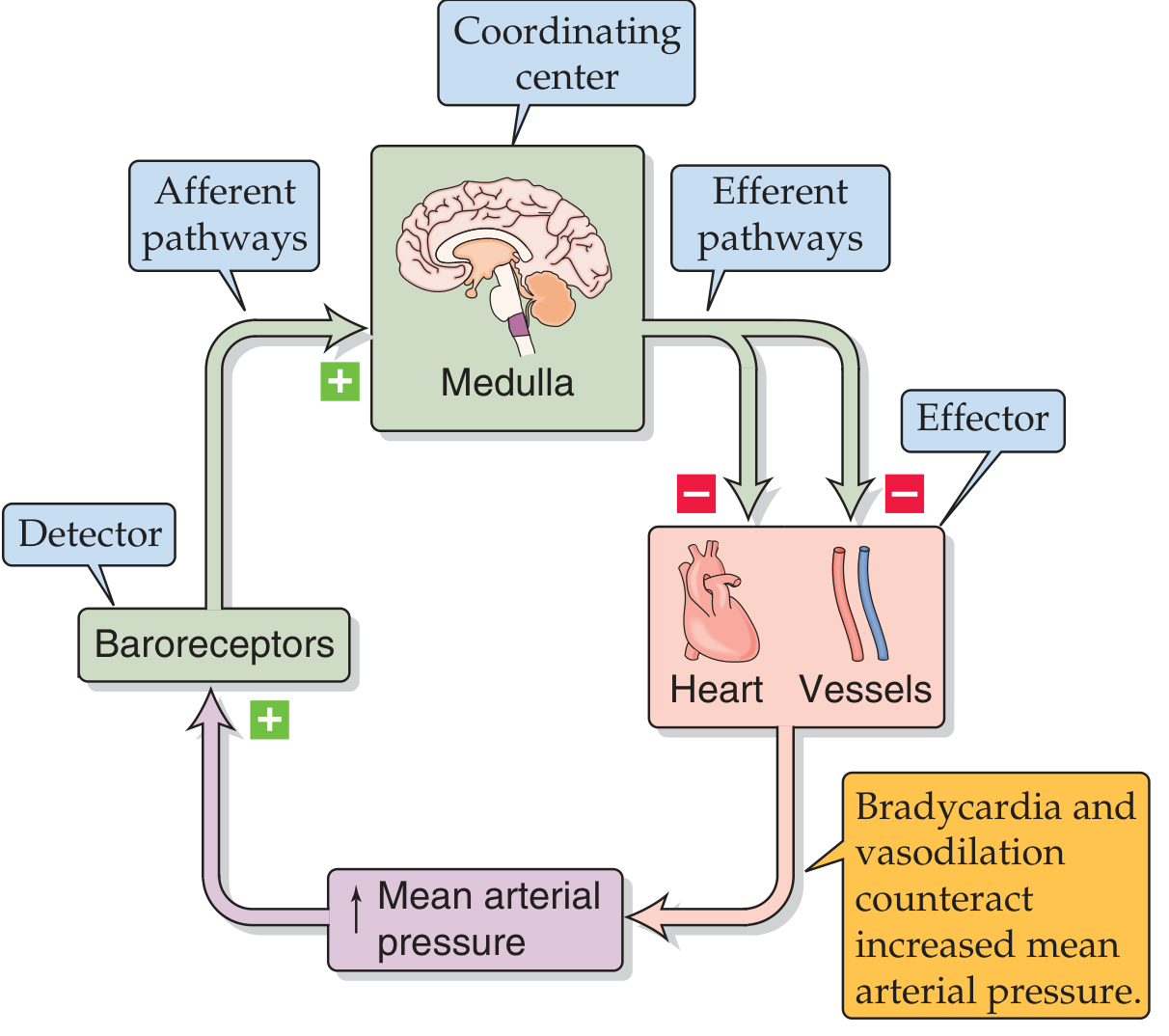
When MAP rises:
- Baroreceptors fire more frequently
- Afferent signals increase to the nucleus tractus solitarius (NTS) in the medulla
- NTS activates the cardioinhibitory center -> increased parasympathetic (vagal) tone -> bradycardia and decreased contractility
- NTS simultaneously inhibits the vasomotor (vasoconstrictor) center -> decreased sympathetic outflow -> vasodilation
- Net result: cardiac output falls, TPR falls -> MAP returns toward normal
When MAP falls:
- Baroreceptor firing decreases
- Parasympathetic tone falls, sympathetic tone rises
- Tachycardia, increased contractility, and vasoconstriction follow
- MAP is restored
The baroreflex responds within 1-2 seconds. Denervation experiments show that without baroreceptors, MAP fluctuates over a range 2.5 times wider than normal throughout the day (Guyton & Hall, p. 228).
Threshold and Sensitivity
- Carotid sinus baroreceptors begin firing at pressures above ~60 mm Hg
- They show maximum sensitivity between 100-180 mm Hg
- Above ~180 mm Hg, they are fully saturated and cannot signal further increases
Low-Pressure (Cardiopulmonary) Baroreceptors
Located in the atria, ventricles, and pulmonary vasculature; they monitor venous filling pressure (preload). Atrial distension reflexively reduces sympathetic tone and ADH secretion (Bainbridge reflex for heart rate). These complement the arterial baroreceptors for volume-pressure homeostasis.
2. Chemoreceptor Reflex (Secondary Mechanism)
Peripheral chemoreceptors located in the carotid bodies (between internal and external carotid arteries) and aortic bodies (under the aortic arch) detect changes in arterial blood gases and pH.
- A fall in PO2, rise in PCO2, or fall in pH stimulates chemoreceptors
- Afferents increase firing to the medulla
- Direct cardiovascular response: vasoconstriction and bradycardia
- However, chemoreceptor stimulation also increases breathing; the increased ventilation then raises PO2 and lowers PCO2, which secondarily causes tachycardia
The net integrated response during hypoxia is usually tachycardia and vasoconstriction, because the ventilatory response overrides the direct cardiac slowing (Medical Physiology, p. 797).
Unlike baroreceptors, chemoreceptors exert a positive drive on the vasomotor center (vasoconstriction), not negative drive. They also contribute a positive drive to the cardioinhibitory center (bradycardia) - identical to the baroreceptors in that regard.
3. Vasomotor Center and Sympathetic Nervous System
The vasomotor center is located bilaterally in the reticular substance of the medulla and lower pons. It has three functional areas (Guyton & Hall, p. 223-224):
| Area | Location | Action |
|---|---|---|
| Vasoconstrictor area (C1) | Anterolateral upper medulla | Sends tonic excitatory impulses to sympathetic preganglionic neurons -> vasoconstriction |
| Vasodilator area | Anterolateral lower medulla | Projects to vasoconstrictor area, inhibits it -> vasodilation |
| Cardioinhibitory area (vagal motor nuclei) | Medial medulla | Sends parasympathetic (vagal) impulses to heart -> decreased HR and contractility |
Normally, the vasomotor center maintains a tonic state of partial vasoconstriction called vasomotor tone, keeping resting peripheral resistance at a level above zero. This tone can be increased or decreased as needed.
Sympathetic stimulation produces:
- Increased heart rate and contractility (via beta-1 receptors)
- Arteriolar vasoconstriction (via alpha-1 receptors) -> increased TPR
- Venous constriction -> increased venous return -> increased preload -> increased cardiac output
- Release of epinephrine and norepinephrine from the adrenal medulla
4. CNS Ischemic Response ("Last-Ditch" Mechanism)
When blood flow to the vasomotor center itself falls critically (MAP < 60 mm Hg), CO2 accumulates locally in the brainstem. This directly and powerfully stimulates vasomotor neurons (Guyton & Hall, p. 231):
- MAP can be driven up to 250 mm Hg within minutes
- It is the most powerful activator of the sympathetic vasoconstrictor system
- Renal arterioles can constrict so severely that urine production ceases entirely
This is NOT a normal day-to-day mechanism. It is an emergency "last-ditch stand" that activates only when MAP drops below 60 mm Hg and becomes maximal at pressures of 15-20 mm Hg. The Cushing reaction is a variant: when CSF pressure rises and compresses cerebral arteries, it triggers the CNS ischemic response, driving MAP above CSF pressure to restore cerebral flow.
5. Rapid-Acting Humoral Mechanisms
While primarily neural in onset, a few hormones act fast enough to contribute to short-term regulation:
| Hormone | Source | BP Effect | Onset |
|---|---|---|---|
| Epinephrine | Adrenal medulla (sympathetic activation) | Vasoconstriction (alpha-1) + tachycardia (beta-1); vasodilation in skeletal muscle (beta-2) | Seconds-minutes |
| Norepinephrine | Sympathetic nerve terminals, adrenal medulla | Vasoconstriction (alpha-1 > beta) | Seconds |
| Angiotensin II | RAAS activation (can begin within minutes) | Potent vasoconstriction + stimulates aldosterone + stimulates central thirst/ADH | Minutes |
| ADH (Vasopressin) | Posterior pituitary | Vasoconstriction at high concentrations; water reabsorption (longer-term) | Minutes |
Epinephrine and norepinephrine reinforce the sympathetic response initiated by the baroreceptor reflex - they are released within seconds of sympathetic activation and sustain the hemodynamic adjustment beyond the duration of direct neural firing.
Summary: Time Hierarchy of Short-Term Mechanisms
| Mechanism | Onset | Primary Action |
|---|---|---|
| Baroreceptor reflex | 1-2 seconds | Adjust HR, contractility, TPR |
| Chemoreceptor reflex | Seconds | Vasoconstriction + ventilatory adjustment |
| Adrenal catecholamine release | 10-30 seconds | Sustain sympathetic effects |
| CNS ischemic response | Seconds (emergency only) | Maximal sympathetic activation |
| Angiotensin II / ADH | Minutes | Vasoconstriction, bridges to long-term |
Long-term regulation (hours to days) is handled by renal mechanisms - primarily through pressure natriuresis and the renin-angiotensin-aldosterone system controlling ECF volume (Costanzo Physiology, p. 169).
Sources:
- Medical Physiology (Boron & Boulpaep), pp. 780-797
- Guyton and Hall Textbook of Medical Physiology, pp. 223-231
- Costanzo Physiology 7th Edition, pp. 169-171
This is a shared conversation. Sign in to Orris to start your own chat.