


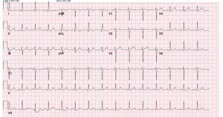
IMG-20261017-WA0007.jpg
IMG-20261017-WA0006.jpg
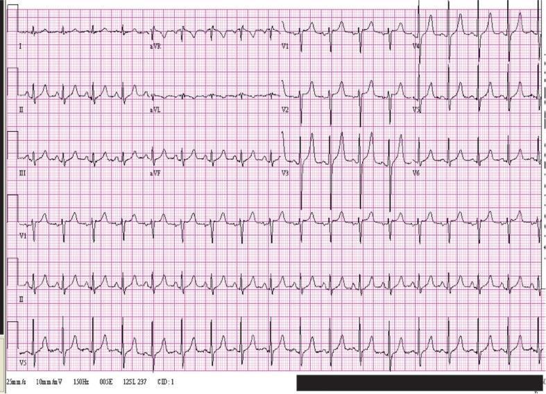
Is this is normal ecg Hyperkalemia or what
Reading File
analyse_media
analyse_media
Quick answer
This ECG does not look fully normal. It appears to show:
- Sinus tachycardia, about 105 bpm
- Prominent tall, peaked T waves, especially across the chest leads (V2-V6)
- Mildly broad QRS reported around 118 ms
Those T waves can be seen with hyperkalemia, although an ECG photo alone cannot confirm potassium level and other causes are possible. I do not see the most advanced hyperkalemia pattern such as absent P waves, marked QRS widening, or a sine-wave appearance, but this still needs prompt assessment.
Action: obtain an urgent blood test for serum potassium, renal function, calcium, magnesium, glucose, and repeat a properly recorded 12-lead ECG. If the person has weakness, palpitations, fainting, chest pain, shortness of breath, kidney disease, is on dialysis, or takes potassium-raising medicines, seek emergency care now. Do not wait for symptoms if hyperkalemia is a real concern.
Can you explain further t wave leads ho lw to suspect in ecg with images
Reading File
Finding Sources
Searching Images
ECG hyperkalemia peaked tented T waves versus normal T wave 12 lead ECG
Searching PubMed
hyperkalemia AND electrocardiography
Searching the Web
hyperkalemia ECG peaked T waves guideline electrocardiogram
Reading File
Reading File
Loading Image
query_images_db
How to assess T waves for possible hyperkalemia
A T wave is the rounded wave immediately after each QRS complex. In hyperkalemia, T waves may become tall, narrow-based, sharply pointed, and symmetric, commonly called tented or peaked T waves.
Which leads to look at
Check all 12 leads, but focus on:
| Lead group | Leads | Why check |
|---|---|---|
| Chest/precordial leads | V2, V3, V4, then V5-V6 | Peaked T waves from hyperkalemia are often easiest to recognize here. |
| Inferior leads | II, III, aVF | Tenting may also be conspicuous in these leads. |
| Rhythm strip | Usually lead II | Best for judging P waves, PR interval, and QRS width. |
Normal vs hyperkalemic T wave
| Feature | Usual normal T wave | T wave suspicious for hyperkalemia |
|---|---|---|
| Shape | Rounded, not perfectly symmetric | Narrow base, pointed top, “tent-shaped” |
| Sides | Often asymmetric | More symmetric: upstroke and downstroke look similar |
| Distribution | May vary from lead to lead | Similar peaked appearance in multiple contiguous leads |
| In chest leads | Usually <5 mm tall | Can be unusually tall, particularly V2-V4 |
| Other ECG findings | Normal P, PR, QRS | P waves flatten or disappear, PR lengthens, QRS widens as toxicity worsens |
A useful visual contrast:
Typical progression when potassium rises
This sequence is common but not reliable enough to rule hyperkalemia in or out:
- Tall, narrow, symmetric peaked T waves
- P waves become small or flat
- PR interval prolongs
- QRS becomes wide
- P waves disappear, severe bradycardia/blocks or a broad “sine-wave” appearance
- Ventricular arrhythmia or cardiac arrest
Emergency medicine reference material describes this classic combination as peaked T waves, then flattened/absent P waves and PR prolongation, followed by QRS widening and potentially a sine-wave pattern. Rosen's Emergency Medicine, p. 2514. A UK Kidney Association guideline notes that a normal T wave is generally <5 mm in precordial leads and <10 mm in limb leads, while hyperkalemic T waves tend to be tall, narrow, sharp, and symmetric.
In the ECG photo you sent
The T waves look prominent and somewhat pointed in the precordial leads, especially approximately V2-V5/V6. That is why hyperkalemia was a concern. However:
- A photographed ECG is not adequate to diagnose hyperkalemia.
- Tall T waves can also occur with early acute coronary occlusion, normal variants, left-ventricular hypertrophy, or technical factors.
- The ECG can be relatively normal even with a dangerous potassium level. Conversely, a peaked-looking T wave does not prove high potassium.
So the deciding test is an urgent serum potassium level, ideally with renal function and a repeat 12-lead ECG. If potassium is high, a sample may be repeated if hemolysis is possible, but ECG changes, symptoms, renal failure, or a high result should be treated as urgent.
Seek emergency assessment now if there is palpitations, fainting, marked weakness, chest pain, breathlessness, reduced urine output, known kidney failure/dialysis, or use of medicines that raise potassium.
Can you explain further ecg images cases how to diagnose hyperkalemia give me image example treatment plans
Reading File
Quick answer
Hyperkalemia on ECG: practical recognition
Hyperkalemia means high blood potassium. The ECG may be an early warning, but it is not sensitive enough to exclude dangerous hyperkalemia. Confirm urgently with a blood potassium test and assess the clinical situation.
ECG examples
Moderate hyperkalemia: tall, narrow, symmetric T waves, especially V2-V4
Normal-looking T waves after treatment
What to look for step by step
1. T waves: the earliest classic clue
A hyperkalemic T wave is usually:
- Tall
- Narrow at the base
- Sharp/pointed at the apex
- Symmetrical: the upstroke and downstroke look similar
- Seen in several leads, often most clearly in V2-V4, but also in II, III, aVF and V5-V6.
A normal T wave is usually more rounded and asymmetric, with a gentler rise and faster descent.
Do not diagnose hyperkalemia based only on a tall T wave. Compare with old ECGs if available and check for changes in more than one lead.
2. P waves and PR interval
As potassium rises, atrial conduction slows:
- P waves become low amplitude or flattened
- PR interval gets prolonged
- P waves can disappear
3. QRS width
As potassium rises further:
- QRS becomes broad, often >120 ms
- Conduction blocks or bradycardia can occur
- The QRS can merge with the T wave into a sine-wave pattern, a pre-arrest emergency.
Common ECG pattern by severity
| ECG stage | Possible ECG features | Clinical meaning |
|---|---|---|
| Early | Peaked, narrow, symmetric T waves | Suspicious but not diagnostic |
| Progressing | Reduced P waves, PR prolongation | Conduction is being affected |
| Severe | Wide QRS, loss of P waves, bradycardia, AV block | Dangerous hyperkalemia likely |
| Pre-arrest | Sine wave, ventricular tachycardia/fibrillation, asystole | Immediate resuscitation-level emergency |
The changes do not always happen in this neat sequence. Someone can have a high potassium with little ECG abnormality, especially with chronic kidney disease. Conversely, dramatic ECG changes can occur at a lower measured potassium if the rise was rapid or there is acidosis, hypocalcemia, or medication effect.
Distinguishing from other tall T waves
| Cause | Typical clue |
|---|---|
| Hyperkalemia | Narrow, tented, symmetric T waves in multiple leads, plus P/PR/QRS abnormalities as it worsens |
| Acute coronary occlusion | Broad-based, bulky "hyperacute" T waves with localized ST changes, often in a coronary territory |
| Normal variant | Stable compared with older ECGs, no PR/QRS abnormality, clinical context reassuring |
| Early repolarization | J-point/ST elevation, often young healthy person, not typically narrow tented waves |
| Left ventricular hypertrophy | High-voltage QRS with secondary ST-T changes |
Treatment plan for clinicians: acute hyperkalemia
This is emergency management, requiring monitoring, repeat ECGs, serial potassium and glucose testing, and treatment of the underlying cause. It is not a home-treatment plan.
When to treat urgently
Urgent emergency treatment is generally indicated for:
- ECG changes attributable to hyperkalemia
- Potassium markedly elevated, often ≥6.5 mmol/L
- Rapidly rising potassium
- Weakness, paralysis, arrhythmia, bradycardia, hypotension
- Renal failure, dialysis dependence, severe acidosis, crush injury, rhabdomyolysis, or tumor lysis syndrome
Treatment priorities: protect, shift, remove
| Priority | Typical intervention | Purpose |
|---|---|---|
| 1. Protect myocardium | IV calcium, such as calcium gluconate or calcium chloride in appropriate monitored settings | Stabilizes cardiac membrane. Does not lower potassium. |
| 2. Shift potassium into cells | IV regular insulin with IV glucose | Rapid temporary lowering of serum potassium |
| Nebulized salbutamol/albuterol | Additional intracellular shift | |
| IV sodium bicarbonate | Consider mainly when significant metabolic acidosis is present | |
| 3. Remove potassium from body | Loop diuretic if kidneys produce urine and volume status permits | Enhances renal excretion |
| Potassium binders | Removes potassium through the gut, but not adequate as sole emergency therapy | |
| Hemodialysis | Fastest definitive removal in severe/refractory hyperkalemia or renal failure |
Monitoring matters: cardiac monitoring, repeat potassium, and frequent bedside glucose checks after insulin are required. Hypoglycemia after insulin treatment is a significant risk.
Immediate practical actions while arranging emergency care
- Put the patient on a cardiac monitor if available.
- Obtain a repeat blood sample for potassium if hemolysis is possible, but do not delay treatment when ECG changes or severe illness are present.
- Review and hold potential potassium-raising agents under clinician direction: potassium supplements, ACE inhibitors, ARBs, spironolactone/eplerenone, trimethoprim, NSAIDs, and others.
- Look for renal failure, acidosis, diabetic ketoacidosis, tissue injury/rhabdomyolysis, or missed dialysis.
For the ECG you shared earlier, the prominent T waves in the chest leads make potassium testing urgent, but an image cannot confirm the diagnosis. A same-day urgent serum potassium, creatinine/eGFR, bicarbonate, glucose, calcium and magnesium, with a repeat 12-lead ECG, is the right next step. If there are symptoms, kidney disease, reduced urine, dialysis, weakness, fainting, or palpitations, go to emergency care now.
This is a shared conversation. Sign in to Orris to start your own chat.