
Patient came with unilateral ear pain What to examine And what to prescribe
otalgia ear pain examination otoscopy
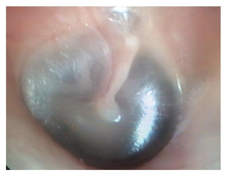
This clinical photograph displays a close-up view of a human tympanic membrane (eardrum) captured during an otoscopic examination. The imaging modality is clinical otoscopy. The tympanic membrane exhibits a generally pearly-gray to slightly translucent appearance in the superior regions, while the inferior portion shows darker, shadowed areas. Key anatomical landmarks are visible, including the manubrium of the malleus extending from the superior periphery toward the umbo at the center of the membrane. The external auditory canal wall surrounding the membrane appears healthy, with a smooth, pinkish-flesh tone and no signs of acute inflammation, erythema, or obstructive cerumen. This image serves as an educational example of a normal otoscopic finding or a baseline clinical assessment in a patient presenting with otalgia, illustrating the standard morphology and positioning of the middle ear structures as seen through the ear canal.
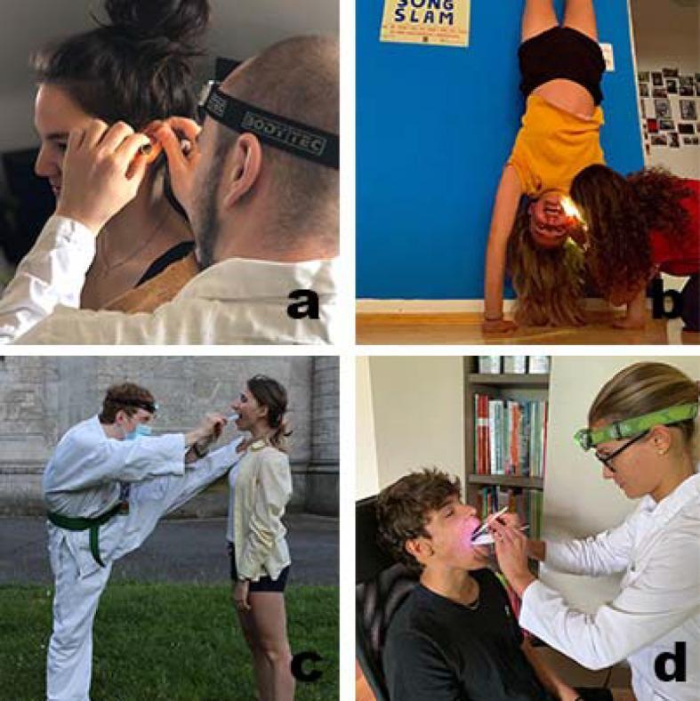
A composite of four clinical photographs (a-d) demonstrating basic Otorhinolaryngology (ENT) examination techniques performed by students. Images (a) and (b) focus on otoscopy, showing the manipulation of the auricle and the use of an ear speculum and head-mounted light source to visualize the external auditory canal and tympanic membrane. Images (c) and (d) depict oral inspections and indirect laryngoscopy, using tongue depressors (spatulas) and laryngeal mirrors to examine the oropharynx. The series highlights the use of portable, disposable instrumentation—including nasal speculums, ear speculums, and mouth spatulas—alongside improvised or standard headlamps for illumination. The variations in settings (from clinical white coats to domestic and outdoor environments) and patient positioning (including a handstand in panel b) illustrate a gamified medical education approach designed for remote practical training. Key educational concepts include proper instrument handling, maintaining a line of sight with a light source, and the anatomical focus of head and neck physical examinations.
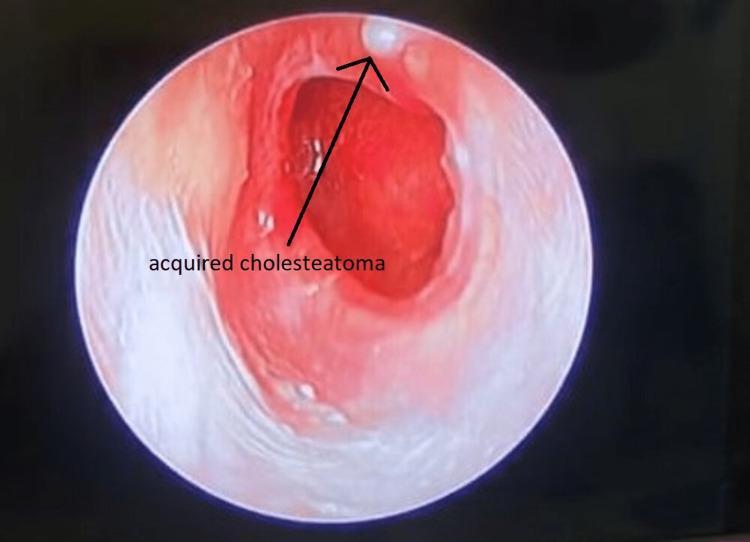
This clinical photograph, likely obtained via otoscopy or endoscopic ear examination, demonstrates an acquired cholesteatoma within the external auditory canal. The view reveals a significant area of pathological change characterized by intense erythema and inflammation of the canal wall and tympanic membrane region. A hallmark finding is the presence of a small, white, keratinous debris mass (cholesteatoma) located at the superior aspect of the canal, indicated by an arrow. The surrounding mucosal tissue appears thickened and irregular, suggesting chronic irritation or infection. The central portion of the field shows a deep red hue and compromised tissue integrity, typical of middle ear pathology extending into the canal. This image is an educational resource for identifying the visual hallmarks of primary acquired cholesteatoma, specifically the retraction pocket formation and the accumulation of squamous epithelial debris in the attic or pars flaccida region.
Unilateral Ear Pain (Otalgia) - Clinical Approach
Understanding the Anatomy First
STEP 1 - History
- Duration and character of pain (sharp/dull/deep)
- Associated discharge (otorrhea), hearing loss, fever, tinnitus, vertigo
- Jaw pain, dental pain, difficulty swallowing, hoarseness
- Trauma (including water sports, cotton bud use)
- Diabetes or immunocompromise (risk for malignant OE)
- Smoking/alcohol history (risk for head and neck cancer)
- Recent URTI
STEP 2 - Examination
2a. Ear Examination (Always First)
| What to Do | What to Look For |
|---|---|
| Inspect the pinna | Erythema, vesicles (Ramsay Hunt), swelling, haematoma, cellulitis, nodule |
| Tragal pressure / pinna traction | Pain = otitis externa |
| Post-auricular region | Swelling/tenderness/erythema = mastoiditis; auricle pushed forward |
| Otoscopy (otoscope) | Canal: oedema, erythema, discharge, foreign body, cerumen impaction |
| Tympanic membrane | Erythema, bulging (AOM); retraction, perforation, cholesteatoma; bullae (bullous myringitis) |
| Hearing (Tuning fork) | Weber/Rinne to differentiate conductive vs sensorineural loss |
2b. If Ear Examination is NORMAL - Examine Referred Sources
| Region | What to Examine | Nerve |
|---|---|---|
| Temporomandibular joint | Tenderness on palpation, jaw deviation, crepitus, bruxism | CN V |
| Teeth/gums | Dental caries, periodontal tenderness, percussion test | CN V |
| Oropharynx | Tonsils (tonsillitis, peritonsillar abscess), posterior pharynx | CN IX |
| Nasopharynx / sinuses | Sinus tenderness, nasal polyps | CN V |
| Neck | Lymphadenopathy, thyroid, cervical spine tenderness | C2/C3 |
| Larynx/hypopharynx | Laryngoscopy if indicated (especially in smokers) | CN X |
| Parotid gland | Swelling, tenderness | CN V |
STEP 3 - Common Diagnoses and Management
1. Acute Otitis Externa (OE)
- Clean the canal (aural toilet/gentle suction or wicking)
- Topical fluoroquinolone drops (e.g. ciprofloxacin/dexamethasone) - first line; safe even if TM status uncertain - Rosen's Emergency Medicine, p. 912
- Avoid aminoglycoside drops if TM perforation is possible (ototoxic)
- Analgesia: paracetamol/ibuprofen
- Keep ear dry; no swimming
- Systemic antibiotics NOT needed in immunocompetent patients with canal-limited disease
Malignant (Necrotizing) OE: In diabetic/immunocompromised patients with persistent, severe OE: CT temporal bone, urgent ENT referral, systemic IV ciprofloxacin or anti-pseudomonal agent.
2. Acute Otitis Media (AOM)
- Analgesia first: paracetamol or ibuprofen (antibiotics do not relieve pain in the first 24 hours)
- Antibiotic of choice: Amoxicillin (high dose 80-90 mg/kg/day in children; 500 mg TDS in adults) for 5-10 days
- Treatment failure or recurrent AOM: Amoxicillin-clavulanate
- Observation strategy (watchful waiting 48-72 hours) is acceptable in mild cases in children >2 years with no complications - Rosen's Emergency Medicine, p. 912
- If tympanostomy tubes present with otorrhoea: topical fluoroquinolone drops
3. Bullous Myringitis
4. Mastoiditis
- CT scan (assess intracranial extension)
- Admit, IV vancomycin (15 mg/kg); add anti-pseudomonal agent if prior antibiotics used
- ENT referral for possible myringotomy or mastoidectomy
5. Referred Otalgia - Common Causes
| Cause | Clue | Management |
|---|---|---|
| TMJ dysfunction | Jaw click, tenderness over joint, bruxism | NSAIDs, dental referral, night guard |
| Dental abscess/caries | Percussion tenderness, visible caries | Dental referral, amoxicillin/metronidazole |
| Tonsillitis | Exudate, pharyngeal erythema, fever | Penicillin V or amoxicillin; consider abscess drainage |
| Cervical spine | Neck stiffness, tenderness over C2/C3 | Physio, NSAIDs, imaging if needed |
| Laryngopharyngeal reflux | Globus, dysphonia | PPI trial |
| Parotitis | Parotid swelling | Hydration, antibiotics if bacterial |
| Head/neck malignancy | Smoker, >40 yr, no otologic finding | Urgent ENT/laryngoscopy referral |
Summary Algorithm
Unilateral Ear Pain
       |
       ├── Otoscopy ABNORMAL
       │       ├── Canal inflamed/discharge → Otitis Externa → Topical fluoroquinolone drops
       │       ├── TM bulging/red → AOM → Amoxicillin + analgesia
       │       ├── TM bullae → Bullous myringitis → Same as AOM
       │       ├── Post-auricular swelling → Mastoiditis → IV antibiotics + ENT
       │       └── White debris/retraction → Cholesteatoma → ENT referral
       │
       └── Otoscopy NORMAL
               ├── Jaw/dental tenderness → TMJ/Dental → Dental referral + NSAIDs
               ├── Pharynx abnormal → Tonsillitis/abscess → Antibiotics
               ├── Neck tenderness → Cervical spine → Physio/imaging
               └── Smoker/no cause found → HEAD AND NECK MALIGNANCY → Urgent ENT laryngoscopy
- Normal ear exam + unilateral otalgia in a smoker or >40 years old
- Facial nerve palsy + ear pain + vesicles (Ramsay Hunt syndrome - needs aciclovir + prednisolone)
- Postauricular swelling/protrusion (mastoiditis)
- Progressive OE in a diabetic (malignant OE)