
Drug or ointment of choice for acute paronychia
acute paronychia treatment
acute paronychia fingernail infection
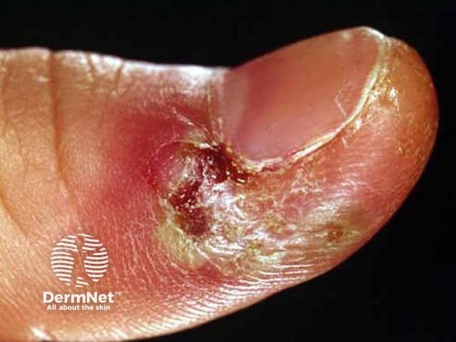
This is a high-resolution clinical photograph in visible light depicting acute herpetic paronychia of the fingernail region. Modality: Clinical photography; technique: close-up, macro view of the nail fold. Anatomical location: digits, paronychia around the nail plate of a fingertip (paronychia at the lateral/lateral nail fold). Appearance: marked erythema and edema of the periungual tissue with surrounding desquamation, crusting and whitish exudate; nearby skin may be tense; central vesicles or pustules may be present but not clearly visible in this frame; the tissue shows vesicular clusters typical of herpes simplex infection; the nail fold fissures and crusts indicate acute inflammatory response; arrangement is unilateral, localized to the lateral nail border. Pathology: HSV-induced vesiculobullous lesions targeting the proximal and lateral nail folds; differential includes bacterial paronychia; differentiation through vesicle groups on erythematous base; clinical features include painful swelling and rapid onset. Diagnostic significance: recognition of herpetic etiology guides antiviral management (acyclovir, valacyclovir) and cautions incision/drainage; may require microbiology confirmation (PCR, Tzanck smear). Clinical use: educational illustration for dermatology training, infectious disease differential, primary care assessment of paronychia; relevant for telemedicine triage; keywords: paronychia, herpes simplex virus, HSV, distal finger infection, periungual edema, vesicles, vesicular lesions, crusting, acute, infectious dermatology.
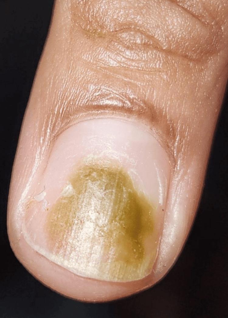
Clinical photograph of a human fingernail demonstrating Chloronychia (green nail syndrome). The image displays a clear, localized green-brown discoloration of the nail plate, primarily concentrated in the central and distal portions. The pigment distribution is non-uniform, featuring varying shades of olive green and yellowish-brown. The proximal nail fold, lunula, and surrounding paronychial skin appear healthy, without visible edema, erythema, or signs of acute paronychia. The nail plate surface remains relatively smooth and intact. This visual finding is classically associated with Pseudomonas aeruginosa infection, often occurring in moist environments or beneath artificial (acrylic) nails where subungual moisture is trapped. The image serves as a teaching tool for dermatological nail disorders and infectious disease identification.
Acute Paronychia: Drug/Ointment of Choice
Causative Organisms
Treatment - By Stage
1. Mild (No Fluctuance / No Abscess)
- Warm soaks (3-4x daily) + elevation -- this is the cornerstone first step
- Topical antibiotic of choice: Mupirocin (Bactroban) ointment -- applied directly to the affected nail fold
- Mupirocin is specifically named in Pfenninger and Fowler's Procedures for Primary Care as the topical agent of choice for paronychia
2. Moderate (Cellulitis Present)
- 1st choice: Anti-staphylococcal penicillins -- cloxacillin or dicloxacillin
- Alternative: Cephalexin (1st-generation cephalosporin)
- If CA-MRSA is suspected: Trimethoprim-sulfamethoxazole (TMP-SMX) or doxycycline
- A 7-day course is standard, or until infection resolves
3. Severe (Fluctuance / Abscess)
- Incision and Drainage (I&D) is the primary treatment -- more important than antibiotics alone
- Digital nerve block is used for anesthesia
- A #11 blade is used to nick the most translucent/fluctuant area
- Post-I&D: warm soaks to keep wound clean
- Routine antibiotics post-I&D are NOT needed unless cellulitis, immunocompromise, or vascular insufficiency is present
Summary Table
| Severity | Treatment of Choice |
|---|---|
| Mild (no abscess) | Warm soaks + mupirocin ointment (topical) |
| With cellulitis | Oral cloxacillin / dicloxacillin / cephalexin (7 days) |
| MRSA suspected | TMP-SMX or doxycycline |
| Fluctuant abscess | Incision & Drainage (antibiotics if cellulitis present) |
| Chronic (>6 wks) | Topical corticosteroids + topical antifungals, avoid wet environments |
Key Points
- Systemic antibiotics are NOT routinely required after drainage unless cellulitis, immunosuppression, or vascular disease is present
- Mupirocin ointment is the specific topical drug named in primary care references
- Culture should be considered in moderate-severe cases to guide therapy (especially for MRSA)
- Herpetic paronychia (grouped vesicles, HSV) is a mimic -- do NOT incise; treat with acyclovir/valacyclovir instead