
Cellulites
cellulitis skin infection lower leg erythema swelling
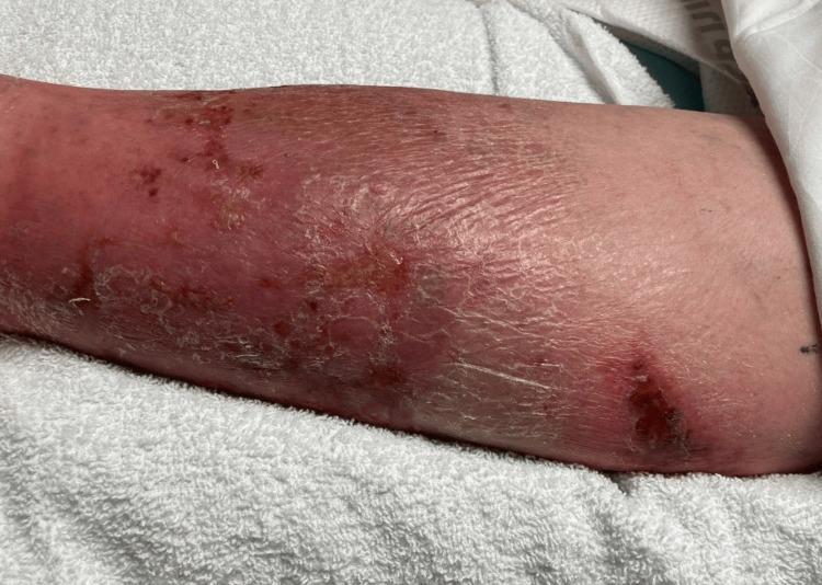
This clinical photograph displays the lower extremity of a patient, demonstrating classic physical examination findings of cellulitis. The skin exhibits diffuse erythema and swelling extending across the anterior and lateral aspects of the leg. The texture is notably compromised, showing signs of desquamation (peeling), xerosis (dryness), and lichenification, particularly in the mid-to-lower portion. Multiple discrete skin lesions are visible, including areas of darker, crusted scabbing and potential ulceration that may serve as a portal of entry for infection. Small petechiae or ecchymotic spots are scattered within the erythematous region. The presentation is highly characteristic of an acute bacterial skin and soft tissue infection (SSTI), emphasizing the visual transition from healthy proximal skin to distal inflamed, edematous, and scaling tissue. This image is an educational resource for identifying inflammatory dermatological conditions and assessing secondary skin changes associated with chronic or severe cellulitis.
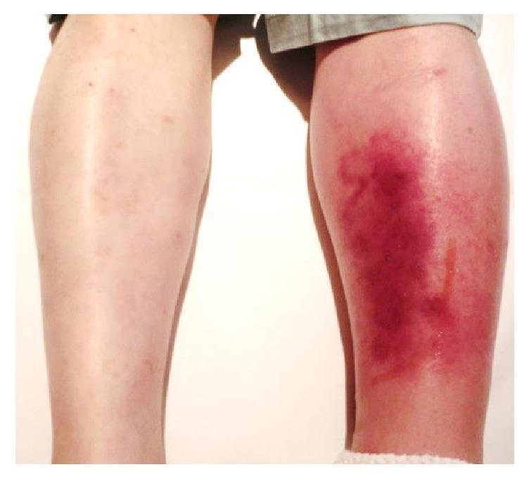
This clinical photograph shows a posterior view of a patient's lower legs, providing a side-by-side comparison between a healthy limb and a symptomatic one. The left lower leg (right side of the image) exhibits classic clinical signs of cellulitis, characterized by significant unilateral edema and diffuse erythema. The affected skin shows a spectrum of inflammatory discoloration, ranging from bright red to deep purplish hues, particularly over the calf region. The skin texture appears tense and glossy due to underlying swelling. A thin, vertical line of serous fluid exudate is visible on the posterior-lateral aspect of the left calf. In contrast, the right lower leg (left side of the image) appears normal with healthy skin tone and no evidence of swelling. This visual demonstrates the acute presentation of a soft tissue infection, useful for teaching the differential diagnosis of unilateral leg swelling and redness, which may include cellulitis, deep vein thrombosis (DVT), or localized vasculitis.
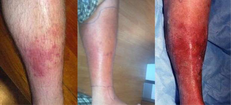
A sequence of three clinical photographs illustrating the progression of cellulitis on the lower leg, categorized by clinical severity. The 'Mild' stage (left) shows a localized, erythematous patch with poorly defined borders and scattered red papules. The 'Moderate' stage (center) demonstrates expanding confluent erythema and edema covering a larger surface area; notably, ink markings are visible on the skin to monitor the spread of infection. The 'Severe' stage (right) depicts intense, deep red to violaceous erythema involving the majority of the limb, accompanied by significant inflammatory swelling, a shiny skin texture suggestive of severe edema, and focal areas of potential skin breakdown or desquamation. This comparison highlights key diagnostic features of skin and soft tissue infections (SSTIs), including changes in color intensity, surface area involvement, and signs of tissue tension associated with worsening bacterial infection.
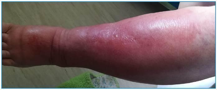
This clinical photograph displays a lateral view of a left lower limb manifesting classic inflammatory signs indicative of cellulitis or severe soft tissue infection. The image captures the leg from the dorsum of the foot to the upper calf/knee area. Key visual features include diffuse, poorly circumscribed erythema that is most intense along the anterior surface of the leg. Marked non-pitting edema (noted clinically as Godet grade III/IV) is evident, resulting in a swollen, tubular appearance of the limb that obscures normal anatomical bony landmarks of the ankle and shin. The skin surface appears tense, taut, and shiny, reflecting significant underlying tissue swelling. No obvious bullae, necrosis, or abscesses are visible in this view, though the dorsum of the foot exhibits exuberant swelling. This material serves as an educational example of acute bacterial skin and soft tissue infection (SSTTIs), demonstrating the cardinal signs of inflammation: rubor (redness) and tumor (swelling), relevant for dermatology and infectious disease specialties.
cellulitis management treatment
Cellulitis
Definition
- Fitzpatrick's Dermatology, Ch. 151
Etiology & Microbiology
| Type | Primary Pathogen(s) |
|---|---|
| Non-purulent cellulitis | Group A Streptococcus (S. pyogenes) - ~75% of cases |
| Purulent cellulitis / abscess | Staphylococcus aureus (including CA-MRSA) |
| Erysipelas | Group A beta-hemolytic streptococci |
| Diabetic foot | S. aureus, streptococci, polymicrobial (gram-negatives, anaerobes in chronic wounds) |
| Bite wounds | Pasteurella multocida (cat), Eikenella (human), polymicrobial |
| Immunocompromised | Gram-negative organisms, fungi, atypical pathogens |
- Andrews' Diseases of the Skin, p. 47 | Rosen's Emergency Medicine, p. 594
Risk Factors
-
Disruption of the skin barrier (wounds, tinea pedis, ulcers, fissures)
-
Lymphedema or impaired lymphatic drainage
-
Venous insufficiency and edema
-
Obesity
-
Diabetes mellitus
-
Immunodeficiency or immunosuppression
-
Prior saphenous vein harvest, lymphadenectomy, or irradiation
-
IV drug use
-
Goldman-Cecil Medicine, p. 233 | Fitzpatrick's Dermatology, Ch. 151
Clinical Features
- Erythema, warmth, swelling, pain/tenderness
- Fever, chills, malaise (systemic features may be absent in mild cases)
- Borders are typically poorly defined and irregular (vs. the raised, sharply demarcated border of erysipelas)
- Lymphangitis - streaks extending proximally, typically seen with streptococcal infection and Pasteurella (bite wounds)
- Vesicles/bullae - form when edema is severe enough to separate epidermal layers
- Pit on pressure - infiltrated, edematous tissue
- Pitting edema; regional lymphadenopathy
- Rosen's Emergency Medicine, p. 598
Diagnosis
-
Clinical diagnosis - laboratory studies including blood cultures, skin biopsies, or aspirates are frequently negative or equivocal
-
Blood cultures are rarely positive in uncomplicated cellulitis
-
If an open wound is present, a wound culture may be informative
-
Bedside ultrasound helps differentiate cellulitis (classic "cobblestone" appearance - hypoechoic fluid between hyperechoic soft tissue) from abscess (essentially anechoic collection)
-
Mark the borders with a pen to monitor spread
-
Tintinalli's Emergency Medicine, p. 514-516 | Andrews' Diseases of the Skin
Differential Diagnosis
| Condition | Distinguishing Feature |
|---|---|
| Stasis dermatitis | No fever/pain, often bilateral, centered over medial malleoli |
| Allergic contact dermatitis | Itchy, not painful; often with clear borders |
| DVT | Unilateral swelling but no skin warmth/erythema pattern |
| Erysipelas | Sharply raised, demarcated border; more superficial |
| Erythema migrans (Lyme) | Red patch, typically less painful; expanding ring |
| Eosinophilic cellulitis | Insect bite history; eosinophilia, not neutrophilia |
| Necrotizing fasciitis | "Pain out of proportion," systemic shock, rapid progression to blue/purple/black |
Management
Outpatient (Non-purulent, No Systemic Toxicity)
- First-line: Dicloxacillin or cephalexin (covers streptococci and MSSA) for 5 days
- If CA-MRSA is suspected (prior MRSA, failed beta-lactam, or purulent features): TMP-SMX, doxycycline, or clindamycin
Inpatient / Systemic Toxicity / Rapid Spread
- IV penicillin G (erysipelas) or IV nafcillin/oxacillin (MSSA)
- IV vancomycin if MRSA suspected or confirmed
- Hospitalize if: systemic sepsis, immunocompromised, face/periorbital, rapid progression, failed oral therapy
Purulent Cellulitis / Abscess
- Incision and drainage is the primary treatment for abscesses
- Small abscesses (<5 cm pus collections) can often be treated with I&D alone
- Add antibiotics covering MRSA for spreading cellulitis around the abscess
Diabetic Foot Infections
-
Add gram-negative and anaerobic coverage for chronic/deep wounds
-
Wound care, possible debridement, revascularization evaluation
-
Rule out osteomyelitis (especially in deep wounds over bony prominences)
-
Andrews' Diseases of the Skin | Tintinalli's Emergency Medicine | Rosen's Emergency Medicine
Recurrent Cellulitis / Prevention
- Treat predisposing conditions (tinea pedis, venous insufficiency, lymphedema)
- Compression therapy to reduce lymphedema
- Long-term prophylactic antibiotics (low-dose penicillin or erythromycin) for patients with frequent recurrences
- Untreated recurrent bouts lead to progressive lymphatic obstruction and chronic lymphedema, ultimately resulting in elephantiasis nostras (permanent hypertrophic fibrosis)
Complications
- Lymphangitis and regional lymphadenopathy
- Bacteremia and metastatic abscesses
- Sepsis (especially in children and immunocompromised patients)
- Progression to necrotizing fasciitis (rapidly spreading necrosis with "pain out of proportion," bullae, and skin color change from red - purple - blue/black)
- Chronic lymphedema / elephantiasis nostras
Key Points Summary
- Non-purulent cellulitis = streptococcal - treat with a beta-lactam (cephalexin)
- Purulent cellulitis/abscess = CA-MRSA until proven otherwise - drain it; add MRSA-active antibiotic
- Diagnosis is clinical; blood cultures are rarely positive
- Mark the borders to track progression
- Ultrasound differentiates cellulitis from abscess (cobblestone sign vs. anechoic collection)
- Always consider necrotizing fasciitis when pain is out of proportion or systemic signs are severe
- A recent network meta-analysis (PMID: 39240378) confirms beta-lactams remain first-line for non-purulent cellulitis, with clindamycin/TMP-SMX for MRSA
Can we give pipzo?
Piperacillin-Tazobactam (Pip-Tazo) in Cellulitis
What Pip-Tazo Covers (Relevant to Cellulitis)
| Organism | Covered? |
|---|---|
| Group A Streptococcus | Yes |
| MSSA | Yes |
| CA-MRSA | No |
| Gram-negatives (Enterobacterales, Pseudomonas) | Yes |
| Anaerobes (B. fragilis, etc.) | Yes |
| Enterococci | Yes |
When Pip-Tazo IS Indicated in Cellulitis
1. Severe Non-Purulent Cellulitis with Systemic Toxicity
- Rapid progression of infection
- Hypotension / hemodynamic instability
- Immunocompromised patient (HIV, chemotherapy, transplant, etc.)
- End-organ damage
"Broad-spectrum antimicrobial coverage (e.g. vancomycin plus piperacillin/tazobactam, imipenem, or meropenem) should be provided for patients with severe non-purulent cellulitis and immunocompromised status, hypotension, or rapid progression."
- Dermatology 2-Volume Set 5e, p. 5128
"Severe cases should be treated with IV vancomycin plus piperacillin-tazobactam."
- Rosen's Emergency Medicine, p. 1243
2. Diabetic Foot Infections (Moderate-Severe)
"Diabetic or decubitus ulcers complicated by cellulitis also require broad-spectrum coverage, such as piperacillin/tazobactam or, in penicillin-allergic patients, metronidazole plus ciprofloxacin."
- Dermatology 2-Volume Set 5e, p. 5128
Katzung's Basic & Clinical Pharmacology lists piperacillin-tazobactam as a drug of first choice for diabetic foot SSTIs (alongside ceftriaxone/cefepime + metronidazole, carbapenem).
3. Orbital / Postseptal Cellulitis
- Tintinalli's Emergency Medicine, p. 72
4. Necrotizing Soft Tissue Infections / Suspected NSTI
- Rosen's Emergency Medicine, Management section
5. Critically Ill / Septic Shock from Skin Source
When Pip-Tazo is NOT Needed (Most Cellulitis)
| Severity | Correct Drug | Why NOT Pip-Tazo |
|---|---|---|
| Mild non-purulent, outpatient | Cephalexin or dicloxacillin PO | Overkill; no gram-negatives involved |
| Moderate non-purulent, IV | Cefazolin or ceftriaxone IV | Adequate streptococcal coverage |
| Mild purulent (MRSA risk), outpatient | TMP-SMX, doxycycline, clindamycin | Pip-tazo does NOT cover MRSA |
- Fitzpatrick's Dermatology, Table 151-5
The Critical Point: Always Pair with Vancomycin
Standard severe cellulitis regimen: Vancomycin IV + Piperacillin-Tazobactam IV
Summary
| Scenario | Pip-Tazo? |
|---|---|
| Mild-moderate routine cellulitis | No |
| Severe / rapidly spreading cellulitis | Yes (+ vancomycin) |
| Immunocompromised with cellulitis | Yes (+ vancomycin) |
| Diabetic foot infection | Yes (monotherapy acceptable in moderate cases) |
| Orbital cellulitis | Yes (one of several options) |
| Suspected necrotizing fasciitis | Yes (+ vancomycin) |
| Purulent cellulitis / abscess (MRSA) | No (pip-tazo misses MRSA) |