
Femoral tringle and sheath
Reading File
The femoral triangle is a depression in the upper thigh formed by muscular boundaries, containing key neurovascular structures. The femoral sheath is a fascial sleeve that encloses the femoral vessels and canal. Let me pull the full details from the medical library.
Searching sources...
Finding Sources
Finding Sources
Reading File
Reading File
Reading File
Reading File
Now let me fetch the key diagrams to include in the answer.
Loading Image
Loading Image
Loading Image
Here is a full, well-structured account of the femoral triangle and femoral sheath, drawn from Gray's Anatomy for Students and the THIEME Atlas of Anatomy.
Femoral Triangle
Definition and Shape
The femoral triangle is a wedge-shaped depression in the upper thigh at the junction between the anterior abdominal wall and the lower limb. Its apex points inferiorly and is continuous with the adductor canal, which carries vessels down the medial thigh and through the adductor hiatus of adductor magnus into the popliteal fossa.
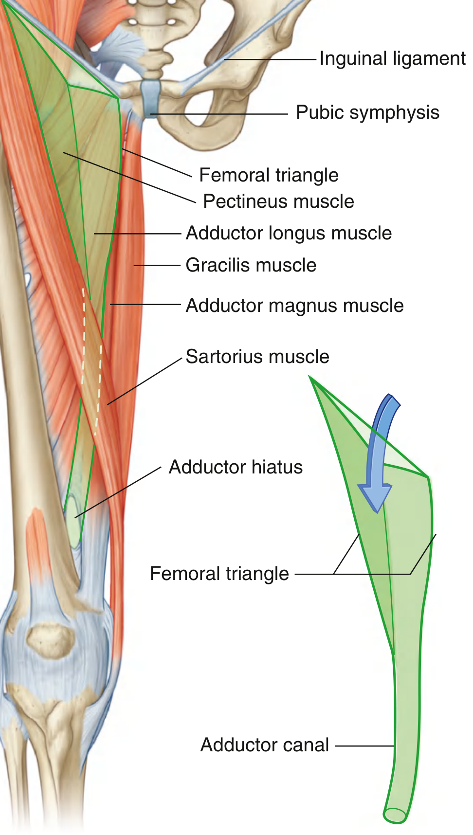
Boundaries
| Boundary | Structure |
|---|---|
| Base (superior) | Inguinal ligament |
| Lateral border | Medial margin of sartorius |
| Medial border | Medial margin of adductor longus |
| Apex | Where sartorius and adductor longus meet (inferiorly) |
| Roof | Fascia lata + skin |
| Floor (medial half) | Pectineus + adductor longus |
| Floor (lateral half) | Iliopsoas |
Contents (Lateral to Medial — mnemonic: NAVL)
From lateral to medial, the major structures within the triangle are:
- N - Femoral Nerve (lateral-most; outside the femoral sheath)
- A - Femoral Artery
- V - Femoral Vein
- L - Lymphatics (in the femoral canal, most medial)
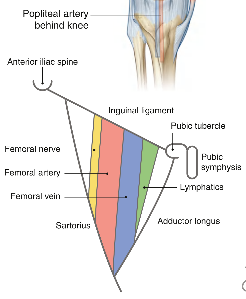
Clinical tip: The femoral artery pulse is palpable just below the inguinal ligament, midway between the anterior superior iliac spine (ASIS) and the pubic symphysis - this is the midinguinal point.
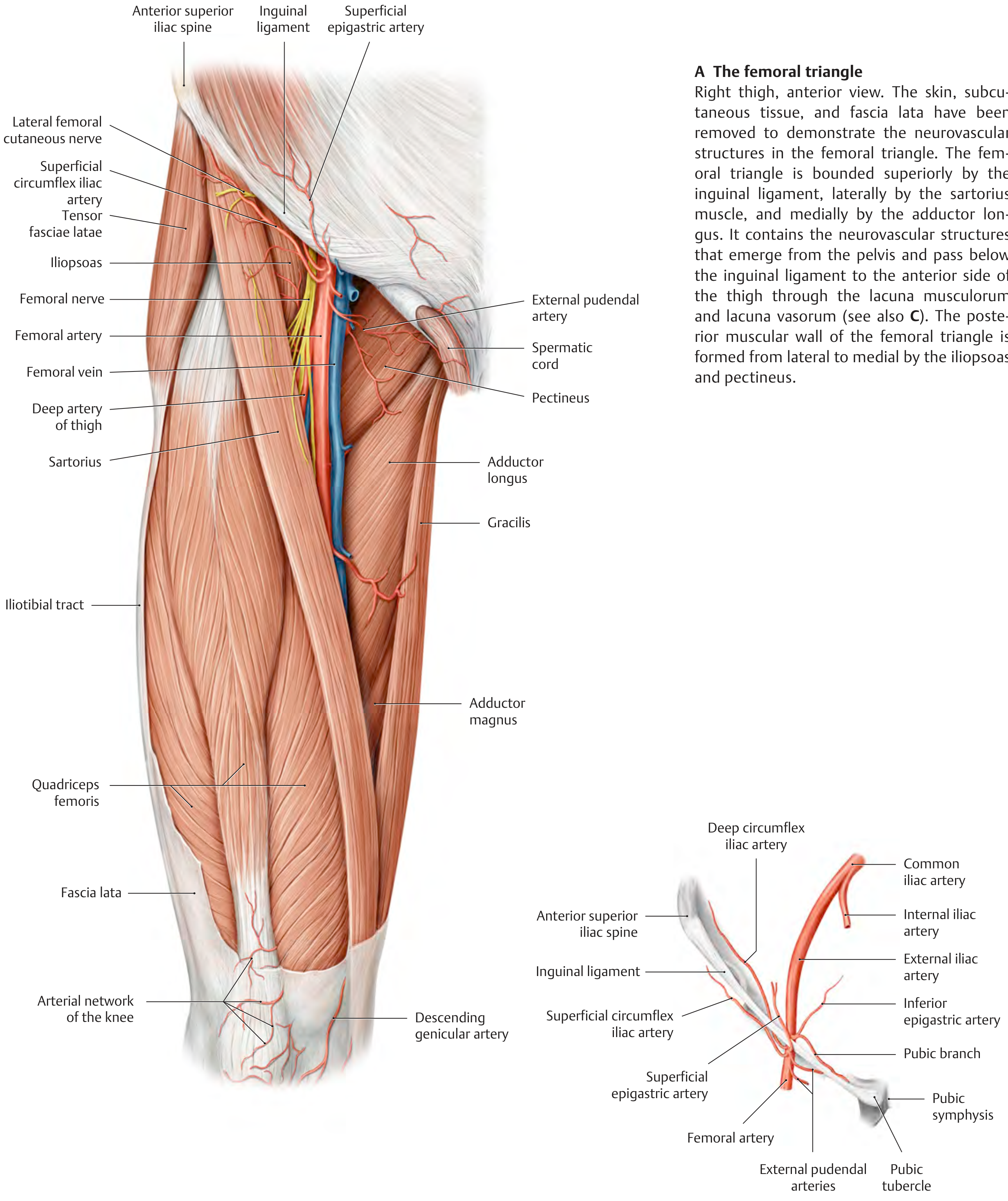
Femoral Sheath
Structure
The femoral sheath is a funnel-shaped fascial sleeve that surrounds the femoral vessels and lymphatics as they pass under the inguinal ligament into the femoral triangle. It is formed by:
- Anteriorly/superiorly: Transversalis fascia (abdominal)
- Posteriorly/superiorly: Iliac fascia
It tapers inferiorly and merges with the connective tissue of the vessels about 3-4 cm below the inguinal ligament.
Important: The femoral nerve is NOT enclosed within the femoral sheath - it lies lateral to the sheath, separated by the iliopsoas fascia.
Three Compartments of the Femoral Sheath
| Compartment | Position | Contents |
|---|---|---|
| Lateral | Lateral | Femoral artery |
| Intermediate | Middle | Femoral vein |
| Femoral canal | Most medial | Lymphatics + areolar tissue (sometimes the Cloquet/Rosenmüller lymph node) |
Femoral Canal
The femoral canal is the medial-most compartment of the sheath. It is conical in shape and clinically important because:
- Its superior opening = the femoral ring
- The femoral ring is a potential weak spot in the abdominal wall
- It is the site of femoral hernias (abdominal contents herniate through the femoral ring into the canal)
- Medially bounded by the lacunar ligament, anteriorly by the inguinal ligament
Femoral Ring Boundaries
| Wall | Structure |
|---|---|
| Anterior | Inguinal ligament |
| Posterior | Pectineal ligament (Cooper's) + pectineus |
| Medial | Lacunar (Gimbernat's) ligament |
| Lateral | Femoral vein (with a thin septum) |
Clinical Relevance
- Vascular access: The femoral artery and vein are the most commonly used large-vessel access points for catheterization - cardiac catheterization, coronary angiography, venous access to the right heart and pulmonary artery all use this route.
- Femoral hernia: More common in women (wider femoral ring). Presents as a lump below and lateral to the pubic tubercle (distinguishing it from an inguinal hernia, which is above and medial to the pubic tubercle). High risk of strangulation.
- Femoral nerve block: Performed just lateral to the femoral artery below the inguinal ligament.
Sources: Gray's Anatomy for Students, pp. 675-676; THIEME Atlas of Anatomy - General Anatomy and Musculoskeletal System, p. 573
This is a shared conversation. Sign in to Orris to start your own chat.