
VT treatment
"ventricular tachycardia" AND management
ventricular tachycardia ECG management algorithm
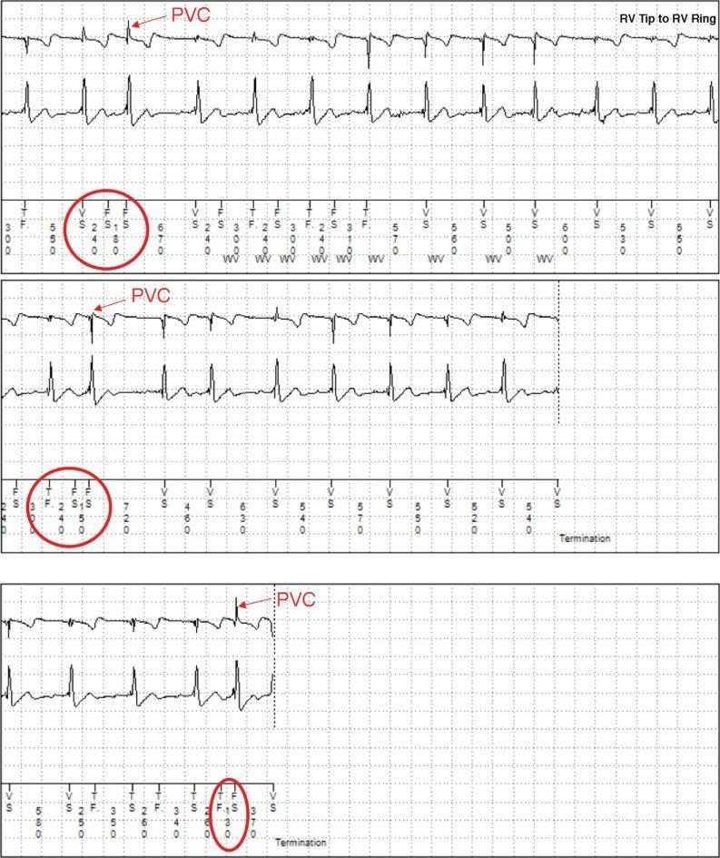
This diagnostic image displays three segments of intracardiac electrogram (EGM) tracings and surface ECG leads, likely recorded from an Implantable Cardioverter Defibrillator (ICD). The primary tracing, labeled 'RV Tip to RV Ring,' illustrates cardiac electrical activity during an episode of non-sustained ventricular tachycardia (NSVT) and frequent premature ventricular contractions (PVCs). Red arrows highlight specific PVCs characterized by wide QRS morphology. The lower portion of each strip includes digital marker annotations common in cardiac rhythm management devices: 'S' (Sensing), 'F' (Fibrillation detection), and 'V' (Ventricular event). The label 'WV' indicates the application of a wavelet discrimination algorithm used to differentiate supraventricular tachycardia from ventricular tachycardia to prevent inappropriate therapy. Numerical values represent R-R and V-V intervals in milliseconds. The segments demonstrate a lead integrity alert (LIA) triggered by rapid R-R intervals (<220 ms) and T-wave oversensing (TWOS) leading to short V-V intervals (<140 ms). The clinical relevance of this image is to demonstrate device-based arrhythmia detection, the use of discrimination algorithms, and the identification of potential sensing artifacts like TWOS in cardiology.
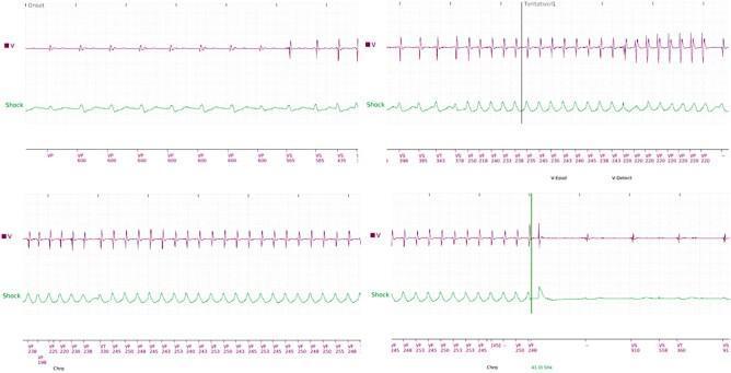
Diagnostic electrocardiographic (ECG) tracings from an implantable cardioverter-defibrillator (ICD) divided into four sequential panels illustrating the management of ventricular arrhythmias. The top row of each panel shows the ventricular intracardiac electrogram (EGM) in red (labeled 'V'), while the bottom row shows the 'Shock' channel in green. The sequence begins (top-left) with a baseline rhythm that transitions into a monomorphic ventricular tachycardia (VT), characterized by a sudden increase in frequency and regular, high-amplitude sharp peaks. The second and third panels (top-right, bottom-left) demonstrate the persistence of this high-frequency ventricular rhythm with associated markers indicating detection. In the final panel (bottom-right), a vertical green marker denotes the delivery of a high-energy defibrillation shock. Following the shock, there is an immediate termination of the tachycardia, evidenced by an abrupt change in the red waveform back to a lower frequency, lower amplitude, and more regular morphology, indicating successful restoration of a stable rhythm. Annotations at the bottom of the strips provide cycle length intervals in milliseconds, confirming the rate changes from VT to a non-tachycardic state.
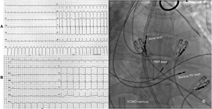
This composite educational image illustrates the management of refractory tachycardia-induced cardiomyopathy. Panel A presents a 12-lead electrocardiogram (ECG) showing atrial fibrillation with a rapid ventricular response (tachyarrhythmia). The tracing is characterized by absent P waves, irregular R-R intervals, and narrow QRS complexes at a rate of approximately 175 bpm. Panel B displays a follow-up ECG after an 'ablate and pace' intervention, specifically showing a more controlled rhythm resulting from atrioventricular (AV) node ablation and subsequent His bundle pacing (HBP). Panel C is a fluoroscopic angiogram in a post-procedural state, depicting the anatomical placement of cardiac pacing leads. Key landmarks include the atrial lead positioned superiorly, the HBP lead situated in the region of the His bundle, and a backup right ventricular (RV) lead. A large, radiopaque extracorporeal membrane oxygenation (ECMO) cannula is visible in the background, crossing the cardiac silhouette. This content demonstrates advanced electrophysiological strategies and mechanical circulatory support in the context of cardiogenic shock and refractory arrhythmia.
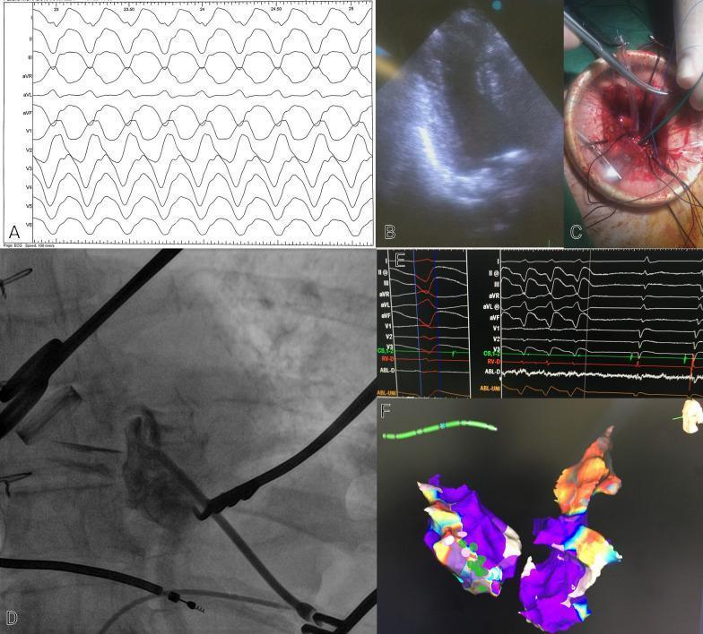
This composite figure illustrates a multidisciplinary approach to managing ventricular tachycardia (VT) via transapical access. Panel A presents a 12-lead electrocardiogram (ECG) tracing showing a wide-complex tachycardia consistent with VT. Panel B is a transthoracic echocardiogram demonstrating an aneurysmal left ventricular (LV) apex. Panel C provides a clinical photograph of a surgical transapical incision in the left thorax, showing stay sutures and surgical instruments utilized for sheath insertion. Panel D displays a fluoroscopic image showing the placement of an ablation catheter within the LV cavity, positioned to avoid contact with metallic prosthetic valves. Panel E contains electrophysiology study (EPS) tracings, including surface leads and intracardiac electrograms (RV-D and ABL-D), showing the termination of VT during radiofrequency (RF) ablation. Panel F exhibits a 3D electroanatomical voltage map of the LV aneurysm and epicardium; purple areas indicate low-voltage scar tissue, while color-coded points signify ablation sites. This collection highlights the integration of cardiac imaging, surgery, and electrophysiology for complex arrhythmia management.
Ventricular Tachycardia (VT) - Treatment
Step 1: Initial Assessment - Hemodynamic Stability
Step 2A: Unstable VT (with pulse) - Immediate Cardioversion
Step 2B: Pulseless VT - ACLS Protocol
Step 3: Stable VT - Pharmacologic Treatment
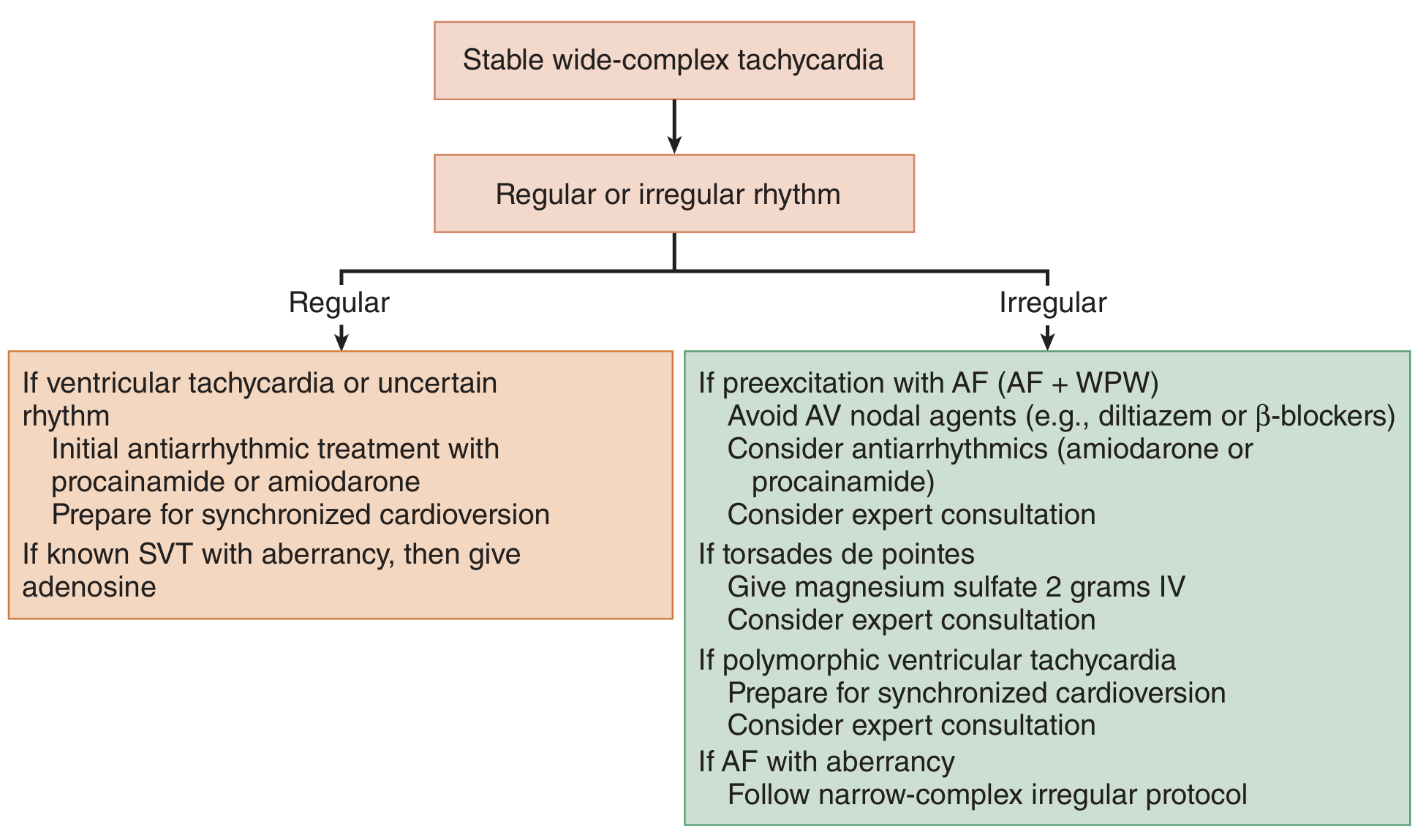
Drug Options for Stable Monomorphic VT
| Drug | Dose | Notes |
|---|---|---|
| Procainamide | 20-50 mg/min IV until arrhythmia suppressed, hypotension, QRS widens >50%, or max 17 mg/kg | Drug of choice for stable VT; superior to amiodarone or lidocaine. Slow infusion required to avoid hypotension. |
| Amiodarone | 150 mg IV over 10 min; then 1 mg/min x 6h, then 0.5 mg/min x 18h | Second-line; preferred in structural heart disease or unknown rhythm |
| Lidocaine | 1-1.5 mg/kg IV bolus; may repeat 0.5-0.75 mg/kg q5-10min; maintenance 1-4 mg/min | Alternative to amiodarone; used especially in ischemic VT |
| Magnesium sulfate | 2 g IV | For torsades de pointes, known hypomagnesemia, or polymorphic VT with prolonged QT |
Procainamide is superior to amiodarone or lidocaine for converting stable VT. If the patient deteriorates during drug therapy, proceed immediately to synchronized cardioversion. - Tintinalli's Emergency Medicine, p. 157
Special Situations
Polymorphic VT / Torsades de Pointes
- IV magnesium sulfate 2 g is first-line
- Overdrive ventricular pacing is the definitive treatment - maintains rate to suppress arrhythmia until the cause of QT prolongation (drug toxicity, electrolyte imbalance) is corrected
- Isoproterenol or phenytoin have been reported in case series but no controlled data
- Remove offending drugs, correct electrolytes (K+, Mg2+)
- Avoid Class IA/III antiarrhythmics that further prolong QT. - Tintinalli's, p. 157
Electrical Storm (Recurrent VT requiring repeated shocks)
- Relieve ischemia (PCI/CABG) - paramount in ischemic VT
- IV esmolol (500 mcg/kg bolus, then 50-200 mcg/kg/min) - short-acting beta-blocker
- IV lidocaine (2-4 mg/min) or IV amiodarone (0.5-1.0 mg/min)
- Intra-aortic balloon pump for hemodynamic support
- Catheter ablation reduces recurrence and may improve survival
- Refractory cases: cardiac sympathetic denervation (left or bilateral), stereotactic radioablation, or renal denervation - Goldman-Cecil Medicine
VT in Acute MI
- Sustained VT with hemodynamic compromise: synchronized cardioversion
- Stable/nonsustained VT: IV amiodarone or lidocaine
- Beta-blockade for polymorphic VT with adrenergic activation ("electrical storm")
- Post-resuscitation from VF/pulseless VT: target normothermia (avoid hyperthermia)
- ICD indicated for VF or hemodynamically significant sustained VT occurring >48 hours after STEMI and not due to a reversible cause. - Goldman-Cecil Medicine
Long-Term / Chronic Management
Implantable Cardioverter-Defibrillator (ICD)
- Primary therapy for survivors of cardiac arrest due to VT/VF
- Indicated for sustained VT with structural heart disease (EF <35%) if not a transient cause
- Reduces mortality better than antiarrhythmic drugs alone
- ~70% of ICD patients with documented VT will receive appropriate therapy over follow-up. - Goldman-Cecil Medicine
Antiarrhythmic Drugs (adjunct to ICD)
- Amiodarone 200-400 mg daily - most effective oral agent; reduces ICD shocks
- Beta-blockers (metoprolol, atenolol, nadolol) - first-line in idiopathic VT, long QT, catecholaminergic polymorphic VT
- Sotalol - alternative; can worsen QT in renal impairment
- Verapamil (SR 180-360 mg/day) - specifically for idiopathic left fascicular VT (verapamil-sensitive VT)
Catheter Ablation
- Cures 80-90% of idiopathic VT (outflow tract, fascicular) with <1% major complication rate - preferred over long-term drug therapy
- For ischemic/structural VT: palliative, reduces ICD shock burden, but does not replace ICD
- Endpoint and epicardial approach used for ARVC (nearly half have epicardial origin)
- Brugada syndrome: epicardial ablation of RVOT substrate can normalize ECG; quinidine 600-900 mg/day in divided doses is the only effective drug. - Goldman-Cecil Medicine
Disease-Specific Long-Term Approach
| Condition | Treatment |
|---|---|
| Idiopathic VT (no structural disease) | Catheter ablation (preferred), beta-blocker, verapamil (fascicular VT) |
| Ischemic cardiomyopathy | ICD + beta-blocker + amiodarone; catheter ablation for recurrent VT |
| Non-ischemic cardiomyopathy | ICD + beta-blocker; amiodarone/sotalol as adjuncts |
| Long QT syndrome | Beta-blocker; ICD if breakthrough; left cardiac sympathetic denervation |
| Catecholaminergic polymorphic VT (CPVT) | Beta-blocker; flecainide adjunct; ICD for refractory cases |
| ARVC | ICD + catheter ablation (endo + epicardial approach) |
| Hypertrophic cardiomyopathy | ICD; amiodarone sometimes useful |
| Brugada syndrome | Quinidine; catheter ablation (epicardial RVOT); ICD for high-risk |
2025 AHA Guideline Update
- Pulseless VT - Defibrillate + CPR immediately
- Unstable VT with pulse - Synchronized cardioversion + sedation
- Stable VT - Procainamide (first choice) > Amiodarone > Lidocaine
- Torsades - Magnesium + overdrive pacing + remove QT-prolonging drugs
- Electrical storm - Beta-blocker (esmolol) + amiodarone + ablation
- Long-term - ICD for structural disease; catheter ablation for idiopathic VT; amiodarone/beta-blockers as adjuncts

What is diagnosis
ECG Monitor Analysis
Monitor Findings
| Parameter | Value |
|---|---|
| Heart Rate | 120 bpm |
| Rhythm | Regular |
| Resp Rate | 17 rpm |
| SpO2 | ⚠️ "!SpO2 No Pulse" (probe not reading) |
| BP | ⚠️ "!NIBP Cuff Loose" (not reading) |
| Previous HR readings | 143 bpm (08:01), 142 bpm (08:03), 95 bpm (08:30) |
ECG Interpretation
- Rate: 120 bpm - tachycardic
- Rhythm: Regular
- QRS morphology: The QRS complexes appear widened (broad morphology visible in both leads), with a somewhat irregular and bizarre appearance
- P waves: Not clearly discernible - likely absent or dissociated
- QRS axis: Appears deviated
Likely Diagnosis
Wide-Complex Tachycardia - most consistent with Ventricular Tachycardia (VT)
- Rate 120 bpm with regular rhythm
- Wide, bizarre-looking QRS complexes
- No clearly identifiable P waves
- Prior higher rates (142-143 bpm) suggest the arrhythmia may be evolving or partially treated
- SpO2 probe showing "No Pulse" - could indicate poor peripheral perfusion (supports hemodynamic compromise)
Differential Diagnosis
| Diagnosis | Likelihood |
|---|---|
| Monomorphic Ventricular Tachycardia | Most likely |
| SVT with aberrant conduction (bundle branch block) | Possible |
| Accelerated idioventricular rhythm | Less likely (rate >100) |
Immediate Action
- Is there a pulse? (SpO2 probe not reading)
- Is the patient hemodynamically stable (BP not reading)?
- If pulseless → CPR + defibrillation immediately
- If unstable with pulse → Synchronized cardioversion
- If stable → IV procainamide or amiodarone; 12-lead ECG for confirmation
⚠️ The "!SpO2 No Pulse" alarm is a critical red flag - this may represent pulseless VT requiring immediate ACLS intervention.
Can we add vaso dopamin adr norad at atime