
What is rooting reflex in child
rooting reflex newborn infant
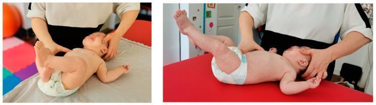
Two clinical photographs demonstrate the application of Stage 1 reflex rolling in Vojta therapy on an infant. The infant is positioned in a supine (decubitus dorsalis) position on a therapy table. The therapist is shown applying specific tactile stimulation and resistance to facilitate innate motor patterns. One hand of the therapist provides resistance at the head, specifically targeting the zygomatic bone, nuchal line, and mastoid tuberosity to inhibit head rotation, while the other hand stimulates the pectoral or chest area. The images capture kinesiological responses, including limb extension and elevation. The clinical focus is on neurorehabilitative intervention for infants with motor development impairments, such as hypertonia or hypotonia following neonatal hypoxia. The infant's facial expression indicates the intense physical effort or sensory input characteristic of this neuromuscular activation therapy. This visual serves as an educational guide for pediatric physical therapy and diagnostic reflex locomotion assessment.
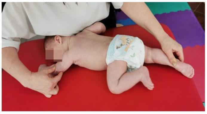
A clinical photograph illustrating a pediatric physical therapy session, specifically demonstrating Vojta therapy and the reflex crawling (Reflexkriechen) technique. The infant is positioned in a prone posture on a stable red treatment surface within a clinical or educational setting. A therapist's hands are visible, applying specific tactile stimuli and directional pressure to trigger global motor patterns. The infant's posture shows asymmetrical limb positioning characteristic of the crawling reflex: one arm is extended while the ipsilateral leg is flexed toward the torso, and the opposite limbs are in a complementary state of extension and flexion. This technique is often used in the rehabilitation of motor development delays and conditions such as muscular torticollis to improve neurological maturation and postural control. The image serves as an educational example of neuro-developmental treatment (NDT) and the assessment of primitive reflexes in infants.
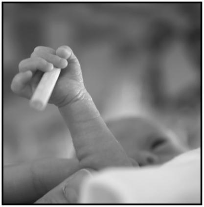
This grayscale clinical photograph depicts the palmar grasp reflex in a preterm infant. The focus is on the infant's hand and forearm as it securely grips a small, smooth cylindrical object. The infant's fingers are flexed around the cylinder, demonstrating the tactile and motor response characteristic of the primitive grasp reflex. The cylinder, approximately 35 mm in length and 6 mm in diameter, is positioned within the palm, with the distal end protruding. In the blurred background, the infant's face is partially visible, indicating the neonatal clinical setting. This image serves as an educational tool for neonatal development, illustrating haptic perception and sensorimotor coordination in premature newborns. It is particularly relevant for pediatric and neonatal research focusing on the maturation of the central nervous system and the evaluation of primitive reflexes as indicators of neurological health.
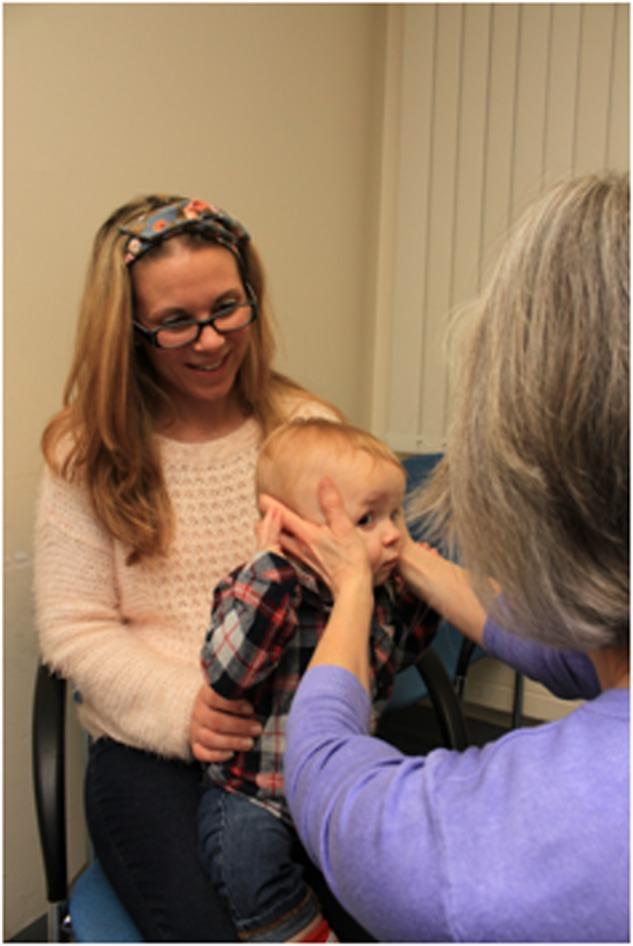
Clinical photograph demonstrating the 'doll's head manoeuvre' (oculocephalic reflex testing) performed on an infant in a pediatric or neuro-ophthalmological setting. The infant is seated on a caregiver's lap, who provides trunk stabilization. A clinical examiner, positioned in front of the infant, gently but firmly holds the child's head with both hands placed laterally over the parietal-temporal regions. The image illustrates the correct positioning for manual head rotation to assess the Vestibulo-Ocular Reflex (VOR). The procedure involves rapid horizontal or vertical head movements while observing the infant's eyes; a normal response is the maintenance of steady gaze or eye movement in the direction opposite to the head rotation. This clinical test is essential for evaluating brainstem function and vestibular system integrity in non-verbal or uncooperative pediatric patients, provided there are no contraindications such as cervical spine instability.
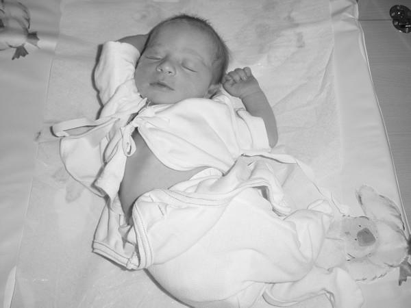
This clinical photograph shows a newborn infant in a supine position, representing standard neonatal care and attire within a maternity ward setting. The infant is dressed in a light-colored, long-sleeved cotton shirt and matching leggings. The shirt is designed with a side-tie closure, currently open at the abdomen to allow for clinical observation or umbilical cord care. The infant appears in a relaxed, sleeping state with closed eyes and arms flexed toward the head, indicating normal neurological tone and comfort. The infant is placed on a flat, padded surface covered with a protective sheet, typical of a nursery changing station or bassinet. A plush toy is visible near the feet. This image serves to illustrate traditional postpartum care practices and the use of loose-fitting infant apparel as an alternative to swaddling in clinical research focused on early infant development and breastfeeding outcomes.
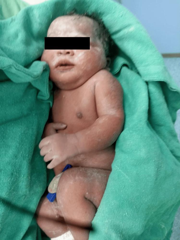
Clinical photograph of a newborn immediately following delivery, likely via cesarean section as indicated by the surgical setting. The infant presents with a healthy reddish skin tone (erythema neonatorum) and is partially covered in vernix caseosa, a protective white, creamy biofilm visible on the face, scalp, and limbs. The newborn is lying in a supine position on a green sterile surgical drape. Anatomical features include a flexed posture of the lower extremities and a blue plastic umbilical cord clamp applied to the umbilical stump. The face shows mild physiological edema common in neonates. This image serves as an educational example of immediate neonatal appearance and the outcome of obstetric anesthetic management. For patient privacy, the eyes are obscured by a black bar.
Rooting Reflex in Children
Definition
How to Elicit It
Purpose and Significance
Age of Appearance and Disappearance
| Feature | Detail |
|---|---|
| Present from | Birth |
| Disappears | 3-4 months of age (normal) |
| In sleep | May persist up to 12 months during sleep |
Classification
| Primitive Reflex | Ages Present |
|---|---|
| Stepping reflex | Birth to 1-2 months |
| Galant reflex | Birth to 2 months |
| Asymmetric tonic neck reflex | Birth to 2-4 months |
| Rooting reflex | Birth to 3-4 months |
| Palmar/plantar grasp reflex | Birth to 3-4 months |
| Moro (startle) reflex | Birth to 4-6 months |
| Babinski reflex | Birth to 12 months |
Clinical Relevance
-
Normal development: Presence of the rooting reflex in a newborn confirms normal brainstem function and neurological integrity. Absence at birth is a warning sign.
-
Persistence beyond expected age: If the rooting reflex does not disappear by 3-4 months, it may indicate a neurological problem such as cerebral palsy or developmental delay - primitive reflexes are normally suppressed by maturing cortical (frontal lobe) pathways.
-
Re-emergence in adults (frontal release sign): The rooting reflex can re-appear in adults with frontal lobe damage - such as bilateral damage to mesiofrontal structures, anterior cerebral artery (ACA) infarction, or diffuse cortical disease. In this context it is called a "frontal release sign" alongside sucking and snout reflexes, indicating loss of normal cortical inhibition. Per Localization in Clinical Neurology, "turning of the mouth toward a round object that approaches the patient's mouth or gently strokes her cheek (rooting reflex)... may reflect impairment of the corticobulbar projection."
Summary
- Textbook of Family Medicine, 9e - Table 21-5 Primitive Neurologic Reflexes
- Kaplan and Sadock's Synopsis of Psychiatry - Developmental Milestones in Infants, p. 2756
- Localization in Clinical Neurology, 8e - Sucking, Snout, Rooting Reflexes
What is grasp reflex in short
Grasp Reflex (Short Answer)
Definition
Two Types
| Type | Stimulus | Response |
|---|---|---|
| Palmar grasp | Press against the palm (between thumb and index finger) | Fingers curl and grip tightly |
| Plantar grasp | Press against the sole | Toes curl downward |
How to Elicit
Age
- Present: From birth
- Disappears: 3-4 months of age (palmar); plantar grasp persists slightly longer
Clinical Significance
-
In newborns: A normal primitive reflex confirming intact neurological function. Its presence at birth and disappearance by 4 months follows normal cortical maturation.
-
Persistence beyond 4 months: Suggests delayed cortical maturation or neurological damage (e.g., cerebral palsy).
-
Re-emergence in adults (frontal release sign): Reappearance of the grasp reflex in adults indicates damage to the contralateral frontal lobe - specifically area 6 (premotor cortex) on the mesial hemisphere. It is seen in frontal lobe lesions, bilateral prefrontal impairment, and conditions like frontotemporal dementia. It reflects loss of cortical inhibition over primitive brainstem circuits.
Per Localization in Clinical Neurology: "Damage to the contralateral area 6, particularly in the mesial aspect of the hemisphere, accounts for the release of the grasp reflex."
- Localization in Clinical Neurology, 8e - Grasp Reflex section
- Textbook of Family Medicine, 9e - Table 21-5 Primitive Neurologic Reflexes
- Kaplan and Sadock's Synopsis of Psychiatry - Developmental Milestones in Infants
Moro reflex
Moro reflex startle reflex newborn infant arms abduct
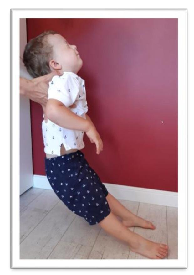
Clinical photograph illustrating the assessment of the Moro reflex in a preschool-aged child. The image depicts a therapist supporting the child's upper torso and head while the child is leaned backward. The child's head is in a position of cervical extension (tilted back) with eyes closed, which is a provocative maneuver to stimulate the vestibular system and trigger the reflex. The upper limbs are observed in a state of abduction or 'throwing out' to the sides, while the lower limbs show knee flexion. This clinical examination is used in pediatric neurology and physiotherapy to assess the integration of primitive reflexes. Persistent, unintegrated Moro reflexes beyond early infancy can indicate developmental delays or neurological maturity issues. The image demonstrates the standard physical therapy test position used to evaluate for residual reflex activity in older children, where compensatory movements like arm flailing or loss of balance are monitored.
This clinical photograph shows a newborn infant in a supine position, representing standard neonatal care and attire within a maternity ward setting. The infant is dressed in a light-colored, long-sleeved cotton shirt and matching leggings. The shirt is designed with a side-tie closure, currently open at the abdomen to allow for clinical observation or umbilical cord care. The infant appears in a relaxed, sleeping state with closed eyes and arms flexed toward the head, indicating normal neurological tone and comfort. The infant is placed on a flat, padded surface covered with a protective sheet, typical of a nursery changing station or bassinet. A plush toy is visible near the feet. This image serves to illustrate traditional postpartum care practices and the use of loose-fitting infant apparel as an alternative to swaddling in clinical research focused on early infant development and breastfeeding outcomes.
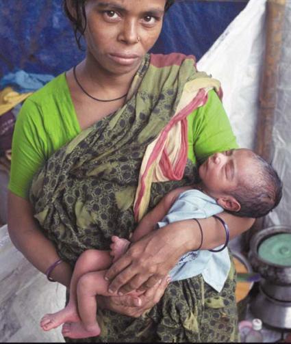
Clinical photograph of a mother holding a newborn infant in a domestic or field setting, representative of maternal and child health studies in South Asian regions like Dhaka, Bangladesh. The mother is shown in a three-quarter view, wearing traditional attire including a green blouse and a floral-patterned shawl. The newborn is positioned in the mother's arms in a resting, supine-to-sidelying posture. The infant has light brown skin, visible scalp hair, and appears stable with no immediate signs of acute respiratory distress or visible cutaneous lesions. From an educational perspective, this image illustrates neonatal care, the postpartum period, and the socio-economic determinants of health in developing urban environments. It serves as a visual reference for community health outreach, breastfeeding support, and pediatric assessment in global health contexts.
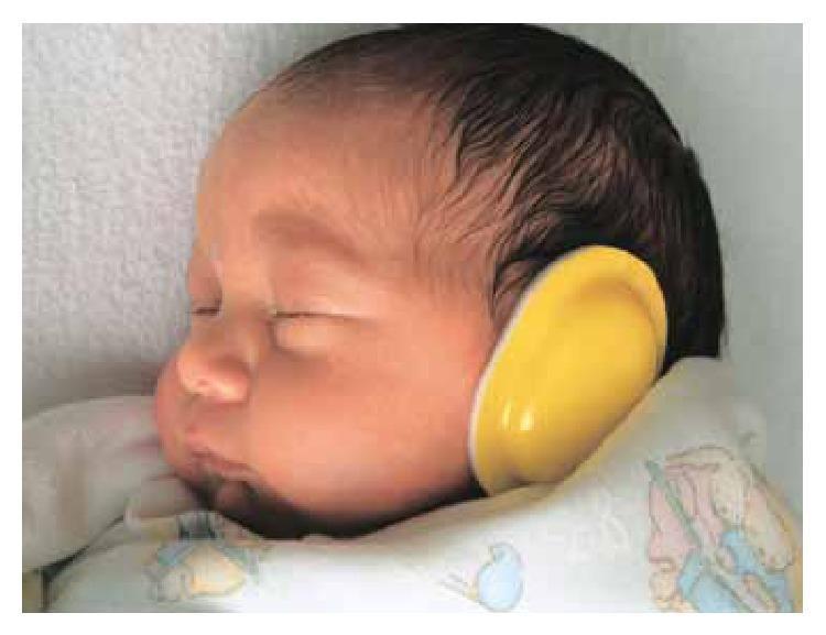
This clinical photograph displays a newborn infant in a resting state within a Neonatal Intensive Care Unit (NICU) setting. The infant is shown in a lateral profile, featuring a fair skin tone and dark, fine terminal hair. A primary visual focus is a yellow, oval-shaped noise-reduction earmuff applied over the external ear. This device is utilized as a non-pharmacological intervention aimed at neuroprotection and developmental care by mitigating high decibel levels in the NICU environment. Such interventions are clinically relevant for reducing stress-induced motor responses, such as tremors and startle reflexes, and for supporting physiological stability and weight gain in neonates. The image demonstrates the practical application of environmental modification protocols designed to protect fragile auditory systems and promote neurodevelopmental maturation in early infancy.
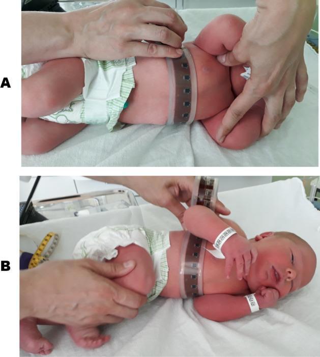
Two-panel clinical photograph (A and B) demonstrating a medical procedure in a neonatal setting. The images show a newborn infant placed in a lateral decubitus position on a clinical examination surface for torso shape measurement. An electronic measuring belt—constructed from a flexible, transparent material with integrated, equidistantly spaced dark sensors—is wrapped circumferentially around the infant’s chest. In panel A, the infant's arms are positioned upwards and away from the body, leaving the chest wall relatively uncompressed. In panel B, the arms are positioned downward against the chest, resting between the mattress and the torso, which visibly compresses and alters the cross-sectional shape of the rib cage. A clinician’s hands are visible, stabilizing the infant’s position and securing the sensor belt for data acquisition. The visual content serves to demonstrate the biomechanical impact of posture and limb position on the compliance and deformation of the neonatal chest wall, relevant for respiratory and anthropometric research in pediatrics and neonatology.
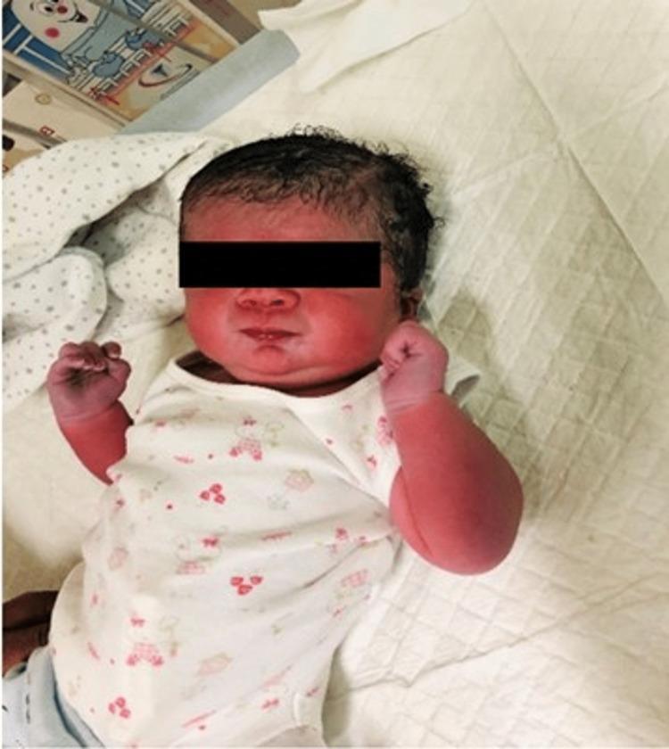
Clinical photograph of a late preterm newborn infant following delivery. The image shows a macrosomic infant (birth weight 3.9 kg) with a prominent reddish hue (plethora) visible on the face, arms, and hands. The infant has dark, thick, curly scalp hair and is shown in a supine position with hands clenched. The facial profile does not exhibit overt dysmorphic features. The clinical context involves an infant of a diabetic mother (IDM), born via cesarean delivery, who subsequently developed complications including respiratory distress, hypocalcemia, and hypoglycemia. This visual serves as an educational example of a newborn affected by gestational diabetes, illustrating the physical appearance of macrosomia and cutaneous plethora without specific congenital anomalies or dysmorphic features.
Moro Reflex (Startle Reflex)
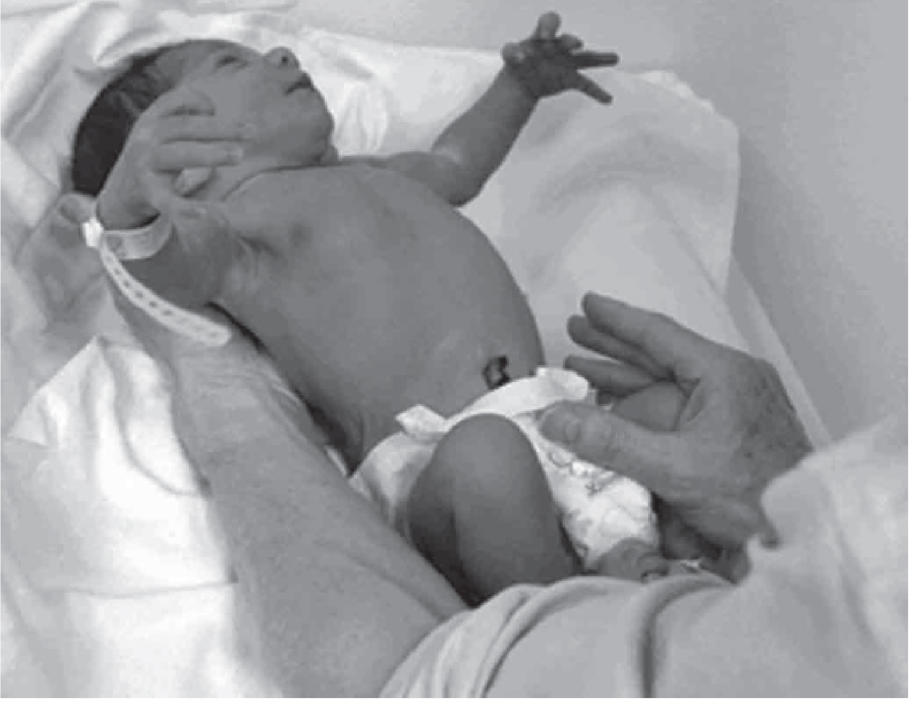
Definition
How to Elicit
- Phase 1 - Abduction/Extension: Both arms abduct and extend outward; fingers splay open (digital extension)
- Phase 2 - Adduction: Arms come back together in a hugging/embracing motion; fists close
- Cry often follows
Age
| Present from | Birth (even in premature infants) |
| Disappears | 4-6 months of age |
Clinical Significance
1. Normal neonatal assessment
2. Asymmetric Moro reflex
- Brachial plexus injury (Erb's palsy - C5/C6; Klumpke's palsy - C7/C8/T1) - the affected arm will not abduct/extend properly
- Fractured clavicle (pain inhibits response on that side)
- Hemiplegia
3. Absent Moro reflex
- Severe birth asphyxia / hypoxic-ischemic encephalopathy
- Severe neurological depression
- Kernicterus (high bilirubin) - associated with shrill cry, decreased tone, and decreased Moro reflex
4. Weak Moro reflex
- Seen in neonatal myasthenia gravis - signs include poor cry, respiratory difficulties, weak suckling, and feeble Moro reflex
5. Persistent Moro reflex beyond 6 months
- Imposable asymmetric tonic neck reflex
- Persistent Moro reflex
- Strong extensor thrust on vertical suspension
- Persistent neck-righting reflex
- Absence of parachute reaction after 11 months
Summary Table
| Feature | Detail |
|---|---|
| Also called | Startle reflex |
| Stimulus | Sudden head drop, loud noise, unexpected stimulus |
| Response | Arms abduct + extend + fingers spread → then adduct back |
| Present | Birth to 4-6 months |
| Absent at birth | Severe neurological depression, birth asphyxia |
| Asymmetric | Brachial plexus injury, clavicle fracture |
| Persists beyond 6 months | Cerebral palsy, severe brain damage |
- Kaplan and Sadock's Synopsis of Psychiatry - Developmental Milestones in Infants (Fig. 32-6)
- Campbell's Operative Orthopaedics, 15e - Diagnosis of Cerebral Palsy
- Textbook of Family Medicine, 9e - Table 21-5 Primitive Neurologic Reflexes
Babinski reflex in short
Babinski Reflex (Short)
Definition
How to Elicit
- Use a blunt instrument (e.g., key or tongue depressor)
- Stroke the lateral (outer) plantar surface of the foot from the heel upward toward the little toe, then medially across the ball of the foot
- Apply mild, not forceful, pressure
Responses
| Response | Big Toe Movement | Meaning |
|---|---|---|
| Normal (adult) | Plantar flexion (curls downward) | Normal corticospinal tract |
| Positive Babinski sign | Dorsiflexion (extends upward) + fanning of other toes | UMN (corticospinal) damage |
| Normal (infant <12 months) | Extension (upward) | Normal - corticospinal tracts not yet myelinated |
Age in Children
- Normal in infants up to 12 months (some say up to 2 years) - upward toe extension is the expected response because the corticospinal tracts are not yet fully myelinated
- Disappears as myelination completes and the Babinski response becomes plantar flexion (downward)
Clinical Significance in Adults
- Spasticity (increased tone)
- Hyperreflexia
- Weakness
- Loss of abdominal reflexes
Alternative Tests (Same Significance as Babinski)
- Chaddock sign - stroke lateral foot below lateral malleolus
- Oppenheim sign - scrape down the shin
- Gordon sign - squeeze the calf
- Triple flexion reflex - pathological spinal reflex (flexion of knee, hip, and dorsiflexion of foot) - same significance as Babinski
"The plantar reflexes, particularly the elicitation of the Babinski sign by stroking the lateral sole of the foot from heel to toe, are an essential part of most examinations. The sign is a dependable marker of damage to the corticospinal system." - Adams and Victor's Principles of Neurology, 12e
- Adams and Victor's Principles of Neurology, 12e - Testing of Reflexes
- Tintinalli's Emergency Medicine - BASIC Neurological Examination
- Kaplan and Sadock's Synopsis of Psychiatry - Developmental Milestones in Infants
Doll's eye reflex in short
Doll's Eye Reflex (Short)
Definition
How to Elicit
- Prerequisite: First rule out cervical spine injury before performing
- Hold the patient's eyes open
- Rotate the head rapidly from side to side (horizontal) or up and down (vertical)
- Observe the direction of eye movement
Responses
| Response | Eye Movement | Interpretation |
|---|---|---|
| Doll's eyes PRESENT (positive) | Eyes move opposite to head rotation (like a doll's fixed eyes) | Brainstem intact - VOR pathways functioning |
| Doll's eyes ABSENT (negative) | Eyes move with the head, or no movement | Brainstem dysfunction - midbrain/pons damage |
| Normal awake patient | Doll's eyes NOT present | Visual fixation and voluntary eye movements suppress the reflex - absence is NORMAL in awake patients |
Key Rule
"Note that in awake patients, doll's eyes are usually not present. This is because visual fixation and voluntary eye movements mask the reflex. Thus, the absence of doll's eyes suggests brainstem dysfunction in the comatose patient but can be normal in the awake patient."
- Neuroanatomy through Clinical Cases, 3e
Neural Pathway
- CN VIII (vestibular input - afferent)
- Brainstem pathways from medulla → pons → midbrain
- CN III, IV, VI (ocular motor - efferent)
- Interconnecting MLF (medial longitudinal fasciculus)
Clinical Significance
| Situation | Doll's Eyes | Meaning |
|---|---|---|
| Coma with intact brainstem | Present | Metabolic/hemispheric cause of coma |
| Coma with brainstem damage | Absent | Structural brainstem lesion |
| Brain death | Absent | Brainstem non-functional |
| Drug overdose | May be absent | Mimic of brainstem damage - but pupils remain reactive |
More Potent Alternative: Caloric (Cold Water) Test
- Irrigate external auditory canal with cold water (head at 30°)
- Normal response in coma: tonic eye deviation toward cold water side
- Mnemonic: COWS - Cold water = eyes deviate to the Opposite side for fast phase (warm = Same side)
- In coma, the fast phase (nystagmus) is often absent; only slow tonic deviation remains
- Harrison's Principles of Internal Medicine, 22e - Brainstem Reflexes
- Neuroanatomy through Clinical Cases, 3e - Cranial Nerves / Vestibulo-ocular reflex
- Bradley and Daroff's Neurology in Clinical Practice - Ocular Motility