
Explain me OCT as three fluid spaces in retina
OCT retina fluid spaces subretinal intraretinal sub-RPE
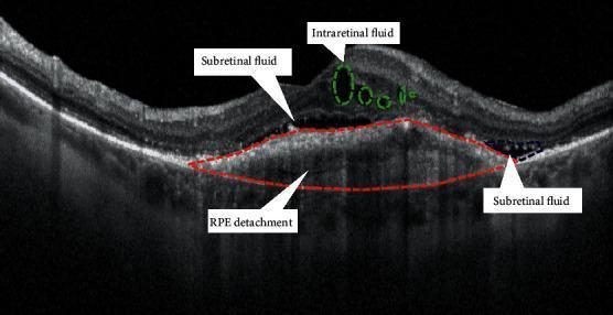
This diagnostic image is a B-scan spectral-domain optical coherence tomography (OCT) of the macula, demonstrating the hallmark signs of exudative (neovascular) age-related macular degeneration (nAMD). The cross-sectional view shows three distinct types of fluid accumulation. First, intraretinal fluid (IRC) is visible as hyporeflective cystoid spaces within the inner retinal layers, outlined by green dashed circles. Second, subretinal fluid (SRF) is present as hyporeflective pockets between the neurosensory retina and the retinal pigment epithelium (RPE), indicated by white arrows. Third, a prominent serous pigment epithelial detachment (PED) is highlighted by a red dashed outline, showing the elevation of the hyperreflective RPE band away from the underlying Bruch's membrane due to sub-RPE fluid. The image illustrates the complex fluid distribution common in type 1 and type 2 macular neovascularization, emphasizing the structural disruption of the retinal layers and the RPE-choriocapillaris complex.
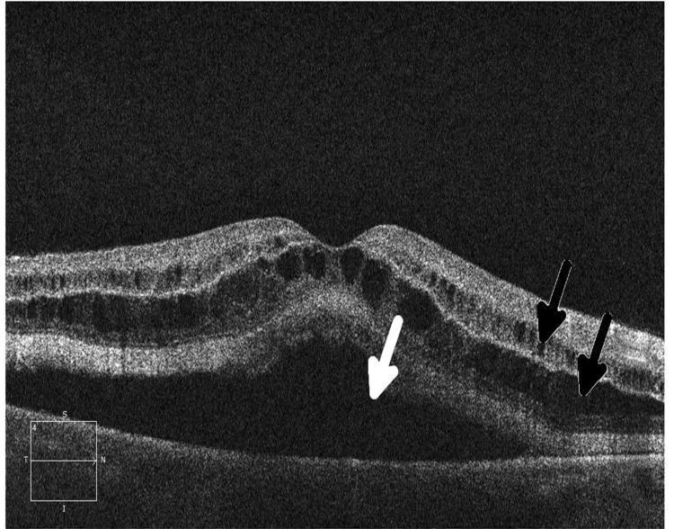
This diagnostic image is a cross-sectional Optical Coherence Tomography (OCT) scan of the human retina, demonstrating drug-induced retinopathy secondary to MEK inhibitor therapy. The scan shows significant disruption of normal macular architecture characterized by both intraretinal and subretinal fluid accumulation. Intraretinal fluid is visible as multiple, hyporeflective (dark), oval-shaped cystic spaces located within the inner retinal layers, leading to increased retinal thickness. Subretinal fluid is identified as a large, homogeneous hyporeflective area beneath the neurosensory retina, causing a serous retinal detachment from the underlying retinal pigment epithelium (RPE). The foveal contour is distorted by the elevation of the neurosensory layers. Black arrows highlight the intraretinal cystic changes, while a white arrow points to the subretinal fluid collection. This imaging is characteristic of MEK inhibitor-associated retinopathy, an important side effect in ophthalmology and oncology for medical students and clinicians to recognize.
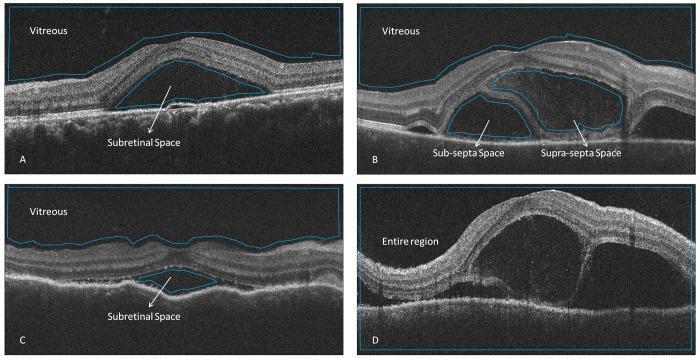
Diagnostic optical coherence tomography (OCT) B-scans of the retina comparing fluid accumulation patterns in Central Serous Chorioretinopathy (CSCR) and Vogt-Koyanagi-Harada (VKH) disease. Panel A (CSCR) and Panel C (VKH without septa) show classic subretinal fluid (SRF) manifesting as hyporeflective spaces between the neurosensory retina and the retinal pigment epithelium (RPE), characterized by a single, continuous fluid pocket. Panel B illustrates VKH with subretinal septa, featuring thin, hyperreflective membranes (septa) that compartmentalize the subretinal space into 'supra-septa' and 'sub-septa' fluid pockets. Panel D demonstrates the segmentation of the 'entire region' for optical intensity analysis. The images highlight key morphological biomarkers in inflammatory uveitis (VKH), specifically the presence of subretinal fibrin or septa, which distinguishes it from the typically non-septated fluid seen in CSCR. The educational focus is on identifying subretinal compartmentalization and assessing the reflectivity (optical intensity) of fluid in different posterior segment pathologies.
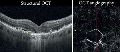
This diagnostic composite includes a structural Optical Coherence Tomography (OCT) cross-section and an OCT Angiography (OCTA) en face scan of the retina. The structural OCT demonstrates signs of choroidal neovascularization (CNV) complicated by retinal pigment epithelium (RPE) atrophy. Key pathological features include intraretinal cystic spaces indicative of edema and subretinal fluid situated above a sub-RPE hyperreflective material. A prominent area of RPE atrophy is marked by increased foveal backscattering of the OCT signal into the deeper choroid. The accompanying OCTA image displays a distinct, round flow signal with a peripheral capillary fringe, confirming active neovascularization. These images illustrate the clinical progression of CNV where secondary atrophy may limit visual outcomes despite anti-VEGF therapy. The content is suitable for ophthalmology students and specialists studying retinal pathologies and multimodal imaging interpretation.
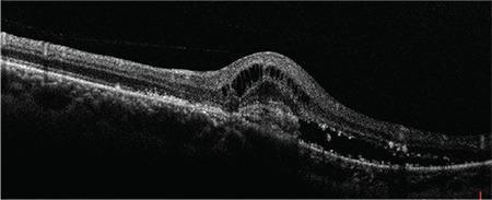
This diagnostic image is an Optical Coherence Tomography (OCT) cross-section of the human retina, demonstrating classic features of active choroidal neovascularization (CNV). The scan reveals a significant elevation of the neurosensory retina caused by subretinal fluid (SRF), appearing as a well-defined hyporeflective space between the photoreceptor layer and the underlying retinal pigment epithelium (RPE). Additionally, multiple hyporeflective intraretinal cystoid spaces are visible, indicating intraretinal fluid (IRF) or edema secondary to vascular leakage. There is an irregular, hyperreflective lesion at the level of the RPE-choriocapillaris complex, consistent with a neovascular membrane. These structural abnormalities disrupt the normal foveal contour and retinal lamination. The findings are characteristic of exudative maculopathy, often associated with conditions such as Presumed Ocular Histoplasmosis Syndrome (POHS) or neovascular age-related macular degeneration. This visual is an essential educational tool for identifying biomarkers of CNV activity and monitoring response to anti-VEGF therapy in ophthalmology.
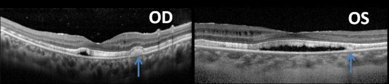
Diagnostic Image: Optical Coherence Tomography (OCT) cross-sections of the right (OD) and left (OS) eyes of a 23-year-old female patient. The images demonstrate several pathological findings characteristic of complex retinal disease. Both scans show a diffused shallow serous neuroretinal detachment and thinning of the retinal nerve fiber layer. There are clear parafoveal intraretinal fluid collections (cystoid spaces) present within the macular retinal layers. Blue arrows indicate discrete, hyperreflective subretinal accumulations located between the sensory retina and the retinal pigment epithelium (RPE). Additionally, both eyes exhibit significant elongation and thickening of the photoreceptor outer segment layer. These features are clinically significant for evaluating conditions involving fluid redistribution, such as uveitis, central serous chorioretinopathy, or secondary effects of angle-closure glaucoma post-surgical intervention. The OCT provides high-resolution visualization of the retinal architecture, allowing for the differentiation between intraretinal edema and subretinal exudative material.
OCT: The Three Fluid Spaces in the Retina
1. Intraretinal Fluid (IRF)
- Hyporeflective (dark) cystoid or oval spaces within the retinal layers
- Causes retinal thickening and disrupts the normal layered architecture
- Can be seen in inner or outer retinal layers depending on the cause
- Also called cystoid macular edema (CME) when the cysts are prominent
- Results from breakdown of the inner blood-retinal barrier (capillary leakage)
- Fluid accumulates in the spaces of Muller cells and Henle's fiber layer
- Diabetic macular edema (most common cause)
- Branch/central retinal vein occlusion
- Post-surgical CME (Irvine-Gass syndrome)
- Uveitis
- Neovascular AMD (Type 2 and Type 3 MNV)
2. Subretinal Fluid (SRF)
- Homogeneous hyporeflective (optically clear) dome-shaped pocket beneath the neurosensory retina
- The neurosensory retina is lifted away from the RPE band
- The RPE band itself remains flat (this distinguishes it from a PED - see below)
- No cystoid changes within the retina
- Results from breakdown of the outer blood-retinal barrier (RPE pump failure or neovascular leakage)
- The neurosensory retina is separated from its nutritional source (choroid), so photoreceptors are at risk
- Central serous chorioretinopathy (CSCR) - the prototype
- Rhegmatogenous retinal detachment (fluid from the vitreous enters through a break)
- Neovascular AMD - Type 2 MNV (neovascularization sits below the NSR but above the RPE, leaking into this space)
- Vogt-Koyanagi-Harada disease (often has subretinal septa partitioning the fluid)
- Choroidal tumors (e.g., choroidal melanoma, metastases)
3. Sub-RPE Fluid (Pigment Epithelial Detachment - PED)
- The RPE band is elevated - it lifts off Bruch's membrane forming a dome or peaked elevation
- The space beneath the lifted RPE can be:
- Optically clear (serous PED): smooth dome, uniform dark interior - due to pure fluid
- Heterogeneous reflectivity (fibrovascular PED / Type 1 MNV): irregular internal reflectivity with vascular elements, often multilobulated - due to neovascular tissue beneath RPE
- Highly reflective (drusenoid PED): contents are drusen material
- Bruch's membrane is usually seen as a thin hyperreflective line at the base
- Type 1 MNV (formerly "occult CNV") is neovascularization that is above Bruch's membrane but below the RPE - it causes a fibrovascular PED. On OCT, this shows elevation of the RPE with heterogeneous internal reflectivity.
- A serous PED in CSCR results from hydrostatic pressure from the choroid pushing fluid under the RPE.
- Neovascular AMD - Type 1 MNV (fibrovascular PED)
- Central serous chorioretinopathy (serous PED)
- Polypoidal choroidal vasculopathy (PCV) - often large serous/hemorrhagic PED
- Drusenoid PED in dry AMD
Quick Comparison Table
| Feature | Intraretinal Fluid (IRF) | Subretinal Fluid (SRF) | Sub-RPE Fluid (PED) |
|---|---|---|---|
| Location | Within NSR layers | Between NSR and RPE | Between RPE and Bruch's membrane |
| OCT appearance | Dark cystoid spaces inside retina | Hyporeflective dome, NSR lifted off RPE | RPE band elevated off Bruch's membrane |
| RPE band | Normal position | Normal position, but lifted with retina | Lifted/elevated |
| NSR integrity | Disrupted (thickened, cysts) | Intact, elevated | Intact, may be slightly elevated |
| Barrier broken | Inner BRB | Outer BRB (RPE pump) | RPE/Bruch's complex |
| Classic disease | Diabetic macular edema | CSCR, rhegmatogenous RD | Neovascular AMD (Type 1 MNV) |
Anatomical Framework to Remember
VITREOUS
    ↓
[NEUROSENSORY RETINA]   ← IRF lives here (within this)
    ↓
--- NSR / RPE interface --- ← SRF lives here (between these two)
    ↓
[RETINAL PIGMENT EPITHELIUM]
    ↓
--- RPE / Bruch's interface --- ← Sub-RPE fluid / PED lives here
    ↓
[BRUCH'S MEMBRANE → CHOROID]
- Kanski's Clinical Ophthalmology, 10th ed., p. 608-609
- Histology: A Text and Atlas (Correlated Cell and Molecular Biology), p. 2405-2406