
Patient has gastritis vomited 2 times given raberprazol has pain throughout abdomen also stool was normal and clear what medicine can be given further to stop pain has extreme pain
gastritis severe abdominal pain treatment antispasmodic analgesic medications clinical management
Gastritis with Severe Abdominal Pain - Medication Management
Important clinical note: "Pain throughout the abdomen" (diffuse abdominal pain) is not typical of simple gastritis alone, which usually causes epigastric/upper abdominal pain. Before adding more medications, it is important to rule out complications like peptic ulcer perforation, pancreatitis, or appendicitis - especially if the pain is severe and diffuse.
Current Medication (Rabeprazole - Already Given)
Medications to Add for Pain Relief
- Syrup Antacid (Aluminium hydroxide + Magnesium hydroxide, e.g., Gelusil, Digene) - 10-20 mL after meals and at bedtime
- Works within minutes by neutralising gastric acid
- Can be given alongside rabeprazole
- Dicyclomine (Dicyclomine HCl) 20 mg orally 3 times a day - reduces smooth muscle spasm
- Hyoscine butylbromide (Buscopan) 10-20 mg orally or IM - fast-acting antispasmodic, very useful for acute cramp-type pain
- Drotaverine (No-Spa) 40-80 mg orally TDS - smooth muscle relaxant, commonly used in India/Asia for gastric pain
- Ondansetron 4-8 mg orally or IV - serotonin antagonist, very effective antiemetic
- Domperidone 10 mg TDS before meals - also a prokinetic, helps gastric emptying and reduces nausea
- Metoclopramide 10 mg - alternative antiemetic/prokinetic (avoid in young patients due to extrapyramidal risk)
- Sucralfate 1 g before meals - coats and protects the gastric mucosa, useful in erosive gastritis
- Pantoprazole + Sodium Alginate combination syrups (e.g., Gaviscon) - adds a protective barrier on top of the mucosa
- Consider testing (urea breath test, stool antigen test)
- If positive: add Amoxicillin 1g + Clarithromycin 500 mg twice daily for 14 days (triple therapy with rabeprazole)
Practical Add-On Regimen Summary
| Drug | Dose | Route | Purpose |
|---|---|---|---|
| Rabeprazole 20 mg | Twice daily (before meals) | Oral | Acid suppression (already given) |
| Antacid syrup | 10-20 mL after meals + bedtime | Oral | Immediate acid neutralization |
| Hyoscine butylbromide (Buscopan) | 10-20 mg TDS or IM | Oral/IM | Acute spasmodic pain relief |
| Domperidone 10 mg | TDS before meals | Oral | Antiemetic + prokinetic |
| Sucralfate 1 g | Before meals (TDS) | Oral | Mucosal protection |
Red Flags - When to Escalate / Investigate Further
- Diffuse/board-like rigidity of the abdomen (suggests perforation)
- Fever with severe pain (suggests infection, peritonitis)
- Sudden onset severe pain (suggests perforation or vascular cause)
- Rebound tenderness (peritoneal irritation)
- Pain radiating to back (pancreatitis - check serum amylase/lipase)
- Haematemesis or black tarry stools (upper GI bleed)
- Shock features (hypotension, tachycardia, cold extremities)
Disclaimer: This information is for clinical guidance only. Prescribing decisions must be made by a licensed physician after physical examination and full clinical assessment of the patient. Always individualise treatment based on patient age, comorbidities, allergies, and severity of presentation.
Pancreatitis
What aspect of pancreatitis do you need help with?
acute pancreatitis CT scan imaging diagnosis
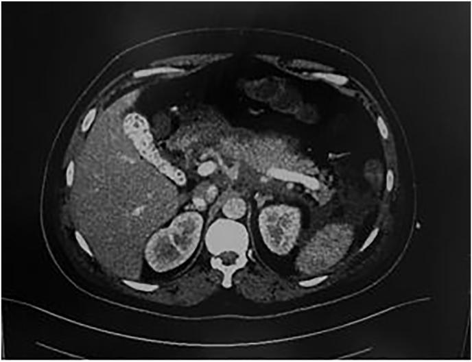
This diagnostic axial contrast-enhanced CT scan of the abdomen demonstrates findings consistent with severe acute pancreatitis. The pancreas exhibits heterogeneous enhancement with significant peripancreatic inflammatory stranding and fat plane blurring, characteristic of acute interstitial edematous pancreatitis progressing toward Balthazar Grade E. Free fluid is noted in the left paracolic gutter. The gallbladder is visible and contains a hyperdense focus, indicating a gallstone (cholelithiasis), suggesting a possible biliary etiology for the pancreatitis. Surrounding anatomical structures including the liver, spleen, and kidneys appear largely unremarkable in size and parenchymal density. This imaging is highly relevant for medical education regarding the radiological diagnosis and grading of acute pancreatitis, illustrating the loss of distinct organ borders due to extensive peripancreatic inflammation and fluid collection.
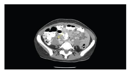
This diagnostic image is an axial computed tomography (CT) scan of the abdomen at the level of the pancreas and kidneys. The vertebral body is visible posteriorly, with the abdominal wall musculature and subcutaneous fat forming the external borders. Internally, the most prominent pathological finding is the presence of multiple hyperdense collections located within the peripancreatic space, indicated by a yellow arrow. These hyperdense areas are consistent with acute hemorrhage within or adjacent to the pancreatic parenchyma, suggesting a diagnosis of hemorrhagic pancreatitis. The pancreas itself appears poorly defined and edematous, accompanied by significant inflammatory fat stranding and haziness in the surrounding retroperitoneal tissue planes. Loops of small bowel with varying amounts of oral contrast and air are visible anteriorly and laterally. The kidneys are visible bilaterally, and the aorta is noted anterior to the vertebral body. This scan demonstrates the imaging features of severe acute pancreatitis, specifically showcasing how high-density blood (hematoma) appears on a non-contrast or portal venous phase CT relative to surrounding inflamed soft tissue.
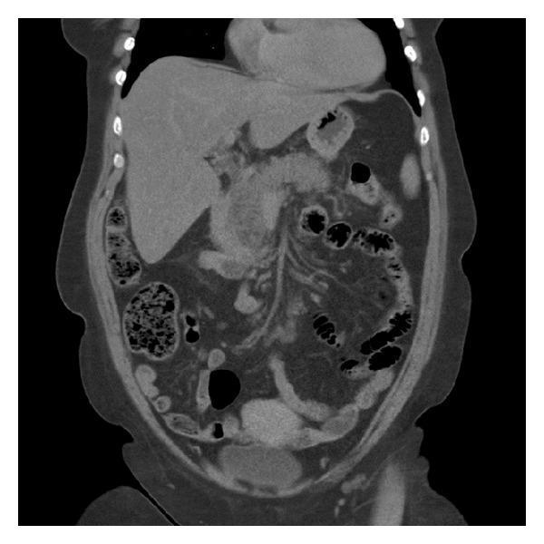
This diagnostic image is a coronal reformatted computerized tomography (CT) scan of the abdomen demonstrating findings consistent with acute pancreatitis. The primary pathological feature is the enlargement and edema of the pancreatic parenchyma, which exhibits heterogeneous density. There is notable blurring of the pancreatic margins with associated peripancreatic fat stranding and minor fluid collections, indicating active inflammatory changes. Anatomical context shows the liver with relatively normal morphology and density. Bowel loops are visible with normal gas distribution patterns. Consistent with the patient's surgical history of cholecystectomy, the gallbladder is absent from its fossa. The image serves as a clinical illustration of the radiologic manifestations of pancreatitis, focusing on organ morphology, parenchymal inflammation, and secondary changes in the surrounding retroperitoneal fat. It is intended for medical education regarding abdominal imaging and the diagnosis of acute gastrointestinal inflammatory conditions.
Acute Pancreatitis - Diagnosis & Investigations
Diagnostic Criteria (Atlanta Revised Criteria 2012)
- Abdominal pain characteristic of acute pancreatitis - acute onset, severe, constant epigastric pain, often radiating to the mid-back
- Serum lipase or amylase >3× upper limit of normal
- Characteristic findings on abdominal imaging (CT/MRI/USG)
If two of the three criteria are met clinically, imaging is not mandatory for diagnosis. - Schwartz's Principles of Surgery, p. 1472
Clinical Features
| Feature | Detail |
|---|---|
| Pain | Severe epigastric/upper abdominal, radiates to back ("band-like"), worse lying down, relieved slightly on leaning forward |
| Nausea/Vomiting | Very common |
| Fever | Low-grade, may be high in infected necrosis |
| Abdominal tenderness | Epigastric tenderness, guarding in upper abdomen |
| Cullen's sign | Periumbilical bruising (retroperitoneal haemorrhage - rare, severe) |
| Grey Turner's sign | Flank bruising (retroperitoneal bleed - rare, severe) |
| Tetany | From hypocalcaemia in severe cases |
Investigations
1. Primary Blood Tests (Most Important)
| Test | Findings | Notes |
|---|---|---|
| Serum Lipase | >3× ULN (diagnostic) | Preferred over amylase - greater sensitivity & specificity. Preferred first-line test. |
| Serum Amylase | >3× ULN | Rises within hours, peaks in 24-72h, returns to normal in 3-5 days. Can be falsely normal in hypertriglyceridaemia. |
| Urine Amylase | Elevated | Stays elevated longer than serum amylase - useful in delayed presentations |
| CBC (FBC) | WBC elevated (leukocytosis) | WBC >16,000 = Ranson criterion |
| Blood Glucose | Elevated (hyperglycaemia) | Due to decreased insulin production |
| Serum Calcium | Low (<8 mg/dL) = bad sign | Saponification of peripancreatic fat |
| Serum LDH | Elevated | Ranson criterion |
| Serum AST/ALT | Elevated | ALT >3× = gallstone aetiology likely |
| BUN/Creatinine | Elevated | Indicates hemoconcentration, third-space fluid loss |
| Haematocrit | Elevated (hemoconcentration) | Fall >10 points in 48h = Ranson criterion |
| Serum CRP | >150 mg/L at 24h = severe | Good severity marker |
| Triglycerides | Check if no gallstone/alcohol cause | Hypertriglyceridaemia is a cause |
| ABG (Blood Gas) | PO₂ <60 mmHg = severe | Watch for ARDS/respiratory failure |
| LFTs + GGT | Bilirubin, ALP raised if biliary cause | |
| Albumin | Low in severe disease |
2. Imaging
Ultrasound Abdomen (First-Line Imaging)
- Should be done in ALL patients with acute pancreatitis
- Primary purpose: identify gallstones (biliary aetiology - commonest cause)
- Can show: oedematous pancreas, peripancreatic fluid, dilated CBD
- Limitation: bowel gas often obscures pancreas in acute setting
CT Abdomen with Contrast (CECT) - Not Routine
- Not required routinely for diagnosis
- Indications for CT:
- Diagnostic uncertainty (criteria not met)
- Assessing complications (necrosis, abscess, pseudocyst)
- No improvement after 48-72 hours of treatment
- Suspected infected necrosis
- Ideally done 48-72 hours after onset (necrosis may not be apparent earlier)
| Grade | CT Findings | Score |
|---|---|---|
| A | Normal pancreas | 0 |
| B | Pancreatic enlargement | 1 |
| C | Pancreatic inflammation + peripancreatic fat changes | 2 |
| D | Single peripancreatic fluid collection | 3 |
| E | Two or more fluid collections OR pancreatic gas | 4 |
- Add necrosis score (0-4) to Balthazar grade for total CTSI
- CTSI 0-3 = mild; 4-6 = moderate; 7-10 = severe
MRCP (MR Cholangiopancreatography)
- Best for evaluating bile duct and pancreatic duct anatomy
- Preferred when CBD stones or duct disruption is suspected without wanting to risk ERCP
- No radiation, no contrast nephrotoxicity
ERCP
- Diagnostic ERCP is not indicated for pancreatitis alone
- Therapeutic ERCP indicated only for: cholangitis or biliary obstruction (CBD stones not clearing)
3. Severity Scoring Systems
Ranson's Criteria
| At Admission | At 48 Hours |
|---|---|
| Age >55 years | Haematocrit fall >10 points |
| WBC >16,000/mm³ | BUN rise >5 mg/dL |
| Blood glucose >200 mg/dL | Serum calcium <8 mg/dL |
| Serum LDH >350 IU/L | Arterial PO₂ <60 mmHg |
| Serum AST >250 U/dL | Base deficit >4 mEq/L |
| Fluid sequestration >6 L |
- <3 criteria: Mild disease, low mortality (<5%)
- 3-6 criteria: Moderate-severe disease
-
6 criteria: Severe, 50% mortality risk
BISAP Score (Bedside Index for Severity in Acute Pancreatitis)
- B - BUN >25 mg/dL
- I - Impaired mental status (GCS <15)
- S - SIRS (systemic inflammatory response syndrome) present
- A - Age >60 years
- P - Pleural effusion on imaging
APACHE II Score
- Score ≥8 at 24 hours = predicted severe pancreatitis
- More complex but widely used in ICU settings
CRP
- Single simple marker: CRP >150 mg/L at 24 hours = similar accuracy to Ranson's for predicting severity
Common Causes (to guide investigations)
| Cause | Investigation |
|---|---|
| Gallstones (most common) | USG abdomen, LFTs, MRCP |
| Alcohol (second most common) | History, GGT |
| Hypertriglyceridaemia | Fasting lipid profile, serum triglycerides |
| Drugs (e.g., azathioprine, thiazides, steroids) | Medication history |
| Trauma / ERCP | History |
| Hypercalcaemia | Serum calcium, PTH |
| Autoimmune | IgG4 levels |
| Idiopathic | Diagnosis of exclusion |
Summary: Minimum Investigation Panel for Suspected Pancreatitis
| Urgency | Tests |
|---|---|
| Immediate | Serum lipase (or amylase), FBC, RFT, LFTs, blood glucose, serum calcium, ABG (if severe), CRP |
| Same day | USG abdomen (gallstones), serum triglycerides, fasting lipid profile |
| If no improvement at 48-72h | CECT abdomen (assess necrosis/complications) |
| If biliary cause suspected | MRCP |
Sources: Rosen's Emergency Medicine, p.3878-3912 | Schwartz's Principles of Surgery, p.1471-1475
Generate management protocol for acute pancreatitis treatment
acute pancreatitis management algorithm flowchart
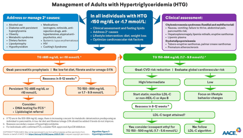
ACUTE PANCREATITIS - MANAGEMENT PROTOCOL
Based on: Rosen's Emergency Medicine, Schwartz's Principles of Surgery, Current Surgical Therapy 14e, Revised Atlanta Classification 2012
STEP 1 - SEVERITY CLASSIFICATION (First 24-48 Hours)
Revised Atlanta Classification 2012
| Grade | Criteria |
|---|---|
| Mild | No organ failure, no local or systemic complications |
| Moderately Severe | Transient organ failure (<48 h) OR local/systemic complications |
| Severe | Persistent organ failure (>48 h) - respiratory, cardiovascular, or renal (Modified Marshall score ≥2) |
Quick Bedside Severity: BISAP Score (within 24h)
- B - BUN >25 mg/dL
- I - Impaired mental status (GCS <15)
- S - SIRS present
- A - Age >60 years
- P - Pleural effusion on imaging
STEP 2 - IMMEDIATE RESUSCITATION (First 6-24 Hours)
A. IV Fluid Resuscitation (Highest Priority)
| Parameter | Recommendation |
|---|---|
| Fluid of choice | Lactated Ringer's (LR) - preferred over Normal Saline |
| Why LR? | NS causes hyperchloraemic metabolic acidosis which activates trypsinogen and worsens pancreatic injury. LR also has anti-inflammatory effects. |
| Initial rate | 5-10 mL/kg/h for first 2 hours (IAP/APA guidelines) |
| Maintenance | 1.5-3 mL/kg/h for next 12-24 hours |
| Total target in 24h | 2-4 litres overall (goal-directed) |
| ACG recommendation | 250-500 mL/h |
- Heart rate < 120/min
- Mean arterial pressure (MAP): 65-85 mmHg
- Urine output: >0.5-1 mL/kg/h
- BUN and haematocrit trending down
Caution: Overly aggressive fluids can cause abdominal compartment syndrome, respiratory failure, and worsen sepsis. Monitor closely.
B. Electrolyte Correction
| Electrolyte | Action |
|---|---|
| Hypocalcaemia | Replace only if ionised Ca²⁺ is low, QT prolonged, or Chvostek/Trousseau signs present. Correct hypomagnesaemia first. |
| Hypomagnesaemia | Replace before calcium |
| Hyperglycaemia | Monitor blood glucose; insulin if needed (hyperglycaemia worsens pancreatic injury and immune function) |
STEP 3 - PAIN MANAGEMENT
Pain is the cardinal symptom and its relief is a clinical priority. No single analgesic has been proven superior. (Schwartz's Principles of Surgery)
Stepwise Analgesic Approach
| Step | Drug | Dose | Notes |
|---|---|---|---|
| Step 1 (Mild pain) | Paracetamol (Acetaminophen) | 1 g IV/oral every 6 hours | First choice; 650 mg/8h if liver disease |
| Step 2 (Moderate pain) | Ketorolac/NSAIDs | Ketorolac 30 mg IV | Avoid in renal impairment, critically ill |
| Step 2 alt | Metamizole (Dipyrone) | 2 g IV every 8 hours | Recommended in Schwartz's for moderate pain |
| Step 3 (Severe pain) | Buprenorphine | 0.3 mg IV every 4 hours | First-line opioid per Schwartz's |
| Opioid sparing | Low-dose Ketamine | 0.1-0.3 mg/kg IV | Good adjunct; reduces opioid requirement |
| Antiemetic | Ondansetron / Metoclopramide | 4-8 mg IV | For nausea/vomiting control |
Morphine: Classically avoided due to theoretical sphincter of Oddi spasm. No clinical trials confirm it worsens pancreatitis, but most guidelines still recommend avoiding it when alternatives are available.
Pentazocine, meperidine, procaine HCl are also effective opioid options.
STEP 4 - NUTRITION
Old Practice vs Current Evidence
| Old (Incorrect) | Current Evidence-Based |
|---|---|
| Keep patient NPO until pancreatitis resolves | Early oral feeding (<24h) if tolerated is recommended |
| Enteral feeding worsens pancreatitis | Enteral feeding is safe and beneficial |
Nutrition Guidelines
| Scenario | Approach |
|---|---|
| Mild pancreatitis, tolerating orally | Start oral diet early (<24h) - low fat, soft diet as tolerated |
| Cannot tolerate oral (nausea/vomiting) | Nasogastric or nasojejunal (NG/NJ) enteral feeding |
| NJ vs NG route | Both are comparable in safety and efficacy |
| If enteral not possible | Total Parenteral Nutrition (TPN) - last resort |
| Why avoid prolonged NPO? | Causes gut mucosal atrophy, bacterial overgrowth, gut translocation, increases infectious complications |
Early enteral nutrition reduces: infectious complications, organ failure severity, need for surgical intervention, and mortality compared to parenteral nutrition. (Current Surgical Therapy 14e)
STEP 5 - ANTIBIOTICS
Key Principle: Prophylactic antibiotics are NOT recommended
| Scenario | Antibiotic Use |
|---|---|
| Mild/moderate sterile pancreatitis | NO antibiotics |
| Severe pancreatitis (SIRS present but no proven infection) | NO prophylactic antibiotics |
| Suspected sepsis / clinically deteriorating | START empirical antibiotics |
| Infected pancreatic necrosis (confirmed by CT/culture) | YES - start antibiotics |
| Extrapancreatic infection (cholangitis, pneumonia, bacteraemia) | YES - treat infection |
Antibiotic Choice for Infected Necrosis
| Drug | Reason |
|---|---|
| Carbapenems (Meropenem, Imipenem) | Best pancreatic penetration - superior to cephalosporins and fluoroquinolones |
| Ciprofloxacin + Metronidazole | Alternative combination |
| Piperacillin-Tazobactam | Alternative |
Probiotics are contraindicated - a multicentre RCT showed more than doubled mortality with probiotics in severe pancreatitis (possible intestinal ischaemia). (Current Surgical Therapy 14e)
STEP 6 - ENDOSCOPIC INTERVENTION (ERCP)
Indications
| Indication | Action |
|---|---|
| Cholangitis with biliary obstruction | ERCP within 24-72 hours (emergency) |
| CBD stone obstruction without cholangitis | ERCP within 72 hours |
| Biliary pancreatitis without obstruction | ERCP NOT routinely required |
| Diagnostic purposes | ERCP NOT indicated - use MRCP instead |
STEP 7 - SURGICAL / INTERVENTIONAL MANAGEMENT
For Necrotising Pancreatitis
| Situation | Action |
|---|---|
| Asymptomatic sterile necrosis / pseudocyst | Conservative management only - these may resorb spontaneously |
| Infected necrosis with clinical deterioration | Urgent percutaneous drainage + antibiotics |
| Infected/symptomatic necrosis, stable patient | Delay intervention to ≥4 weeks to allow collection to "wall off" |
| Symptomatic sterile necrosis >8 weeks | Consider drainage (endoscopic/percutaneous/surgical) |
Drainage Options (in order of preference)
- Endoscopic drainage (EUS-guided, e.g., cyst-gastrostomy) - least invasive, requires walled-off collection
- Percutaneous image-guided drainage - for immature or inaccessible collections
- Video-assisted retroperitoneal debridement (VARD) or minimally invasive surgery
- Open necrosectomy - highest risk, last resort; mortality 75% if done in first 2 weeks
Surgical mortality: 75% if <2 weeks, 45% at 2-4 weeks, 8% if >30 days from onset. Always delay if clinically possible. (Current Surgical Therapy 14e)
Cholecystectomy (for Gallstone Pancreatitis)
- Mild disease: Laparoscopic cholecystectomy before discharge or within 2-4 weeks (same admission preferred)
- Severe disease: Delay cholecystectomy; perform after full recovery
STEP 8 - MONITORING
Parameters to Track
| Parameter | Frequency | Target |
|---|---|---|
| Heart rate | Continuous | <120/min |
| Blood pressure / MAP | Continuous | MAP 65-85 mmHg |
| Urine output | Hourly | >0.5-1 mL/kg/h |
| Blood glucose | 4-6 hourly | 140-180 mg/dL (in ICU) |
| Serum BUN, creatinine | Daily | Trending down |
| Haematocrit | Daily | Trending down (falling Hct = adequate fluids) |
| Serum calcium | Daily | >8 mg/dL |
| ABG / SpO₂ | As needed | PO₂ >60 mmHg |
| CRP | Every 48h | Trending down |
| Serum amylase/lipase | Not useful for monitoring severity - do not repeat routinely |
STEP 9 - ICU ADMISSION CRITERIA
- Persistent organ failure >48h (respiratory: PaO₂ <60 / renal: creatinine >170 / cardiovascular: BP <90 despite fluids)
- BISAP score ≥3 or Ranson score ≥3
- APACHE II score ≥8
- Necrotising pancreatitis on CT
- Ongoing haemodynamic instability despite resuscitation
- Need for vasopressors
SUMMARY FLOWCHART
Suspected Acute Pancreatitis
         |
Confirm: ≥2 of 3 criteria (pain + lipase/amylase >3x ULN + imaging)
         |
     SEVERITY?
    /         \
  MILD        MODERATELY SEVERE / SEVERE
   |                    |
Oral diet        IV LR fluid resuscitation
Early oral        (5-10 mL/kg/h → 1.5-3 mL/kg/h)
feeding          Pain control (Paracetamol → Opioids)
Monitor          Antiemetics
Discharge        NPO only if vomiting severely → NG/NJ feed
when              |
tolerated    Monitor BUN/Hct/UO/HR/MAP
              |
         BILIARY CAUSE?        INFECTED NECROSIS?
              |                       |
         ERCP if                Carbapenems + delay
         cholangitis/           intervention to ≥4 weeks
         obstruction            (percutaneous drain if deteriorating)
              |
         Cholecystectomy
         before discharge
DRUGS SUMMARY TABLE
| Drug | Dose | Purpose |
|---|---|---|
| Lactated Ringer's IV | 5-10 mL/kg/h → 1.5-3 mL/kg/h | Fluid resuscitation (first choice) |
| Paracetamol | 1 g IV/PO q6h | Analgesia (first line) |
| Metamizole | 2 g IV q8h | Moderate pain |
| Buprenorphine | 0.3 mg IV q4h | Severe pain (opioid of choice) |
| Ketamine (low dose) | 0.1-0.3 mg/kg IV | Opioid-sparing analgesia |
| Ondansetron | 4-8 mg IV q8h | Antiemetic |
| Meropenem/Imipenem | Standard dosing | Infected necrosis only |
| Insulin | Sliding scale/infusion | If hyperglycaemia persistent |
| Norepinephrine | Titrate to MAP ≥65 mmHg | Vasopressor if hypotensive |
Sources: Rosen's Emergency Medicine (9th Ed), pp. 3874-3900 | Schwartz's Principles of Surgery (11th Ed), pp. 1471-1475 | Current Surgical Therapy (14th Ed) | Revised Atlanta Classification 2012 (Banks et al., Gut)
Disclaimer: This protocol is a clinical reference guide. All treatment decisions must be individualised based on the patient's clinical state, comorbidities, local institutional guidelines, and physician judgement.