
Tickets details
- Travel or flight tickets
- Event or concert tickets
- Support or service tickets
- Ticket booking details you want me to organize
Rickets
rickets AND treatment
WHO rickets vitamin D calcium diagnosis treatment children guideline
- Most common cause: vitamin D deficiency and/or too little calcium intake.
- Signs: delayed growth, bone pain, muscle weakness, widened wrists/ankles, bowed legs, frontal bossing, or a “rachitic rosary” along the ribs.
- Diagnosis: clinical assessment, blood tests such as calcium, phosphate, alkaline phosphatase and 25-hydroxyvitamin D, plus X-rays of the wrist or knee.
- Treatment: depends on the cause. Nutritional rickets is generally treated with vitamin D plus adequate calcium, while renal, genetic, or phosphate-wasting rickets needs specialist-directed treatment. The global consensus guidance recommends vitamin D with calcium for nutritional rickets, but children should be assessed before treatment because the correct regimen varies by age and cause.
Mbbs theory
rickets AND treatment
Global Consensus Recommendations Prevention Management Nutritional Rickets diagnosis treatment calcium 500 mg vitamin D 2000 IU
rickets wrist radiograph metaphyseal cupping fraying widening
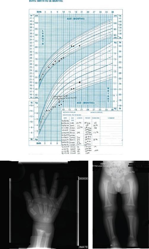
Educational composite image illustrating the clinical and radiological manifestations of nutritional rickets and subsequent recovery. The top panel features a pediatric growth chart (Birth to 36 months) documenting length and weight percentiles. It shows an initial period of growth failure followed by significant catch-up growth following therapeutic intervention with vitamin D and calcium. The bottom-left panel is a diagnostic radiograph of a pediatric hand and wrist, demonstrating classic rachitic changes including metaphyseal fraying and cupping of the distal radius and ulna, along with similar fraying at the distal aspects of the second through fifth metacarpals. The bottom-right panel displays a radiograph of the lower extremities, showing bilateral metaphyseal widening, cupping, and fraying at the distal femurs and proximal/distal tibiae. These findings are characteristic of impaired mineralization at the growth plates. The material serves as an educational resource for pediatric endocrinology and radiology, focusing on the diagnosis of metabolic bone disease and the monitoring of treatment efficacy in nutritional rickets.
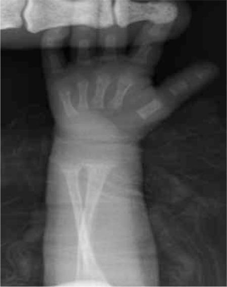
Anteroposterior (AP) pediatric wrist radiograph demonstrating classic radiological signs of rickets, specifically metabolic bone disease of prematurity (MBDP). The primary findings are located at the distal metaphyses of the radius and ulna. These include significant metaphyseal widening (splaying), fraying (loss of sharp cortical definition), and cupping of the metaphyseal margins. The growth plates (physes) appear abnormally widened. The carpal centers are not yet ossified, consistent with neonatal or early infant developmental age. These skeletal changes reflect inadequate mineralization of the osteoid at the zone of provisional calcification. The image serves as a key diagnostic reference for identifying rickets-like changes in a clinical pediatric or neonatal setting.
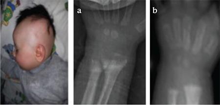
This composite figure presents clinical and radiological findings in a pediatric patient with Hereditary Vitamin D-Resistant Rickets (HVDRR). The left panel is a clinical photograph of an infant showing near-total alopecia, a hallmark feature of certain VDR gene mutations. Panels (a) and (b) are diagnostic radiographs of the wrist and distal forearm. Radiograph (a) demonstrates classic baseline rachitic changes, including significant cupping, fraying, and widening of the distal radial and ulnar metaphyses, alongside generalized osteopenia and an obscured provisional zone of calcification. Radiograph (b) shows the same anatomical region following medical management with high-dose calcitriol, calcium, and cinacalcet. This follow-up image exhibits marked interval improvement, characterized by increased bone density (remineralization) and a significant reduction in metaphyseal fraying and cupping, indicating partial healing of the skeletal rickets. The series serves as an educational comparison of disease manifestation and therapeutic response in metabolic bone disease.
https://pmc.ncbi.nlm.nih.gov/articles/PMC4880117
Rickets
Definition
Etiology
1. Nutritional rickets, most common
- Vitamin D deficiency
- Inadequate dietary calcium intake
- Limited sunlight exposure
- Exclusive breast-feeding without vitamin D supplementation in an at-risk infant
2. Defective vitamin D absorption or metabolism
- Malabsorption: celiac disease, chronic diarrhea, pancreatic insufficiency
- Chronic liver disease
- Chronic kidney disease, with impaired formation of active vitamin D
- Drugs increasing vitamin D metabolism, for example anticonvulsants
3. Vitamin D dependent or resistant rickets
- Vitamin D-dependent rickets type 1: deficiency of renal 1-alpha-hydroxylase, CYP27B1 mutation
- Vitamin D-dependent rickets type 2: vitamin D receptor resistance
- X-linked hypophosphatemic rickets: renal phosphate wasting, commonly due to a PHEX mutation
4. Other phosphate-wasting states
- Renal tubular acidosis
- Fanconi syndrome
- Hereditary hypophosphatemic rickets
- Tumor-induced phosphate wasting, rare in children
Pathogenesis of nutritional rickets
- Vitamin D deficiency or low calcium intake reduces intestinal calcium absorption.
- Serum calcium is initially maintained by increased parathyroid hormone, PTH.
- PTH increases bone resorption and urinary phosphate loss.
- Hypophosphatemia and inadequate calcium-phosphate product prevent mineralization of osteoid and the growth plate.
- The growing metaphyses become widened, irregular, cupped, and frayed.
- Weight bearing on soft bones produces deformities such as bow legs or knock knees.
Clinical features
General
- Irritability, sweating, poor growth
- Delayed motor milestones
- Hypotonia and proximal muscle weakness
- Bone pain, especially in lower limbs
- Growth retardation and short stature
Skull
- Craniotabes, soft occipital or parietal bones
- Frontal bossing
- Delayed closure of anterior fontanelle
- Delayed dentition and enamel defects
Chest
- Rachitic rosary: prominent costochondral junctions
- Harrison sulcus
- Pigeon chest deformity
Limbs
- Widening of wrist and ankle
- Bow legs, genu varum, or knock knees, genu valgum
- Coxa vara
- Bone tenderness
- Pathological fractures may occur in severe disease
Spine and pelvis
- Kyphosis or scoliosis
- Pelvic deformity in longstanding disease
Neuromuscular manifestations of hypocalcemia
- Tetany
- Carpopedal spasm
- Seizures
- Laryngospasm, rarely
Investigations
Biochemical tests
| Test | Typical nutritional vitamin D-deficiency rickets |
|---|---|
| Serum calcium | Normal or low |
| Serum phosphate | Low |
| Alkaline phosphatase, ALP | Markedly raised |
| 25-hydroxyvitamin D | Low |
| Parathyroid hormone, PTH | Raised |
| Serum magnesium | May be low |
| Renal function, liver function | To identify secondary causes |
X-ray findings
- Generalized osteopenia
- Widening of growth plate
- Metaphyseal cupping, fraying, and splaying
- Delayed ossification of epiphyses
- Bowing of long bones
- Looser zones or pseudofractures in severe disease
Differential diagnosis
- Physiological genu varum or genu valgum
- Blount disease
- Skeletal dysplasia
- Osteogenesis imperfecta
- Hypophosphatasia
- Renal osteodystrophy
- Hypophosphatemic rickets
- Metaphyseal dysplasia
Management
A. Nutritional rickets
| Age | Vitamin D daily for 90 days | Maintenance |
|---|---|---|
| <3 months | 2,000 IU/day | 400 IU/day |
| 3-12 months | 2,000 IU/day | 400 IU/day |
| 1-12 years | 3,000-6,000 IU/day | 600 IU/day |
| >12 years | 6,000 IU/day | 600 IU/day |
- Ensure calcium intake of about 500 mg/day through diet or supplementation in most children with nutritional rickets.
- Oral treatment is generally preferred.
- Reassess clinical status, calcium, phosphate, ALP, and vitamin D response after about 3 months.
- Continue maintenance vitamin D and improve dietary calcium intake.
B. Treat the specific cause
- Chronic kidney disease: active vitamin D analogues, phosphate management, and renal specialist care.
- Vitamin D-dependent rickets: calcitriol or alfacalcidol with calcium, under pediatric endocrinology supervision.
- Hypophosphatemic rickets: phosphate replacement plus active vitamin D, or targeted therapy such as burosumab in selected X-linked hypophosphatemia cases.
- Malabsorption or liver disease: higher or modified vitamin D treatment and management of the underlying illness.
C. Orthopedic care
- Most deformities improve after medical correction if treatment is early.
- Bracing is usually not required for early, mild deformity.
- Persistent severe deformity after metabolic healing may need guided growth or corrective osteotomy.
Prevention
- Promote adequate maternal vitamin D and calcium nutrition.
- Exclusive breast-fed infants should receive routine vitamin D supplementation according to local pediatric guidance.
- Ensure age-appropriate dietary calcium.
- Encourage safe sunlight exposure where appropriate.
- Screen high-risk groups: darker skin, low sun exposure, malabsorption, chronic liver or kidney disease, anticonvulsant therapy, and poor dietary intake.
MBBS exam conclusion
Bronchitis and litis
acute bronchitis AND management
acute bronchitis acute laryngitis adult management antibiotics guideline CDC
Bronchitis and Laryngitis - MBBS Theory Notes
1. Acute Bronchitis
Definition
Etiology
- Influenza virus
- Parainfluenza virus
- Rhinovirus
- Coronavirus
- RSV
- Adenovirus
- Mycoplasma pneumoniae
- Chlamydophila pneumoniae
- Bordetella pertussis
Clinical features
- Cough, initially dry and later with sputum
- Cough usually lasts 1-3 weeks and may persist up to 4 weeks
- Low-grade fever
- Malaise, sore throat, coryza
- Chest discomfort or burning sensation
- Rhonchi or wheeze may be heard
- Mild dyspnea can occur
Diagnosis
- Fever 38°C or more
- Respiratory rate 24/min or more
- Pulse 100/min or more
- Focal crepitations, bronchial breathing, or signs of consolidation
- Elderly, immunocompromised, or seriously ill patient
Treatment
- Rest and adequate fluids
- Antipyretic or analgesic: paracetamol
- Honey may help cough in children over 1 year and adults
- Short-term bronchodilator only if wheeze or bronchospasm is present
- Antitussives may be used selectively for troublesome dry cough
Complications
- Secondary bacterial pneumonia, uncommon
- Exacerbation of asthma or COPD
- Persistent post-infectious cough
2. Laryngitis
Definition
Types
- Acute laryngitis
- Chronic laryngitis
Causes
Acute laryngitis
- Viral upper respiratory infection, most common
- Excessive voice use or shouting
- Smoking and inhaled irritants
- Gastroesophageal reflux
- Rarely bacterial or fungal infection
Chronic laryngitis
- Smoking
- Chronic voice abuse
- Laryngopharyngeal reflux
- Chronic sinus or respiratory infection
- Tuberculosis, fungal infection, or syphilis, less common
- Laryngeal malignancy, especially in smokers
Clinical features
- Hoarseness of voice
- Aphonia, loss of voice
- Sore throat or throat irritation
- Dry cough
- Frequent throat clearing
- Pain on speaking
- Low-grade fever and coryza in viral cases
Diagnosis
- Hoarseness persists beyond 3 weeks
- Recurrent episodes occur
- There is smoking history
- Dysphagia, neck mass, hemoptysis, weight loss, or ear pain is present
- Malignancy or vocal-cord lesion is suspected
Management
Acute laryngitis
- Voice rest: avoid shouting and whispering
- Warm fluids and hydration
- Avoid smoking, alcohol, dust, and irritants
- Steam or humidification may provide symptomatic relief
- Treat reflux if present
- Paracetamol or NSAID for pain and fever, when appropriate
Chronic laryngitis
- Stop smoking
- Voice hygiene and speech therapy when indicated
- Treat reflux disease
- Investigate persistent hoarseness with laryngoscopy
- Treat specific infections or lesions according to the cause
Differences between acute bronchitis and laryngitis
| Feature | Acute bronchitis | Acute laryngitis |
|---|---|---|
| Site | Bronchi | Larynx and vocal cords |
| Main symptom | Cough | Hoarseness |
| Sputum | May be present | Usually absent or minimal |
| Voice change | Usually absent | Characteristic |
| Common cause | Viral lower respiratory infection | Viral upper respiratory infection or voice strain |
| Main treatment | Supportive cough care | Voice rest and supportive care |
| Antibiotics | Usually not required | Usually not required |
Red-flag symptoms
Bronchiolitis
bronchiolitis AND treatment
NICE bronchiolitis children diagnosis management oxygen hydration no antibiotics bronchodilators guideline
infant bronchiolitis chest radiograph hyperinflation peribronchial thickening
Bronchiolitis - MBBS Theory Notes
Definition
Etiology
Common viral causes
- Respiratory syncytial virus, RSV - most common
- Rhinovirus
- Parainfluenza virus
- Influenza virus
- Adenovirus
- Human metapneumovirus
- Coronavirus
Risk factors for severe disease
- Age less than 3 months
- Prematurity
- Congenital heart disease
- Chronic lung disease of prematurity
- Neuromuscular disease
- Immunodeficiency
- Poor feeding or dehydration
- Exposure to tobacco smoke
Pathogenesis
- Virus infects bronchiolar epithelial cells.
- There is epithelial necrosis, edema of the bronchiolar wall, mucus hypersecretion, and cellular debris.
- Narrow infant bronchioles become partially or completely obstructed.
- Partial obstruction produces air trapping and hyperinflation.
- Complete obstruction may lead to atelectasis.
- The child develops tachypnea, wheeze, crackles, increased work of breathing, and hypoxemia.
Clinical features
Typical progression
- Rhinorrhea
- Nasal blockage
- Mild fever
- Cough
- Poor feeding
- Tachypnea
- Wheeze
- Fine inspiratory crackles
- Chest retractions
- Nasal flaring
- Grunting
- Difficulty feeding
- Apnea, especially in young or premature infants
- Cyanosis in severe disease
Physical examination
- Tachypnea
- Subcostal and intercostal retractions
- Hyperinflated chest
- Diffuse wheeze
- Fine crackles
- Prolonged expiration
- Reduced air entry in severe disease
- Signs of dehydration
Diagnosis
Investigations
| Investigation | Finding or purpose |
|---|---|
| Pulse oximetry | Detect hypoxemia |
| Chest X-ray | Not routine. May show hyperinflation, peribronchial thickening, patchy atelectasis |
| Viral testing | Usually not required, may help cohort hospitalized patients |
| Blood gas | Severe respiratory distress, suspected respiratory failure |
| CBC, CRP, blood culture | Only if bacterial infection or sepsis is suspected |
Severity assessment
Features suggesting severe bronchiolitis
- Apnea
- Cyanosis
- Severe chest recession, grunting, or exhaustion
- Respiratory rate more than 70/min
- Inability to feed or markedly reduced intake
- Clinical dehydration
- Persistent oxygen saturation below local threshold, commonly <90-92%
- Altered sensorium or poor responsiveness
Management
A. Mild disease - home care
- Nasal saline drops and gentle suction if nasal secretions interfere with feeding
- Continue breastfeeds or give small frequent feeds
- Maintain hydration
- Antipyretics such as paracetamol for fever or discomfort
- Avoid tobacco smoke exposure
- Educate parents about danger signs and when to return
B. Indications for hospital admission
- Apnea or a history of apnea
- Significant respiratory distress
- Hypoxemia
- Inadequate oral intake or dehydration
- Young infant, especially under 3 months
- Prematurity or serious comorbidity
- Caregiver unable to safely observe the child at home
C. Hospital management
- Oxygen for persistent hypoxemia
- Nasal suction when secretions impair breathing or feeding
- Nasogastric fluids or intravenous fluids if oral intake is inadequate
- High-flow nasal oxygen or CPAP for worsening respiratory distress
- Mechanical ventilation if impending or established respiratory failure
Treatments not routinely recommended
- Antibiotics, unless bacterial infection is suspected
- Salbutamol or other bronchodilators
- Nebulized adrenaline
- Corticosteroids
- Chest physiotherapy
- Hypertonic saline in routine emergency treatment
- Montelukast
Complications
- Apnea
- Respiratory failure
- Dehydration
- Secondary bacterial infection, uncommon
- Atelectasis
- Rarely, pneumothorax
- Recurrent wheeze after an episode, though bronchiolitis itself does not equal asthma
Differential diagnosis
- Bronchial asthma or viral-induced wheeze
- Pneumonia
- Foreign body aspiration
- Congestive cardiac failure
- Pertussis
- Congenital heart disease
- Sepsis
- Gastroesophageal reflux with aspiration
Bronchiolitis versus bronchitis
| Feature | Bronchiolitis | Acute bronchitis |
|---|---|---|
| Main site affected | Bronchioles, small airways | Bronchi, large airways |
| Usual age group | Infants and children <2 years | Older children and adults |
| Common cause | RSV and other respiratory viruses | Usually respiratory viruses |
| Main symptoms | Tachypnea, wheeze, crackles, retractions, feeding difficulty | Cough, sputum, mild fever |
| Treatment | Supportive care, oxygen/fluids if required | Symptomatic treatment |
| Antibiotics | Usually not indicated | Usually not indicated |
Short exam answer
Salbutamol receipe details child <4 yr
preschool asthma AND salbutamol
GINA 2025 children 5 years and younger salbutamol dose spacer acute wheeze
Salbutamol for a child under 4 years
Preferred route: inhaler with spacer and face mask
- Give 4 puffs, one puff at a time through the spacer.
- Let the child take 5-6 normal breaths after each puff.
- For moderate or severe wheeze, it may be repeated every 20 minutes, up to 3 doses in the first hour, while obtaining medical assessment.
- If the child improves, further use must follow a clinician-provided asthma/wheeze action plan.
Nebulized salbutamol
- 2.5 mg nebulized salbutamol is the standard acute dose for children aged 5 years and younger in GINA guidance.
- It may be repeated every 20 minutes up to 3 times in the first hour for moderate/severe exacerbation, with reassessment.
Side effects
- Fast heartbeat or palpitations
- Tremor or shakiness
- Excitability, restlessness
- Headache
- With repeated high doses: low potassium and, rarely, abnormal heart rhythm
Seek urgent medical care now if the child has
- Blue lips, pauses in breathing, drowsiness, or exhaustion
- Severe chest indrawing/retractions, grunting, or inability to speak/cry/feed
- No improvement after initial salbutamol doses
- Need for 4 puffs again within 4 hours, or repeated need for salbutamol
- First episode of wheeze in an infant, particularly below 1 year