All nerves in the human body....in detail... for exam... for 15M...in simple sentences and point wise.... give simple digrams photos... reference from bdc or wishram singh
Reading File
Finding Sources
Finding Sources
Finding Sources
Reading File
Reading File
Finding Sources
Reading File
Finding Sources
Reading File
Reading File
Loading Image
Loading Image
Loading Image
Loading Image
Loading Image
I now have all the key diagrams and textbook content needed. Let me compile the comprehensive, exam-ready answer.
ALL NERVES IN THE HUMAN BODY - Exam Notes (15 Marks)
Reference: Gray's Anatomy for Students (BDC / Vishram Singh style)
OVERVIEW - CLASSIFICATION OF NERVES
The nervous system is divided into:
- Central Nervous System (CNS) - Brain + Spinal cord
- Peripheral Nervous System (PNS) - All nerves outside the CNS
PNS nerves are further divided into:
- Cranial Nerves (12 pairs) - exit the skull
- Spinal Nerves (31 pairs) - exit the vertebral column
- Autonomic Nerves - sympathetic + parasympathetic
PART 1 - CRANIAL NERVES (12 Pairs)
Key fact: All 12 pairs exit through foramina or fissures in the cranial cavity. All originate from the brain EXCEPT CN XI (accessory), which also has a spinal component.
MNEMONIC for Names:
"Oh Oh Oh To Touch And Feel Very Good Velvet. Ah Heaven!"
(Olfactory, Optic, Oculomotor, Trochlear, Trigeminal, Abducens, Facial, Vestibulocochlear, Glossopharyngeal, Vagus, Accessory, Hypoglossal)
MNEMONIC for Type (Sensory/Motor/Both):
"Some Say Marry Money But My Brother Says Big Brains Matter More"
(S, S, M, M, B, M, B, S, B, B, M, M)
CN I - OLFACTORY NERVE
- Type: Purely Sensory (Special Afferent - smell)
- Origin: Olfactory mucosa of nasal cavity
- Exit: Cribriform plate of ethmoid bone
- Function: Sense of smell
- Lesion: Anosmia (loss of smell) - common in basal skull fracture
CN II - OPTIC NERVE
- Type: Purely Sensory (Special Afferent - vision)
- Origin: Retinal ganglion cells
- Exit: Optic canal
- Function: Vision
- Lesion: Blindness in one eye (ipsilateral); AFFERENT defect in pupil reflex
CN III - OCULOMOTOR NERVE
- Type: Motor (GSE + GVE/parasympathetic)
- Origin: Midbrain (anterior to cerebral aqueduct)
- Exit: Superior orbital fissure
- Motor function: Moves eyeball (SR, MR, IR, IO muscles); raises eyelid (levator palpebrae)
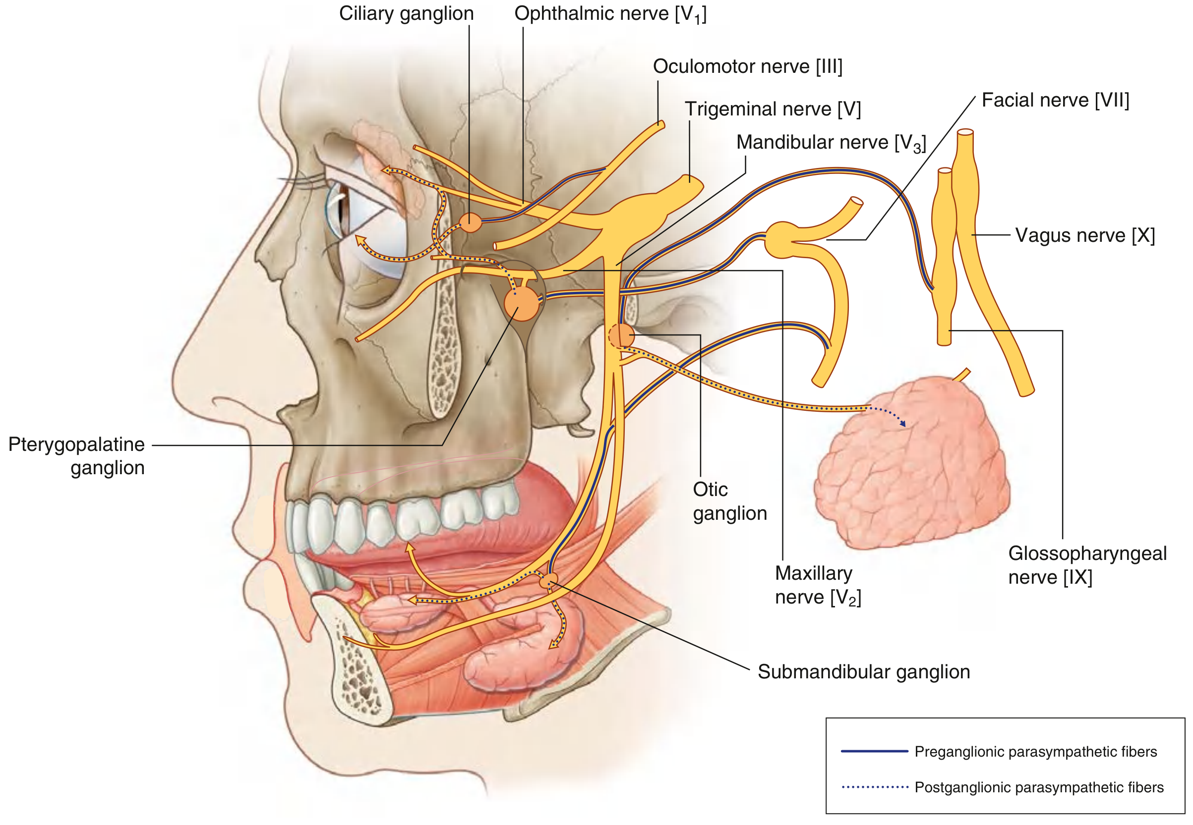
- Parasympathetic: Constricts pupil (sphincter pupillae) + ciliary muscle for accommodation
- Ganglion: Ciliary ganglion
- Lesion: Ptosis, "down and out" eye, dilated pupil (CN III palsy)
CN IV - TROCHLEAR NERVE
- Type: Purely Motor (GSE)
- Origin: Midbrain (dorsal surface - only cranial nerve to exit from the back of brainstem)
- Exit: Superior orbital fissure
- Function: Superior oblique muscle - depresses, intorts, abducts the eye
- Lesion: Diplopia on looking downward (e.g., going down stairs); head tilt to opposite side
CN V - TRIGEMINAL NERVE (Largest cranial nerve)
- Type: Both (Sensory + Branchial Motor)
- Origin: Pons
- Three divisions:
| Division | Exit | Sensory area |
|---|---|---|
| V1 - Ophthalmic | Superior orbital fissure | Forehead, cornea, upper nose |
| V2 - Maxillary | Foramen rotundum | Cheek, upper lip, upper teeth |
| V3 - Mandibular | Foramen ovale | Lower jaw, chin, tongue (anterior 2/3 general), lower teeth |
- Motor (V3 only): Muscles of mastication (temporalis, masseter, pterygoids)
- Ganglion: Trigeminal (Gasserian/semilunar) ganglion
- Lesion: Loss of corneal reflex; loss of facial sensation; jaw deviates to lesion side; Trigeminal Neuralgia (tic douloureux)
CN VI - ABDUCENS NERVE
- Type: Purely Motor (GSE)
- Origin: Pons (floor of 4th ventricle)
- Exit: Superior orbital fissure
- Function: Lateral rectus muscle (ABDucts the eye)
- Lesion: Medial squint (convergent strabismus); cannot abduct eye
CN VII - FACIAL NERVE
- Type: Both + Parasympathetic (mixed)
- Origin: Pons
- Exit: Internal acoustic meatus → facial canal → stylomastoid foramen
- Functions:
- Motor: Muscles of facial expression
- Parasympathetic: Lacrimal gland (via pterygopalatine ganglion); submandibular + sublingual glands (via submandibular ganglion)
- Sensory: Taste from anterior 2/3 of tongue (via chorda tympani)
- Lesion - LMN (Bell's palsy): Entire face paralyzed on same side (cannot close eye, drooping of mouth corner)
- Lesion - UMN (cortical stroke): Only lower face paralyzed (forehead spared - bilateral cortical representation)
CN VIII - VESTIBULOCOCHLEAR NERVE
- Type: Purely Sensory (Special Afferent)
- Two parts:
- Cochlear nerve: Hearing
- Vestibular nerve: Balance and head position
- Origin: Inner ear
- Exit: Internal acoustic meatus
- Lesion: Sensorineural deafness; vertigo; tinnitus
CN IX - GLOSSOPHARYNGEAL NERVE
- Type: Both + Parasympathetic
- Origin: Medulla
- Exit: Jugular foramen
- Functions:
- Motor (branchial): Stylopharyngeus muscle
- Sensory: Posterior 1/3 of tongue (taste + general); pharynx; carotid sinus/body
- Parasympathetic: Parotid gland (via otic ganglion)
- Lesion: Loss of taste from posterior tongue; loss of gag reflex (afferent limb)
CN X - VAGUS NERVE (Longest cranial nerve)
- Type: Both + Parasympathetic (most extensive distribution)
- Origin: Medulla
- Exit: Jugular foramen
- Distribution: Descends into thorax and abdomen
- Functions:
- Motor: Pharynx + larynx (speech, swallowing)
- Parasympathetic: Heart (slows rate), lungs (bronchoconstriction), gut (peristalsis) down to splenic flexure
- Sensory: Viscera (thorax + abdomen), skin near ear
- Lesion: Hoarseness, dysphagia, loss of gag reflex (efferent limb); uvula deviates away from lesion
CN XI - ACCESSORY NERVE
- Type: Purely Motor (Branchial Efferent)
- Unique: Only cranial nerve with a spinal root (C1-C5)
- Exit: Jugular foramen
- Functions:
- Cranial root: Joins vagus to supply larynx/pharynx
- Spinal root: Sternocleidomastoid + Trapezius muscles
- Lesion: Cannot turn head to opposite side (SCM); dropped shoulder (trapezius weakness)
CN XII - HYPOGLOSSAL NERVE
- Type: Purely Motor (GSE)
- Origin: Medulla
- Exit: Hypoglossal canal
- Function: All intrinsic + extrinsic muscles of tongue (except palatoglossus which is CN X)
- Lesion - LMN: Tongue deviates TOWARD lesion side (paralyzed side pushes it); wasting + fasciculations
- Lesion - UMN: Tongue deviates AWAY from lesion side
QUICK REFERENCE TABLE - All 12 Cranial Nerves
| CN | Name | Type | Exit | Key Function | Lesion Sign |
|---|---|---|---|---|---|
| I | Olfactory | S | Cribriform plate | Smell | Anosmia |
| II | Optic | S | Optic canal | Vision | Blindness |
| III | Oculomotor | M+PS | SOF | Eye movement, pupil constriction | Ptosis, "down + out" eye, dilated pupil |
| IV | Trochlear | M | SOF | Superior oblique | Diplopia looking down |
| V | Trigeminal | S+M | SOF/FR/FO | Face sensation, mastication | Loss of corneal reflex |
| VI | Abducens | M | SOF | Lateral rectus | Medial squint |
| VII | Facial | S+M+PS | Stylomastoid foramen | Facial expression, taste (ant 2/3 tongue) | Bell's palsy |
| VIII | Vestibulocochlear | S | IAM | Hearing + Balance | Deafness, vertigo |
| IX | Glossopharyngeal | S+M+PS | Jugular foramen | Taste (post 1/3 tongue), parotid | Loss of gag (afferent) |
| X | Vagus | S+M+PS | Jugular foramen | Heart, lungs, gut | Hoarseness, dysphagia |
| XI | Accessory | M | Jugular foramen | SCM + Trapezius | Dropped shoulder |
| XII | Hypoglossal | M | Hypoglossal canal | Tongue movements | Tongue deviates to lesion side |
(SOF = Superior Orbital Fissure; FR = Foramen Rotundum; FO = Foramen Ovale; IAM = Internal Acoustic Meatus; PS = Parasympathetic; S = Sensory; M = Motor)
PART 2 - SPINAL NERVES (31 Pairs)
Total Count:
- Cervical: 8 pairs (C1-C8)
- Thoracic: 12 pairs (T1-T12)
- Lumbar: 5 pairs (L1-L5)
- Sacral: 5 pairs (S1-S5)
- Coccygeal: 1 pair (Co1)
Key Rule for Cervical Nerves:
- C1-C7 exit ABOVE their respective vertebra.
- C8 exits BETWEEN C7 and T1 vertebrae.
- T1 onwards exit BELOW their vertebra.
PART 3 - NERVE PLEXUSES
A. CERVICAL PLEXUS
Formation: Anterior rami of C1 to C4
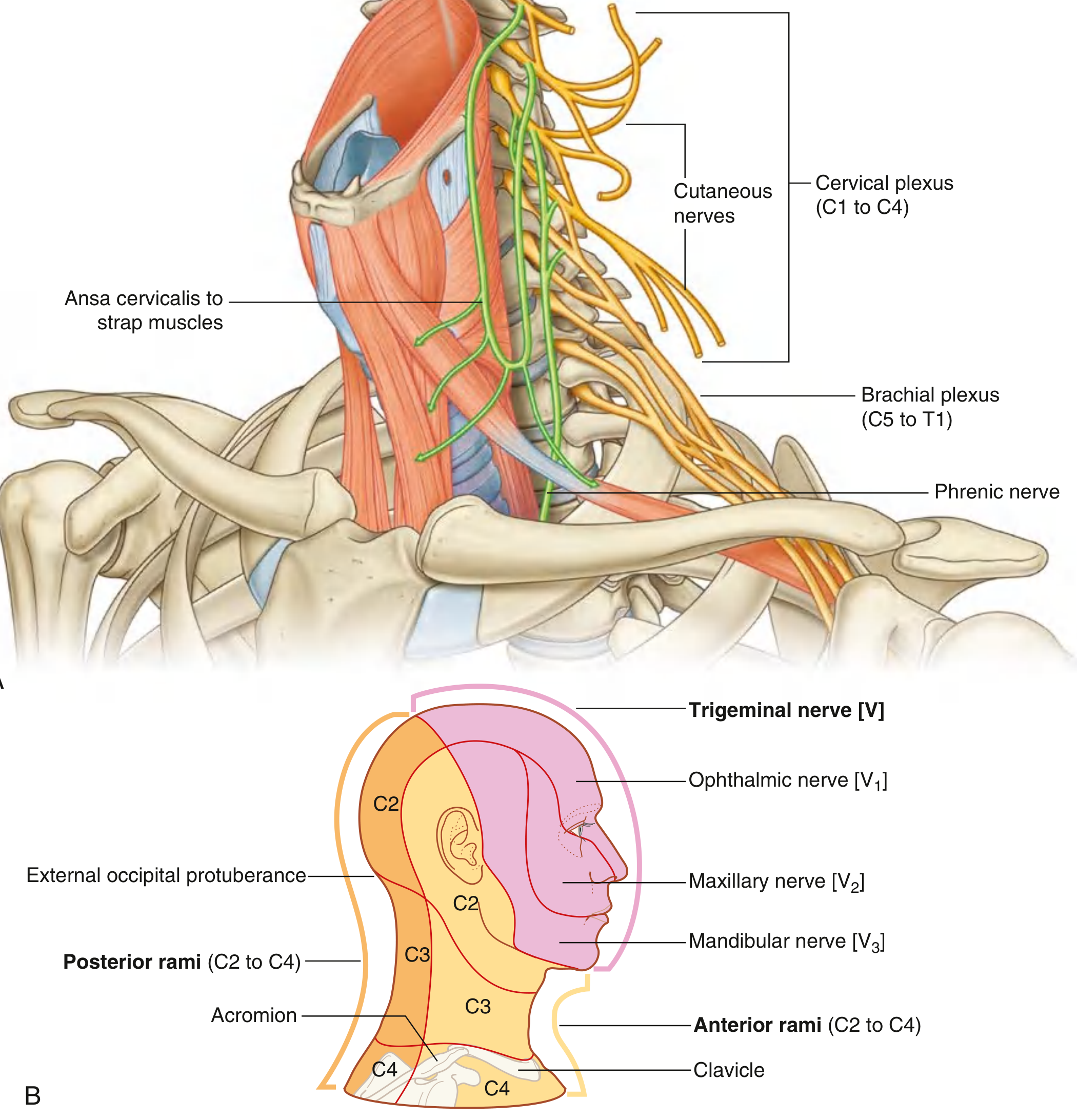
Main branches:
| Branch | Root | Function |
|---|---|---|
| Lesser occipital | C2 | Skin behind ear |
| Great auricular | C2, C3 | Skin over parotid + mastoid |
| Transverse cervical | C2, C3 | Skin of anterior neck |
| Supraclavicular | C3, C4 | Skin over shoulder + clavicle |
| Ansa cervicalis | C1, C2, C3 | Strap muscles of neck |
| Phrenic nerve | C3, C4, C5 | Diaphragm (motor + sensory) |
Phrenic nerve memory: "C3, 4, 5 keeps the diaphragm alive!"
B. BRACHIAL PLEXUS
Formation: Anterior rami of C5, C6, C7, C8, T1
Structure (mnemonic: "Real Texans Drink Cold Beer"):
- Roots → Trunks → Divisions → Cords → Branches
Trunks:
- Superior trunk = C5 + C6
- Middle trunk = C7 (alone)
- Inferior trunk = C8 + T1
Divisions: Each trunk divides into anterior + posterior division.
Cords (named by relation to axillary artery):
- Lateral cord (anterior divisions of superior + middle trunks)
- Medial cord (anterior division of inferior trunk)
- Posterior cord (all 3 posterior divisions)
Main Terminal Branches:
| Nerve | Cord | Roots | Main Motor Function | Sensory | Injury |
|---|---|---|---|---|---|
| Musculocutaneous | Lateral | C5,C6,C7 | Biceps, brachialis, coracobrachialis | Lateral forearm skin | Loss of elbow flexion |
| Median | Lateral + Medial | C6-T1 | Thenar muscles, finger flexors, pronators | Lateral palm + fingers 1-3 | "Ape hand" deformity, carpal tunnel |
| Ulnar | Medial | C8, T1 | Intrinsics (interossei, hypothenar), ring + little finger flexors | Medial palm + fingers 4-5 | "Claw hand" |
| Radial | Posterior | C5-T1 | Triceps, extensors of wrist + fingers | Posterior arm, dorsum of hand | Wrist drop |
| Axillary | Posterior | C5, C6 | Deltoid, teres minor | Deltoid area skin (regimental badge area) | Loss of shoulder abduction |
Brachial Plexus Injury Types:
- Erb's palsy (C5, C6): "Waiter's tip" position - arm adducted, medially rotated, forearm pronated
- Klumpke's palsy (C8, T1): Intrinsic hand muscles paralyzed - claw hand; Horner's syndrome if T1 root avulsed
C. LUMBAR PLEXUS
Formation: Anterior rami of L1-L3, most of L4 (also receives T12 contribution)
- Forms within the substance of psoas major muscle
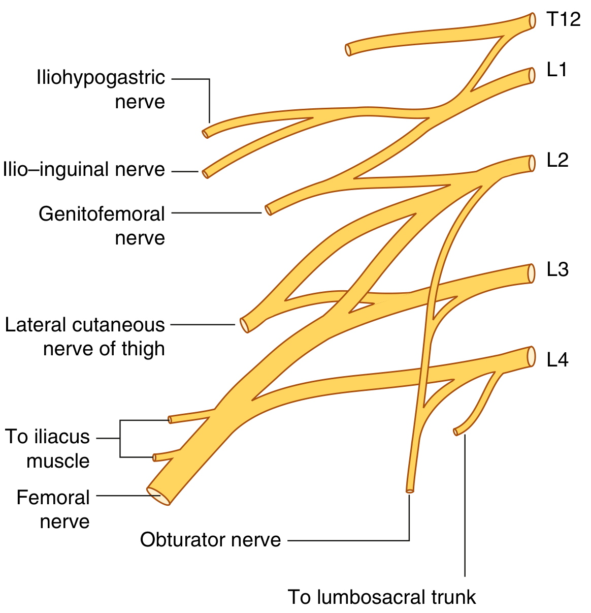
Branches:
| Nerve | Root | Motor | Sensory |
|---|---|---|---|
| Iliohypogastric | L1 | Int. oblique + transversus | Posterolateral gluteal + pubic skin |
| Ilio-inguinal | L1 | Int. oblique + transversus | Upper medial thigh, anterior scrotum/labia |
| Genitofemoral | L1, L2 | Cremaster (genital branch) | Anterior scrotum/labia; upper thigh (femoral branch) |
| Lateral cutaneous nerve of thigh | L2, L3 | None | Anterior + lateral thigh to knee |
| Obturator | L2-L4 | Medial compartment of thigh (adductors) | Medial thigh |
| Femoral | L2-L4 | Anterior compartment thigh (quadriceps), iliacus | Anterior thigh + medial leg (via saphenous nerve) |
Key Femoral nerve injury: Cannot extend knee; loss of patellar reflex; sensory loss on anterior thigh + medial leg.
D. SACRAL PLEXUS
Formation: Lumbosacral trunk (L4 + L5) + anterior rami of S1-S4
- Lies on piriformis muscle (anterior surface) on pelvic wall
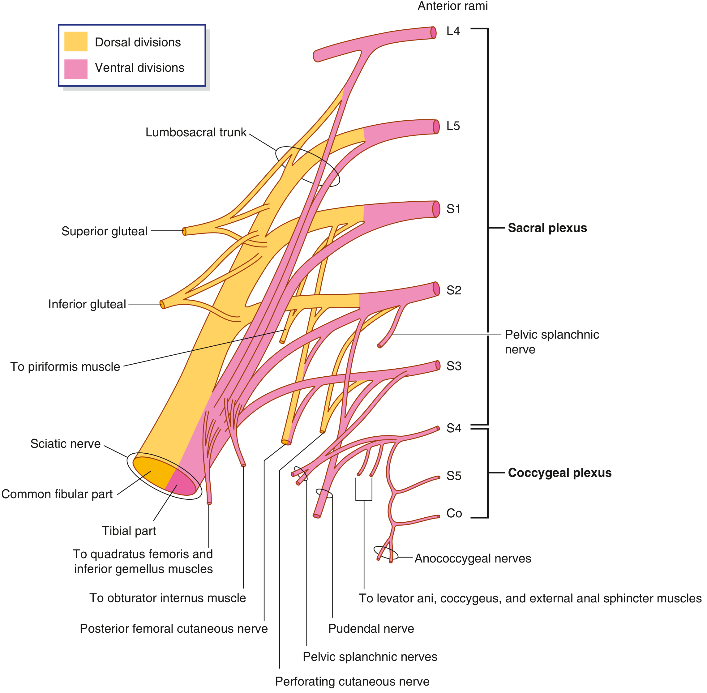
Branches:
| Nerve | Root | Motor | Sensory / Notes |
|---|---|---|---|
| Sciatic nerve | L4-S3 | All muscles below knee; hamstrings | Posterior thigh; entire leg and foot |
| - Tibial division | L4-S3 | Posterior compartment of leg; plantar foot muscles | Sole of foot |
| - Common fibular (peroneal) | L4-S2 | Anterior + lateral compartments of leg | Dorsum of foot |
| Superior gluteal | L4-S1 | Gluteus medius, minimus, tensor fascia lata | None (pure motor) |
| Inferior gluteal | L5-S2 | Gluteus maximus | None |
| Posterior femoral cutaneous | S1-S3 | None | Posterior thigh + perineum |
| Pudendal | S2-S4 | Perineal muscles, external anal + urethral sphincters | Perineum, external genitalia |
| Pelvic splanchnic | S2-S4 | Parasympathetic to pelvic viscera + descending colon | Visceral sensation |
Sciatic nerve key facts:
- Largest nerve in the body
- Exits through greater sciatic foramen, below piriformis
- Divides into tibial + common fibular (usually at apex of popliteal fossa)
- Tibial nerve injury: Foot inverted + plantarflexed ("claw foot"); loss of sensation on sole
- Common fibular nerve injury: Foot drop; cannot dorsiflex or evert foot; loss of sensation on dorsum of foot
PART 4 - AUTONOMIC NERVOUS SYSTEM NERVES
SYMPATHETIC ("Fight or Flight")
- Origin: Thoracolumbar (T1-L2) - also called thoracolumbar outflow
- Preganglionic: Short; synapse in paravertebral or prevertebral ganglia
- Postganglionic: Long; travel with blood vessels or spinal nerves
- Effects:
- Heart: Increases rate + force
- Pupils: Dilation (mydriasis)
- Bronchi: Dilation
- Gut: Decreases peristalsis
- Sweat glands: Increases sweating
PARASYMPATHETIC ("Rest and Digest")
- Origin: Craniosacral outflow
- Cranial part: CN III, VII, IX, X
- Sacral part: S2, S3, S4 (pelvic splanchnic nerves)
- Preganglionic: Long; synapse in ganglia near or in target organ
- Postganglionic: Short
4 Parasympathetic Ganglia in the Head:
| Ganglion | Nerve | Gland supplied |
|---|---|---|
| Ciliary | CN III | Sphincter pupillae + ciliary muscle (eye) |
| Pterygopalatine | CN VII (via greater petrosal) | Lacrimal gland, nasal glands |
| Submandibular | CN VII (via chorda tympani) | Submandibular + sublingual glands |
| Otic | CN IX | Parotid gland |
PART 5 - IMPORTANT INDIVIDUAL NERVES (High Yield)
Radial Nerve
- Root: C5-T1 (posterior cord of brachial plexus)
- Injury sites:
- Axilla (crutch palsy): All triceps, wrist, finger extension lost
- Spiral groove of humerus (Saturday night palsy): Triceps spared; wrist drop
- Elbow: Posterior interosseous nerve - finger drop only
Median Nerve
- Root: C6-T1 (lateral + medial cords)
- Injury sites:
- Elbow (carpal tunnel syndrome): Thenar wasting; sensory loss over thumb, index, middle, lateral half ring finger
- "Ape hand" deformity - thumb cannot oppose
- LOAF muscles spared in ulnar injury but affected in median: Lateral 2 lumbricals, Opponens pollicis, Abductor pollicis brevis, Flexor pollicis brevis
Ulnar Nerve
- Root: C7-T1 (medial cord)
- Injury at elbow (medial epicondyle):
- Claw hand (more prominent in ring + little fingers)
- Loss of intrinsic hand muscles (except LOAF)
- Sensory loss on medial 1.5 fingers + medial palm
Sciatic Nerve
- Root: L4-S3 (largest nerve in body)
- Common injury: Hip fracture/dislocation, buttock injection (avoid upper medial quadrant)
- Loss of all muscles below knee; hamstrings affected too
Phrenic Nerve
- Root: C3, C4, C5
- Motor to diaphragm
- Injury: Diaphragm paralysis on same side; elevated hemidiaphragm on X-ray
PART 6 - SPINAL NERVE ROOTS - REFLEXES (Exam High Yield)
| Reflex | Root | Nerve |
|---|---|---|
| Biceps jerk | C5, C6 | Musculocutaneous |
| Supinator/brachioradialis | C5, C6 | Radial |
| Triceps jerk | C7 | Radial |
| Knee jerk (patellar) | L3, L4 | Femoral |
| Ankle jerk (Achilles) | S1, S2 | Tibial |
| Plantar reflex | S1 | Tibial |
| Cremasteric | L1, L2 | Genitofemoral |
| Anal reflex | S3, S4 | Pudendal |
PART 7 - DERMATOMES (Key ones for Exam)
| Level | Area |
|---|---|
| C4 | Shoulder (cape area) |
| C6 | Thumb |
| C7 | Middle finger |
| C8 | Little finger |
| T4 | Nipple |
| T10 | Umbilicus |
| L1 | Groin |
| L3 | Knee (medial) |
| L4 | Medial leg |
| L5 | Dorsum of foot + big toe |
| S1 | Sole of foot + little toe |
| S4, S5 | Perianal (saddle area) |
EXAM TIPS SUMMARY
- 12 cranial nerves = "Some Say Marry Money But My Brother Says Big Brains Matter More" (S/S/M/M/B/M/B/S/B/B/M/M)
- Brachial plexus = Roots-Trunks-Divisions-Cords-Branches = C5 to T1
- Erb's palsy (C5,C6) = "Waiter's tip" | Klumpke's (C8,T1) = Claw hand
- Radial nerve = Wrist drop | Ulnar = Claw hand | Median = Ape hand
- Femoral nerve = Cannot extend knee | Sciatic = Foot drop + loss of sensation below knee
- Phrenic nerve = C3,4,5 keeps the diaphragm alive
- Parasympathetic ganglia in head = Ciliary, Pterygopalatine, Submandibular, Otic
- Vagus (CN X) = longest cranial nerve; Trigeminal (CN V) = largest cranial nerve
- CN IV (Trochlear) = only CN to exit from dorsal midbrain
- CN IV lesion = cannot go down stairs (diplopia looking down)
Reference: Gray's Anatomy for Students (equivalent to BD Chaurasia & Vishram Singh syllabus content), Tables 8.4, 8.5, and Figs. 8.14, 8.15, 7.52, 4.177, 5.62
This is a shared conversation. Sign in to Orris to start your own chat.