
Supports of hip joint
Reading File
This is a broad anatomical topic. Now searching the medical library for content on hip joint supports.
Finding Sources
Finding Sources
Gray's Anatomy for Students and S Das have dedicated hip joint sections. Let me read both simultaneously.
Reading File
Reading File
Finding Sources
Good content from Gray's. Now let me get the specific "supports" (capsule, ligaments, muscles) content from Gray's and also check the THIEME Atlas which is great for anatomy.
Reading File
Reading File
Excellent - I have very rich content from Gray's Anatomy. Let me fetch the key ligament diagram images and also get the remaining text about muscle support and nerve supply.
Reading File
Loading Image
Loading Image
I now have comprehensive content. Let me also fetch the synovial membrane and blood supply images for completeness.
Loading Image
I now have all the information needed for a comprehensive answer. Here is the full breakdown of the supports of the hip joint:
Supports of the Hip Joint
The hip joint is a multiaxial ball-and-socket synovial joint between the head of the femur and the acetabulum of the pelvic bone. It is designed primarily for stability and weight-bearing. Its supports can be classified into: bony/articular, labral, capsular, ligamentous, and muscular.
1. Bony Support (Articular Surfaces)
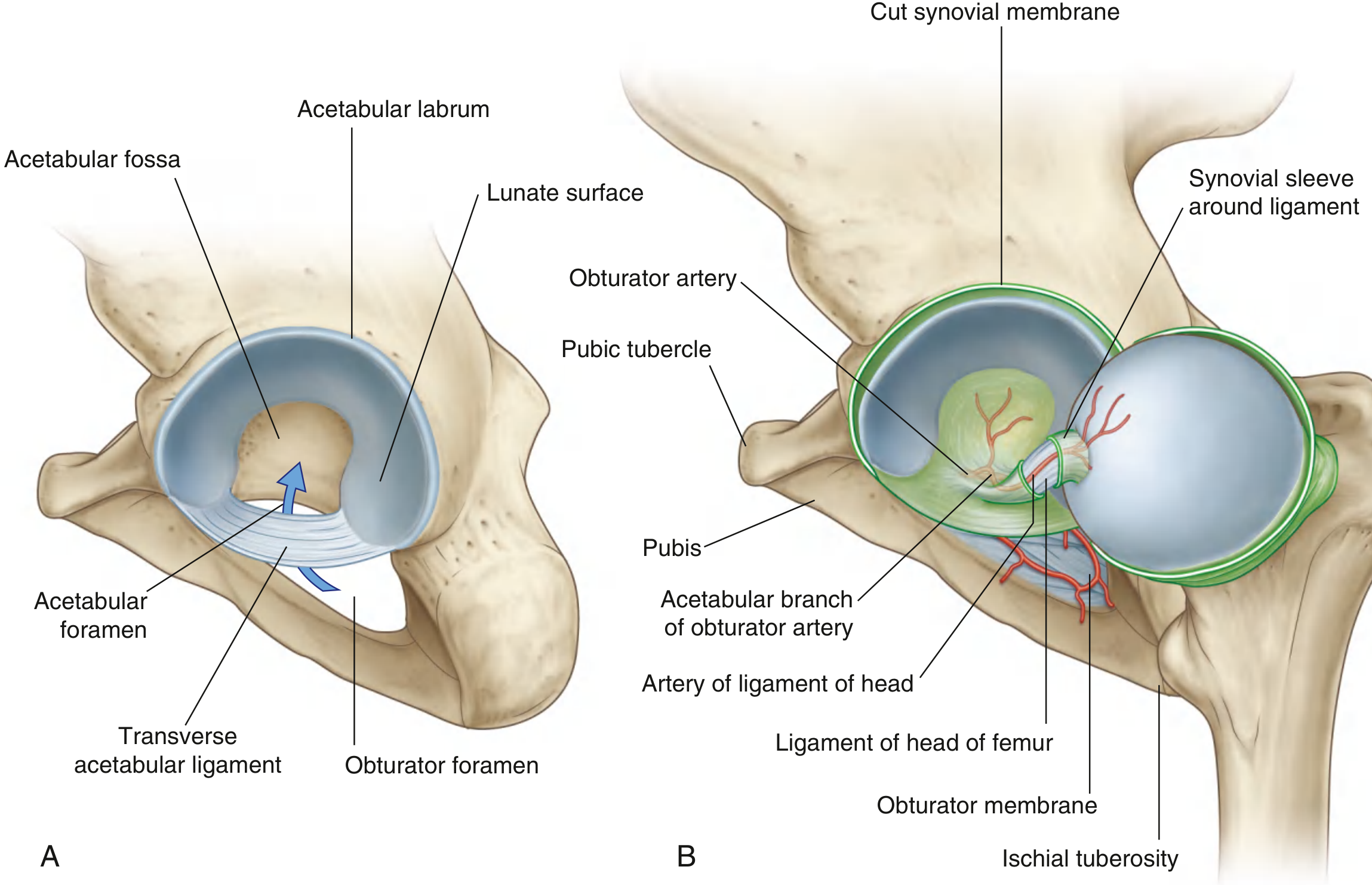
- The lunate surface of the acetabulum (covered by hyaline cartilage, broadest superiorly) receives the spherical head of the femur.
- The acetabulum almost entirely encompasses the femoral head, contributing substantially to inherent bony stability.
- The nonarticular acetabular fossa contains loose connective tissue and the ligament of the head.
2. Acetabular Labrum
- A fibrocartilaginous collar that deepens the acetabulum and grips the femoral head, further improving stability.
- Inferiorly, it bridges the acetabular notch as the transverse acetabular ligament, converting the notch into a foramen.
3. Joint Capsule (Fibrous Membrane)
- A strong, thick fibrous membrane that encloses the joint.
- Medial attachment: margin of the acetabulum, transverse acetabular ligament, and adjacent margin of the obturator foramen.
- Lateral attachment: intertrochanteric line anteriorly; just proximal to the intertrochanteric crest posteriorly (so the posterior femoral neck lies intracapsular).
- The capsule contains zona orbicularis fibers (circular fibers that form a collar around the femoral neck, helping grip the neck).
4. Synovial Membrane
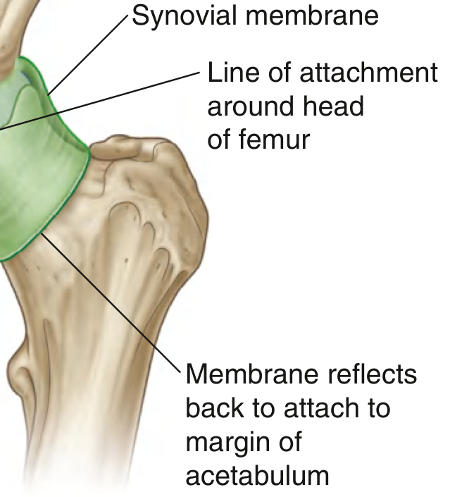
- Lines the fibrous membrane and attaches to the margins of articular surfaces.
- Forms a tubular sleeve around the ligament of the head of femur.
- From its attachment at the margin of the femoral head, it covers the femoral neck before reflecting onto the fibrous membrane - this is clinically important because it carries retinacular vessels supplying the femoral head.
5. Ligaments
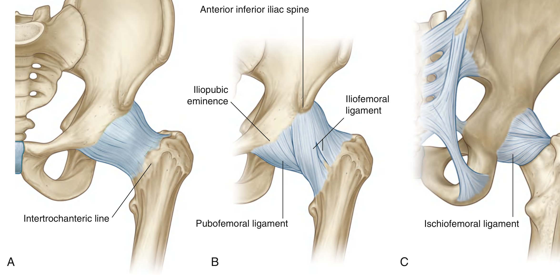
Three major extracapsular ligaments reinforce the fibrous membrane. Their fibers are oriented in a spiral fashion so they become taut in extension, stabilizing the joint and reducing the muscular effort needed to maintain an upright posture.
a. Iliofemoral Ligament (Ligament of Bigelow)
- Strongest ligament in the body - Y-shaped (inverted Y)
- Position: Anterior to the hip joint
- Proximal attachment: Ilium between the anterior inferior iliac spine (AIIS) and the margin of the acetabulum
- Distal attachment: Along the intertrochanteric line of the femur (thicker superiorly and inferiorly, giving the Y shape)
- Function: Resists extension and lateral rotation; prevents the pelvis from tilting backward during standing (reduces muscle effort)
b. Pubofemoral Ligament
- Position: Anteroinferior to the hip joint
- Proximal attachment: Iliopubic eminence, adjacent bone, and obturator membrane
- Distal attachment: Blends with the fibrous membrane and deep surface of the iliofemoral ligament
- Function: Limits abduction and extension
c. Ischiofemoral Ligament
- Position: Posterior to the hip joint (weakest of the three)
- Medial attachment: Ischium, just posteroinferior to the acetabulum
- Lateral attachment: Greater trochanter, deep to the iliofemoral ligament
- Function: Limits extension and medial rotation
d. Ligament of the Head of Femur (Ligamentum Teres)
- A flat band of delicate connective tissue inside the joint
- Attaches from the fovea on the femoral head to the acetabular fossa, transverse acetabular ligament, and margins of the acetabular notch
- Limited mechanical role in adults; carries a branch of the obturator artery (contributing to blood supply of the femoral head in children)
6. Muscle Support
Muscles around the hip provide dynamic stabilization and are the most important active supports:
| Group | Key Muscles | Action |
|---|---|---|
| Anterior (flexors) | Iliopsoas, rectus femoris, sartorius | Flex the hip; iliopsoas also stabilizes the joint anteriorly |
| Posterior (extensors) | Gluteus maximus, hamstrings | Extend and stabilize posteriorly |
| Abductors | Gluteus medius, gluteus minimus | Stabilize pelvis in single-leg stance (prevent Trendelenburg) |
| Short external rotators | Piriformis, obturator internus/externus, gemelli, quadratus femoris | Lie close to the capsule, acting like a "rotator cuff of the hip" |
The short external rotators (piriformis, obturator internus and externus, superior and inferior gemelli, quadratus femoris) lie directly against the posterior capsule and provide additional posterior support, analogous to the rotator cuff of the shoulder.
7. Vascular Supply
Articular branches from multiple arteries form a pericapsular network:
- Medial and lateral circumflex femoral arteries (main supply to femoral head)
- Obturator artery (acetabular branch via ligamentum teres)
- Superior and inferior gluteal arteries
- First perforating branch of the deep femoral artery
8. Nerve Supply (Hilton's Law)
The hip joint is innervated by articular branches from:
- Femoral nerve
- Obturator nerve
- Superior gluteal nerve
- Nerve to quadratus femoris
This explains why hip pathology often refers pain to the knee (shared femoral and obturator supply).
Sources: Gray's Anatomy for Students, pp. 661-664 | S Das: A Manual on Clinical Surgery, 13th ed.
This is a shared conversation. Sign in to Orris to start your own chat.