
What is the treatment plan for Anterior cruciate ligament tear in its mid and femoral attcahment fibers
"anterior cruciate ligament" AND "reconstruction" AND "femoral attachment"
anterior cruciate ligament tear reconstruction femoral attachment surgery
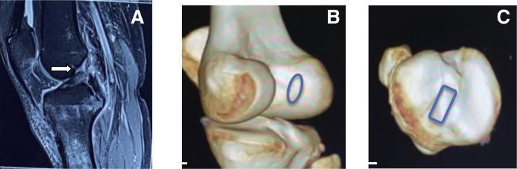
This composite figure provides a diagnostic and anatomical assessment of an anterior cruciate ligament (ACL) injury in a right knee. Panel A is a sagittal proton density fat-suppressed (PDFS) MRI demonstrating a full-thickness ACL tear; a white arrow points to the area of ligamentous discontinuity and high signal intensity at the femoral attachment site. Panels B and C present 3D-CT reconstructions highlighting the native footprint anatomy for preoperative planning. Panel B shows the femoral footprint on the lateral femoral condyle, delineated by a blue oval indicating its natural morphology. Panel C provides a superior view of the proximal tibia, where a blue rectangle marks the rectangular shape and orientation of the tibial footprint. These images illustrate the correlation between diagnostic MRI findings and the 3D anatomical landmarks necessary for anatomic ACL reconstruction.
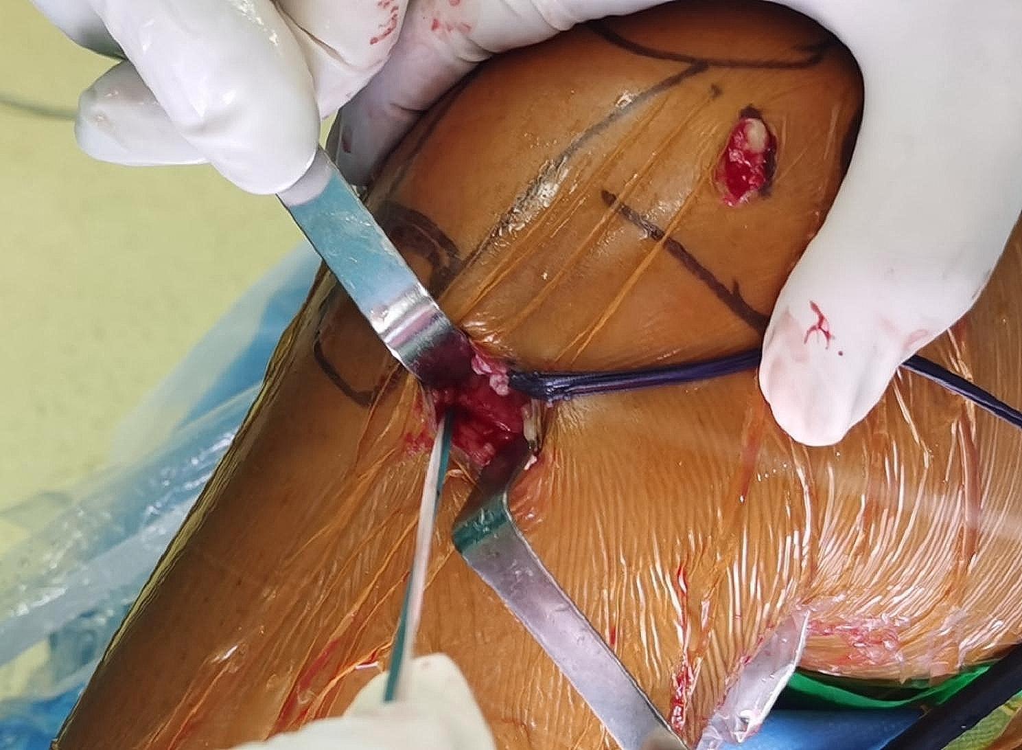
This clinical photograph captures an intraoperative view of an anterior cruciate ligament (ACL) reconstruction or augmentation procedure focusing on the distal tibial attachment site. The surgical field demonstrates a primary incision site near the tibial tuberosity, held open by metallic retractors to expose subcutaneous tissue and the graft stump. A secondary, smaller 1 cm incision is visible superiorly, likely corresponding to the femoral tunnel exit or an accessory portal. High-tensile traction sutures (Ethibond No. 2, visible in blue and white) are utilized to secure the distal end of the graft, forming a loop structure as part of a specialized fixation technique. The skin is prepped with an orange-tinted iodine-based antiseptic and covered with a transparent surgical drape. Surgical marking pen lines delineate anatomical landmarks such as the patella and joint line. The image illustrates a critical step in ligamentous graft tensioning and fixation within orthopedic surgery, highlighting the integration of the graft with the native tibial footprint.
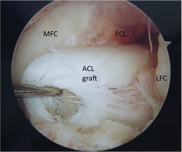
This clinical photograph shows an intra-operative arthroscopic view of the femoral notch in a left knee, specifically highlighting a failed anterior cruciate ligament (ACL) reconstruction. The central focus is a white, fibrous ACL graft which exhibits clear signs of rupture. The graft fibers are visibly frayed, disorganized, and disrupted, with the primary tear located near the distal tibial attachment site. The anatomical context is provided by several labeled landmarks: the medial femoral condyle (MFC) and lateral femoral condyle (LFC) appear as smooth, light-colored cartilaginous surfaces forming the walls of the notch. Posterior to the damaged ACL graft is the posterior cruciate ligament (PCL), which is covered by a vascularized, translucent synovial membrane. A surgical probe is visible on the left side, reflecting the clinical assessment of the graft's integrity. This image serves as a diagnostic example of mechanical failure in orthopedic sports medicine, specifically illustrating post-operative graft complications and the arthroscopic appearance of ligamentous fraying.
Treatment Plan for ACL Tear: Midsubstance vs. Femoral Attachment Fibers
1. Initial Assessment and Decision Framework
- Younger age
- Preinjury hours of sports participation
- Degree of anterior instability (measured by KT-1000 arthrometer)
2. Nonoperative Management (Both Tear Types)
- Aggressive rehabilitation program
- Activity level counseling
- Functional knee bracing (controversial - has not been shown to significantly reduce reinjury rates if the patient returns to high-level sports)
3. Midsubstance ACL Tears
Why Primary Repair Historically Failed
Modern Primary Repair - Resurgence with Augmentation
| Factor | Details |
|---|---|
| Timing | Acute repairs (within 3 weeks of injury) produce better outcomes for load, stiffness, laxity, and rerupture rate |
| Biologic enhancement | Bone marrow access via drilling/microfracture improves healing potential |
| Internal bracing | Suture augmentation lowers failure rate from 10.7% to 7.0% |
| BEAR procedure | Bridge-Enhanced ACL Repair - suture repair augmented with a proprietary scaffold placed between torn ends; RCT showed no difference in pain, effusion, or failure rate vs. autograft hamstring reconstruction |
| PRP | Beneficial only when combined with a structural scaffold |
| Skeletally immature patients | Best outcomes with repair |
Reconstruction for Midsubstance Insufficiency
| Graft | Advantages | Disadvantages |
|---|---|---|
| Bone-patellar tendon-bone (BPTB) autograft | Fastest bone tunnel incorporation; preferred for early return to sport | Anterior knee pain, patellar fracture risk, higher arthritis rates at 5-7 yrs |
| 4-strand hamstring autograft | Similar strength to native ACL | Less stiff; harvest risks knee flexion weakness, saphenous nerve injury |
| Quadriceps tendon autograft | Robust graft with bone block option | Patellar fracture risk |
| Allograft | No donor site morbidity | Higher rupture rate in younger/active patients; slower tunnel incorporation; infection risk (1:6 million for HIV) |
- Anatomic reconstruction targets the 10 o'clock (or 2 o'clock in left knee) position, centering the graft in the middle of the femoral ACL footprint
- A more horizontal graft position reduces rotational instability
- Anterior placement of the femoral tunnel causes flexion loss - the most important technical error to avoid
4. Femoral Attachment Fiber Tears
4a. Soft-Tissue Femoral Avulsion (No Bone Fragment)
- Repair of the ligament back to a freshened femoral bed has been described with success
- However, long-term results of isolated femoral attachment repair are limited
- Internal bracing augmentation significantly improves success rates
- The over-the-top augmentation technique preserves the femoral footprint: the tibial tunnel is placed at the anteromedial edge of the tibial footprint to minimize disruption of the tibial attachment
4b. Bony Femoral Avulsion
- Avulsions typically occur from the tibial insertion, not the femoral origin
- Rarely, disruption from the femoral origin avulses a bone fragment (described in skiers)
- When it occurs: repair back to a freshened femoral bed - acute fixation appropriate
4c. Surgical Repair Technique for Femoral Attachment (Over-the-Top Method)
- Augmenting tissue passed through the posterior capsule high in the intercondylar notch
- Tissue passed over the lateral femoral condyle (over-the-top orientation)
- Secured to the lateral aspect of the distal femur with staples or screw/spiked washer
- Tibial tunnel placed at the anteromedial edge of the ACL tibial footprint to protect the tibial attachment
5. Bony Tibial Avulsion (Separate Entity - for completeness)
- Arthrotomy and exposure of the tibial ACL attachment
- Deepen the crater in the tibia
- Remove any blood clot/debris from the crater before reduction (clot left in place causes residual laxity)
- Pull the bone fragment (on the end of the ligament) into the crater to restore ligament tension
- Pass sutures through transosseous drill holes, tie over bone anteriorly - OR use screw fixation through the fragment into the bed (no significant outcome difference between screw vs. suture fixation in Lysholm scores)
- Apply controlled motion brace at the degree of knee flexion where fragment is best reduced
- 3 weeks: flexion 0-90° in brace + isometric quad/hamstring exercises
- 6 weeks: discontinue crutches
- 8 weeks: full active and passive range of motion achieved
- 3+ months: progressive resistance exercises
6. Extraarticular Procedures (Adjuncts)
- Iliotibial band tenodesis (MacIntosh or MacIntosh-Losee technique): creates a restraining band from the lateral femoral epicondyle to Gerdy's tubercle
- When used alone: high failure rate (under 76% success); does not stop AP tibial translation beyond 2 years
- Currently used primarily as an adjunct to intraarticular ACL reconstruction for severe anterolateral instability
7. Postoperative Rehabilitation (Post-Reconstruction)
| Phase | Key Elements |
|---|---|
| Early | Early motion with emphasis on extension; early weight bearing encouraged |
| Safe exercises | Hamstring-dominated; active ROM 35-90° flexion; closed kinetic chain (foot planted) with compressive loading |
| Avoid (first 6 weeks) | Open kinetic chain extension near full extension (increases ACL graft stress) |
| Bracing | Postoperative bracing not proven beneficial except in downhill skiers |
| Rehabilitation pace | No outcome difference between accelerated vs. non-accelerated programs |
Key Takeaway Summary
| Tear Location | Primary Treatment | Surgical Approach |
|---|---|---|
| Midsubstance | Reconstruction (standard); primary repair + augmentation in acute/young patients | Intraarticular reconstruction with BPTB/hamstring/quadriceps/allograft; anatomic femoral tunnel |
| Femoral attachment (soft tissue) | Primary repair preferred (better healing potential); augmentation with over-the-top technique | Preserve femoral footprint; over-the-top augmentation; tibial tunnel at anteromedial footprint edge |
| Femoral attachment (bony avulsion) | Acute repair - reattach fragment to freshened femoral bed | Suture or screw fixation back to femoral bone |
| Tibial avulsion | Bony repair (most common avulsion type) | Suture through transosseous holes or screw through fragment; restore ligament tension |