
PDA
Reading File
Finding Sources
Searching PubMed
"patent ductus arteriosus"
Searching the Web
patent ductus arteriosus overview CDC
Reading File
PDA = Patent Ductus Arteriosus
It is a congenital heart condition in which the ductus arteriosus, a normal fetal vessel connecting the aorta to the pulmonary artery, fails to close after birth.
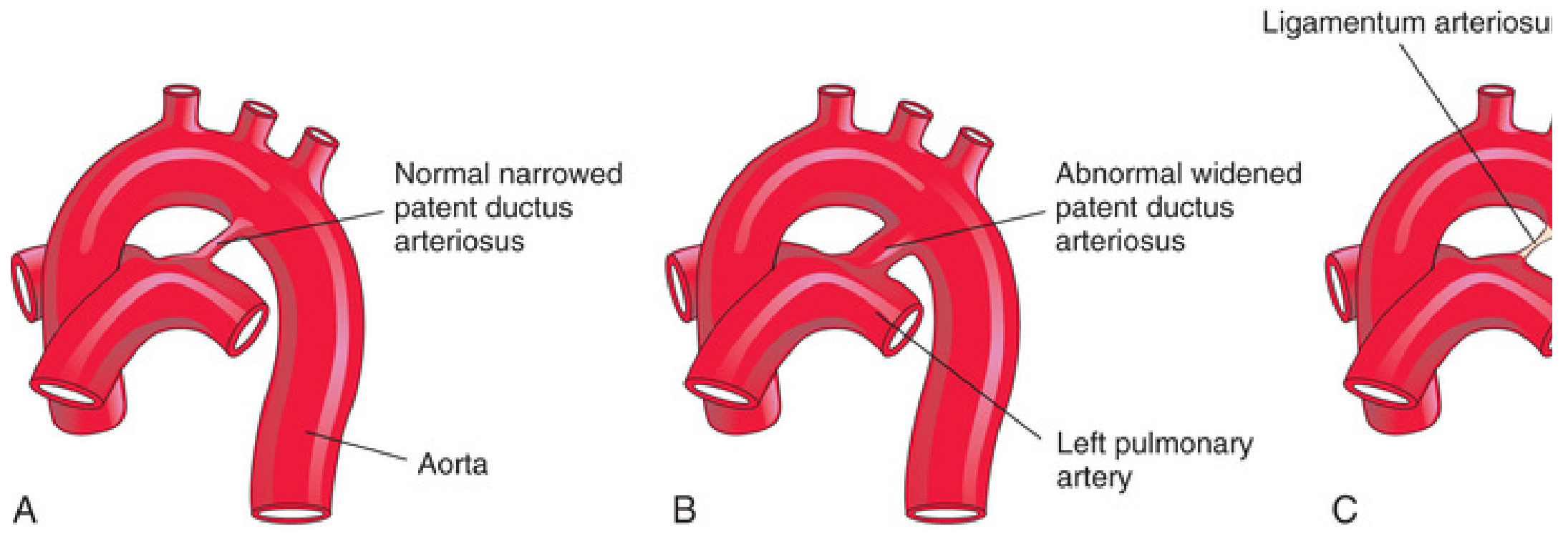
Normal physiology
Before birth, the ductus diverts blood away from the fetal lungs. It normally functionally closes within the first few days of life and later becomes the ligamentum arteriosum. The Developing Human: Clinically Oriented Embryology, p. 880.
What happens in PDA
After birth, aortic pressure exceeds pulmonary arterial pressure, so blood flows:
Aorta → pulmonary artery
This produces a left-to-right shunt, increasing pulmonary blood flow and causing left atrial and left ventricular volume overload.
This produces a left-to-right shunt, increasing pulmonary blood flow and causing left atrial and left ventricular volume overload.
Risk factors / associations
- Prematurity, especially very-low-birth-weight infants
- Maternal rubella infection early in pregnancy
- Hypoxia or respiratory distress syndrome
- High altitude
- Female sex is more commonly affected
The Developing Human: Clinically Oriented Embryology, pp. 880-882.
Clinical features
- Small PDA: often asymptomatic
- Continuous, “machinery-like” murmur, typically in the left infraclavicular area
- Bounding peripheral pulses and wide pulse pressure
- Large PDA: tachypnea, feeding difficulty, poor weight gain, recurrent respiratory infections, or heart failure
Complications
- Pulmonary hypertension
- Heart failure
- Infective endarteritis/endocarditis
- Eisenmenger syndrome in untreated large PDA, with shunt reversal and cyanosis
Diagnosis
- Echocardiography with Doppler confirms the connection and assesses shunt size and cardiac effects.
- Chest radiograph and ECG may show pulmonary plethora and left-sided enlargement in significant PDA.
Treatment
- Preterm symptomatic PDA: conservative care may be considered; pharmacologic closure with ibuprofen, indomethacin, or sometimes acetaminophen, depending on the clinical setting and contraindications.
- Persistent PDA in term infants/children: usually closed by transcatheter device closure.
- Surgery: reserved when catheter closure is not feasible or in selected infants.
A recent meta-analysis suggests pharmacologic PDA treatment in preterm infants involves trade-offs rather than a uniformly beneficial outcome, so management should be individualized (2025 Bayesian meta-analysis). The American Heart Association overview notes that most people do well after PDA repair.
Murmurs in congenital heart defects
Reading File
Finding Sources
Finding Sources
Finding Sources
Searching PubMed
"congenital heart disease" AND auscultation
Searching the Web
congenital heart defects murmurs auscultation clinical table
Finding Sources
Finding Sources
Finding Sources
Murmurs in common congenital heart defects
| Defect | Typical murmur | Best heard | Other key finding |
|---|---|---|---|
| ASD | Ejection systolic flow murmur, not from the septal hole itself | Left upper sternal border (LUSB) | Wide, fixed split S2 |
| VSD | Harsh pansystolic/holosystolic murmur | Left lower sternal border (LLSB) | Small restrictive VSD may be louder and have a thrill; large VSD may have a softer murmur if pulmonary pressure is high |
| PDA | Continuous “machinery” murmur | Left infraclavicular area / LUSB | Bounding pulses and wide pulse pressure |
| Pulmonary stenosis (PS) | Crescendo-decrescendo ejection systolic murmur | LUSB | Ejection click; delayed/soft P2 in severe disease |
| Aortic stenosis (AS) | Crescendo-decrescendo ejection systolic murmur | Right upper sternal border (RUSB) | Radiates to carotids; ejection click may occur |
| Coarctation of aorta | Systolic murmur, often heard posteriorly | Left interscapular back and left sternal border | Weak/delayed femoral pulses, arm hypertension, lower limb hypotension |
| Tetralogy of Fallot (TOF) | Harsh ejection systolic murmur due to RV outflow obstruction | Left upper or mid-sternal border | Cyanosis, single S2. More severe pulmonary stenosis can give a softer murmur because there is less flow |
| Atrioventricular septal defect (AVSD) | Pansystolic murmur if AV valve regurgitation or VSD component; flow murmur may be present | LLSB | Often associated with Down syndrome |
| Tricuspid atresia | Often a pansystolic VSD murmur or ejection murmur from pulmonary stenosis | LLSB/LUSB | Cyanosis, single S2 |
| Transposition of great arteries (TGA) | May have no significant murmur | Variable | Severe cyanosis soon after birth; murmur appears if there is VSD, pulmonary stenosis, or another associated lesion |
| Truncus arteriosus | Systolic ejection murmur with possible early diastolic murmur from truncal-valve regurgitation | Upper left sternal border | Single loud S2, heart failure from pulmonary overcirculation |
| Total anomalous pulmonary venous return (TAPVR) | Ejection systolic pulmonary flow murmur | LUSB | Fixed split S2 in unobstructed TAPVR; obstructed TAPVR can be critically ill with little murmur |
High-yield pattern
- Pansystolic murmur at LLSB: think VSD
- Ejection systolic murmur + fixed wide split S2: think ASD
- Continuous murmur + bounding pulses: think PDA
- Ejection systolic murmur at LUSB: think pulmonary stenosis or increased pulmonary flow, such as ASD
- Cyanosis + ejection systolic murmur: think TOF
- Cyanotic newborn with little or no murmur: consider TGA or another duct-dependent critical lesion
Important exam principle
The loudness of a murmur reflects turbulent flow and pressure gradient, not simply the size or severity of the anatomical defect. Thus:
- A small VSD can be very loud because the pressure gradient is high.
- A very large VSD or advanced pulmonary hypertension may make the murmur softer.
- In severe TOF, reduced flow across the narrowed RV outflow tract can reduce murmur intensity.
A harsh, pansystolic murmur, a grade 3/6 or louder murmur, an abnormal S2, a thrill, cyanosis, poor feeding, tachypnea, or weak femoral pulses should prompt echocardiographic assessment. The AAFP review on pediatric murmurs summarizes these concerning findings. Textbook findings also note that ASD shunt flow itself is usually silent, while the audible murmur is typically from increased flow across the pulmonary valve; VSD characteristically causes a holosystolic murmur. Harrison’s Principles of Internal Medicine, 22e, Chapter 44.
This is a shared conversation. Sign in to Orris to start your own chat.