
• Pancreas – aetiology, clinical features, investigations, treatment & complications of acute & chronic pancreatitis
Reading File
Now let me search the medical library for pancreatitis content in parallel:
Finding Sources
Finding Sources
Excellent finds. Now let me read the best sources in parallel - Sleisenger & Fordtran (the gold standard GI textbook), Pye's Surgical Handicraft, and Robbins Pathology:
Reading File
Reading File
Reading File
Let me now read the main Sleisenger & Fordtran acute pancreatitis sections (block 15) and the Pye's surgical text continuation, plus the Robbins pathology for acute pancreatitis morphology:
Reading File
Reading File
Finding Sources
Bailey & Love is a key surgical textbook. Let me read its acute pancreatitis section and also the Robbins pathology for acute pancreatitis morphology:
Reading File
Finding Sources
Reading File
Reading File
Reading File
Reading File
Reading File
Reading File
Now let me fetch the key images and read the Bailey & Love chronic pancreatitis + severity scoring sections:
Reading File
Loading Image
I now have comprehensive material from multiple authoritative sources. Let me also grab the severity scoring criteria and the chronic pancreatitis section from Bailey & Love:
Reading File
Reading File
I now have enough comprehensive data from multiple authoritative textbooks. Let me compile the full detailed response:
Pancreatitis: Acute & Chronic - Complete Clinical Overview
ACUTE PANCREATITIS
Definition
Acute pancreatitis (AP) is a sudden inflammatory condition of the pancreas resulting from autodigestion by inappropriately activated pancreatic enzymes. The process ranges from mild interstitial oedema to severe haemorrhagic necrosis.
Aetiology
The mnemonic "GET SMASHED" covers the main causes:
| Category | Examples |
|---|---|
| Gallstones | Most common cause overall (40-60%); transient/sustained impaction at the ampulla of Vater |
| Ethanol | Second most common; direct acinar toxicity + sphincter of Oddi spasm |
| Trauma | Blunt abdominal injury; post-ERCP pancreatitis (1-3% of procedures) |
| Steroids | Corticosteroids, azathioprine, L-asparaginase, valproic acid |
| Mumps / viruses | EBV, coxsackievirus, HIV-related |
| Autoimmune | IgG4-associated (lymphocytic sclerosing pancreatitis) |
| Scorpion venom | Tityus trinitatis |
| Hyperlipidaemia / Hypercalcaemia | Hypertriglyceridaemia (chylomicron-induced ischaemia); hyperparathyroidism |
| ERCP | Duct disruption and enzyme extravasation |
| Drugs | Statins (weak link), gliptins, 6-mercaptopurine |
Hereditary pancreatitis - rare; mutations in the cationic trypsinogen gene (PRSS1); patients develop AP in teens, progress to chronic pancreatitis, and carry ~40% lifetime risk of pancreatic cancer.
Structural causes include pancreas divisum (most common anatomic aberration), ampullary tumours, and sphincter of Oddi dysfunction.
No more than 20% of cases should be classified as truly idiopathic after thorough workup. - Bailey and Love's Short Practice of Surgery, 28th Edition
Pathogenesis
Three converging pathways lead to acinar cell injury and enzyme activation:
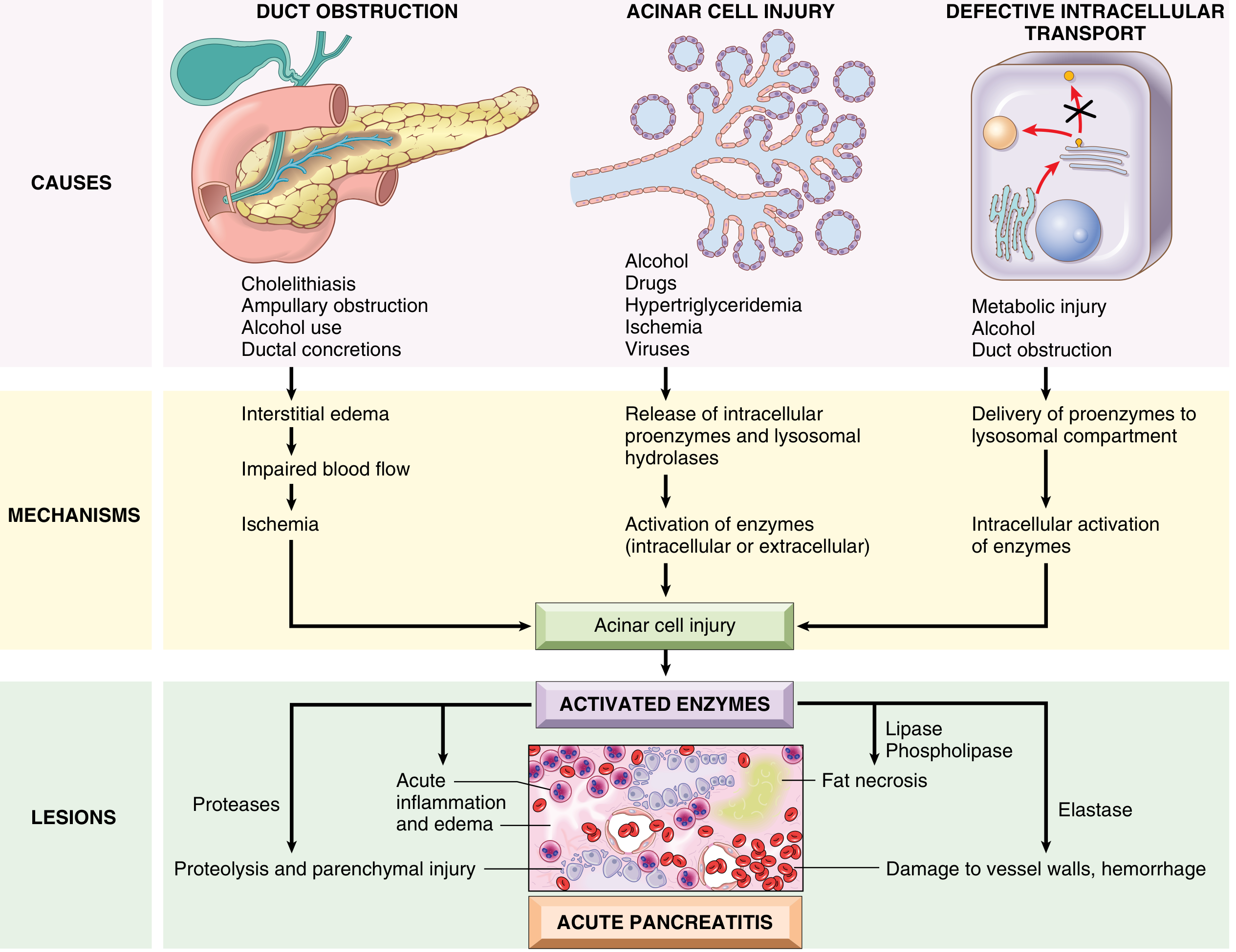
FIG. 15.2 Proposed pathogenesis of acute pancreatitis - Robbins & Kumar Basic Pathology
- Pancreatic duct obstruction - gallstone/biliary sludge impaction raises intraductal pressure -> interstitial oedema -> vascular insufficiency -> ischaemic acinar injury
- Primary acinar cell injury - alcohol, hypertriglyceridaemia, ischaemia, drugs, and viruses directly injure acinar cells -> release of intracellular proenzymes and lysosomal hydrolases
- Defective intracellular transport - proenzymes co-package with lysosomal hydrolases -> intracellular trypsin activation -> lysosomal rupture -> enzyme cascade
Alcohol-specific mechanisms include: (a) transient increase in exocrine secretion + sphincter of Oddi contraction; (b) oxidative stress with direct acinar membrane damage; (c) delivery of proenzymes to lysosomal compartment with intracellular trypsin activation; (d) protein-rich secretions forming ductal plugs.
Once trypsin is activated, it triggers a cascade: trypsinogen -> trypsin activates all other proenzymes (chymotrypsin, elastase, phospholipase A2, kallikrein) -> proteolysis, haemorrhage, fat necrosis, and systemic inflammatory response syndrome (SIRS). - Robbins & Kumar Basic Pathology
Clinical Features
Symptoms:
- Pain - cardinal symptom; epigastric, severe, constant, reaching maximum within minutes; radiates to the back in ~50% of cases; some patients gain relief by leaning forward (classic "pancreatitis posture")
- Nausea and vomiting (does not relieve pain - distinguishes from peptic ulcer)
- Anorexia
Signs:
- Epigastric tenderness and guarding
- Abdominal distension from ileus
- Fever, tachycardia
- Grey Turner's sign - bruising in the flanks (haemorrhagic pancreatitis; ~1-2 days after onset)
- Cullen's sign - periumbilical bruising (retroperitoneal haemorrhage tracking to umbilicus)
- Jaundice (if gallstone aetiology with CBD obstruction)
- Hypotension / shock in severe cases
Both Grey Turner's and Cullen's signs indicate severe haemorrhagic necrotising pancreatitis and carry a poor prognosis.
Investigations
Three key questions to answer (Bailey & Love):
- Is the diagnosis of acute pancreatitis correct?
- How severe is the attack?
- What is the aetiology?
Biochemical:
- Serum amylase - >5× upper limit of normal is diagnostic; peaks within hours of onset; returns to near normal within 48h (may miss peak if bloods taken late); also elevated in peptic ulcer perforation, bowel obstruction, cholecystitis
- Serum lipase - more sensitive and specific than amylase; remains elevated longer (>3 days); preferred where available
- Urinary amylase:creatinine clearance ratio - useful if serum levels have normalised
- Peritoneal aspirate - prune-juice fluid with extremely high amylase (>10,000 units) in haemorrhagic necrosis
Severity markers:
- CRP >150 mg/L at 48h = severe attack indicator
- FBC (WBC, haematocrit), U&E, LFTs, calcium, glucose, LDH, ABG, albumin, triglycerides
Imaging:
- Plain AXR - not diagnostic but shows: "sentinel loop" (gas-filled jejunal loop in upper abdomen - considered pathognomonic), "colon cut-off sign" (transverse colon gas abruptly stops), widened C-loop of duodenum
- CXR - pleural effusion (usually left-sided, indicates severity), ARDS pattern
- Ultrasound (within 24h of all admissions) - detects gallstones and CBD dilatation; pancreas may be oedematous; identifies peripancreatic fluid collections
- Contrast-enhanced CT - not needed in mild disease; indicated when: (a) diagnostic uncertainty; (b) severe attack to distinguish interstitial from necrotising pancreatitis; (c) suspected abscess (gas within fluid collection); (d) failure to improve within 72h. CT may underestimate necrosis extent in first 72h. Severity graded by the CT Severity Index (Balthazar grading A-E)
- MRCP/MRI - non-invasive assessment of CBD stones and pancreatic duct; avoids radiation
- EUS - detects microlithiasis and CBD stones; helps select patients for ERCP
Severity Scoring:
Revised Atlanta Classification (2013):
| Grade | Criteria |
|---|---|
| Mild | No organ failure; no local/systemic complications |
| Moderately Severe | Transient organ failure (<48h) AND/OR local/systemic complications without persistent organ failure |
| Severe | Persistent organ failure (>48h); single or multiple organ failure |
Ranson Criteria (score ≥3 at 48h = severe):
| On Admission | Within 48 hours |
|---|---|
| Age >55 years | Haematocrit fall ≥10% |
| WBC >16×10⁹/L | BUN rise >5 mg/dL despite fluids |
| Blood glucose >11 mmol/L | PaO₂ <8 kPa (60 mmHg) |
| LDH >350 U/L | Serum calcium <2.0 mmol/L |
| AST >250 U/L | Base deficit >4 mmol/L |
| Fluid sequestration >6 litres |
Glasgow (Imrie) Criteria (score ≥3 within 48h = severe):
- Age >55 years; WBC >15×10⁹/L; glucose >10 mmol/L; LDH >600 U/L or AST >200 U/L
- Urea >16 mmol/L (no response to fluids); PaO₂ <8 kPa; Ca²⁺ <2.0 mmol/L; albumin <32 g/L
Mnemonic for Glasgow: PANCREAS - PaO₂, Age, Neutrophils, Calcium, Renal urea, Enzymes, Albumin, Sugar
Other systems: APACHE II, SOFA, SAPS, MODS, Modified Marshall. - Bailey and Love's Short Practice of Surgery, 28th Edition
Treatment
Mild AP - conservative management:
- Fluid resuscitation - aggressive IV crystalloid (Ringer's lactate preferred); guided by urine output, HR, CVP
- Analgesia - opiate analgesia (pethidine traditionally favoured; morphine now acceptable); NSAIDs adjunct
- Antiemetics
- Nutritional support - early oral/enteral feeding when tolerated; prolonged NBM not justified physiologically. Enteral > parenteral nutrition (fewer infections, lower cost). Nasogastric vs nasojejunal feeding is equivalent, though NJ preferred if gastric retention
- Antibiotics - NOT indicated prophylactically in mild disease; reserved for infected necrosis
- No drug therapy or CT needed in mild disease
Severe AP - aggressive management:
- High-dependency / ICU admission with invasive monitoring
- Aggressive fluid resuscitation with close monitoring
- ERCP - for gallstone pancreatitis with cholangitis or biliary obstruction (urgent); early routine ERCP does not improve outcomes in the absence of obstruction
- Early laparoscopic cholecystectomy (within 3 days) in mild biliary pancreatitis improves outcomes
- Prophylactic antibiotics - controversial; no proven mortality benefit; currently NOT recommended routinely
Management of complications:
Infected pancreatic necrosis:
- Diagnose by CT-guided fine-needle aspiration (FNA) for culture
- Treatment: step-up approach - percutaneous/endoscopic drainage first, then minimally invasive necrosectomy if drainage fails
- Surgical necrosectomy options: closed continuous lavage (Beger), closed drainage, open packing, or planned relaparotomy (Bradley)
- CT-guided percutaneous drainage + antibiotics (carbapenems penetrate pancreatic tissue well)
Pseudocysts:
- Allow 4-6 weeks to mature and encapsulate
- Endoscopic transgastric or endoscopic ultrasound-guided cystgastrostomy (if in apposition to stomach wall)
- Percutaneous drainage or surgical cystgastrostomy/cystjejunostomy
Abdominal compartment syndrome, ARDS, AKI, DIC - managed in ICU
Outcomes: Overall mortality remains 10-15%. Mortality in mild disease <1%; in severe necrotising disease with multi-organ failure up to 30-40%. Before discharge, aetiology must be determined to prevent recurrence. - Bailey and Love's Short Practice of Surgery, 28th Edition
Complications of Acute Pancreatitis
Local:
| Complication | Features |
|---|---|
| Peripancreatic fluid collections | Early (<4 weeks); usually resolve spontaneously |
| Acute pancreatic necrosis | Sterile or infected; identified on contrast CT (non-enhancing areas) |
| Pancreatic abscess | Gas within fluid collection on CT; requires drainage |
| Pseudocyst | After >4 weeks; fluid collection with defined wall; devoid of solid debris; result of ductal leak or liquefied fat necrosis |
| Walled-off necrosis (WON) | After >4 weeks; encapsulated necrotic collection; sterile or infected |
| Splenic vein thrombosis | Left-sided (sinistral) portal hypertension |
| Haemorrhage | Erosion of vessels; pseudoaneurysm formation |
Systemic:
| System | Complication |
|---|---|
| Respiratory | Pleural effusion (left-sided), atelectasis, ARDS |
| Renal | Acute tubular necrosis, AKI |
| Cardiovascular | Hypotension, shock, pericardial effusion |
| Metabolic | Hypocalcaemia (calcium saponification in fat necrosis), hyperglycaemia, hypoalbuminaemia |
| Coagulation | DIC |
| GI | Paralytic ileus, stress ulcers, bowel ischaemia |
| Endocrine | Transient or permanent diabetes (islet destruction) |
CHRONIC PANCREATITIS
Definition
Chronic pancreatitis is a progressive inflammatory disease characterised by irreversible destruction of pancreatic tissue, leading to loss of exocrine function (malabsorption) and, eventually, endocrine function (diabetes mellitus). - Bailey and Love's Short Practice of Surgery, 28th Edition
Incidence: 2-10 new cases per 100,000/year (higher in parts of southern India: 100-200/100,000). Male:female ratio 4:1. Mean age of onset ~40 years.
Aetiology
The TIGAR-O classification organises causes:
| Category | Examples |
|---|---|
| Toxic-metabolic | Alcohol (most common; 60-70% of cases in the West), smoking, hypercalcaemia, hyperlipidaemia, drugs |
| Idiopathic | Early-onset (<20yr), late-onset (>50yr), tropical (southern India/Africa) |
| Genetic | PRSS1 (cationic trypsinogen), SPINK1 (trypsin inhibitor), CFTR mutations - hereditary pancreatitis |
| Autoimmune | IgG4-associated (Type 1); ISGP4/AIP criteria; responds to steroids |
| Recurrent acute pancreatitis | Post-necrotic, recurrent biliary |
| Obstructive | Pancreas divisum, sphincter of Oddi dysfunction, ductal stricture, tumour |
Only 5-10% of alcoholics develop chronic pancreatitis (suggesting additional genetic/metabolic susceptibility). - Robbins & Kumar Basic Pathology
Pathology / Morphology
Gross: Hard, fibrotic gland; extremely dilated ducts; visible calcific concretions (ductal stones); may have a mass in the head mimicking carcinoma.
Microscopic:
- Parenchymal fibrosis
- Reduced acinar number and size (acinar loss - constant feature)
- Variable ductal dilatation; ductal epithelium atrophied, hyperplastic, or squamous metaplasia; ductal concretions
- Chronic inflammatory infiltrate around lobules and ducts
- Relative early sparing of islets of Langerhans, which eventually become embedded in sclerotic tissue, fuse, appear enlarged, and finally disappear
Autoimmune pancreatitis (AIP): Dense lymphocyte and IgG4+ plasma cell infiltrate; "swirling" periductal fibrosis; venulitis (lymphocytic sclerosing pancreatitis). - Robbins & Kumar Basic Pathology
Clinical Features
Pain - most common and dominant symptom:
- Continuous or episodic, often severe and refractory
- Epigastric, radiating to the back; accentuated by meals and alcohol
- May be so severe the patient becomes narcotic-dependent
- Later in disease, pain may actually diminish as gland "burns out"
Exocrine insufficiency (develops after >90% gland destruction):
- Steatorrhoea - bulky, foul-smelling, pale, floating stools with fat globules; fat malabsorption precedes carbohydrate malabsorption
- Weight loss and malnutrition
- Fat-soluble vitamin deficiency (A, D, E, K)
Endocrine insufficiency:
- Diabetes mellitus ("type 3c" or pancreatogenic diabetes) - islet destruction; may precede steatorrhoea
Other features:
- Jaundice (biliary duct compression by fibrotic head)
- Repeated bouts mimicking acute pancreatitis
- Nausea, anorexia
- May be entirely silent until exocrine/endocrine failure develops
Investigations
Biochemistry:
- Serum amylase/lipase - often normal in established chronic disease (no functional acinar tissue)
- Glucose tolerance test - abnormal in >50% of patients with chronic pancreatitis/steatorrhoea
- Faecal elastase-1 (non-invasive exocrine function test; <200 µg/g = exocrine insufficiency)
- Lundh test meal - gold standard exocrine function test; reduced trypsin and amylase output post-meal
- Serum IgG4 - elevated in autoimmune pancreatitis
- Tumour markers - CA 19-9, CEA to exclude pancreatic carcinoma (important differential)
- Fat-soluble vitamin levels; vitamin B12
Faecal studies:
- Faecal fat (>7g/day = steatorrhoea)
- Faecal elastase-1 - practical first-line test
Imaging:
- Plain AXR - pancreatic calcification (pathognomonic when present, seen in ~30% of cases; especially in alcoholic CP and tropical pancreatitis)
- Ultrasound - ductal dilatation, calcification, pseudocysts, parenchymal changes
- CT scan - best for overall assessment; shows fibrosis, ductal dilatation, calcification, pseudocysts, vascular complications; excludes malignancy
- MRCP - non-invasive ductal imaging; shows "chain of lakes" appearance of irregular dilated main pancreatic duct
- ERCP - invasive; "chain of lakes" ductal morphology; allows therapeutic intervention (stenting, stone extraction); risk of post-ERCP pancreatitis
- EUS - most sensitive for parenchymal and ductal changes; allows FNA; detects early chronic pancreatitis
- CECT - staging; identifies mass lesions, vascular involvement
Treatment
Medical (conservative):
- Abstinence from alcohol - essential; slows progression
- Cessation of smoking
- Analgesia - stepped approach; paracetamol -> weak opioids -> strong opioids (risk of dependence); adjuncts: pregabalin, duloxetine
- Pancreatic enzyme supplementation - with all meals and snacks (lipase-containing enteric-coated preparations); reduces steatorrhoea; may reduce pain (negative feedback on CCK)
- Diet - low-fat, high-protein, high-carbohydrate; small frequent meals; medium-chain triglycerides if severe fat malabsorption (absorbed directly without lipase)
- Fat-soluble vitamins supplementation (A, D, E, K) and vitamin B12
- Micronutrient therapy - methionine, vitamins C and E, selenium (may reduce pain and slow progression)
- PPI/H2 blocker - reduce gastric acid to optimise enzyme function
- Treat diabetes mellitus - insulin usually needed (brittle due to glucagon deficiency also)
- Steroids - only in autoimmune pancreatitis (Type 1 AIP responds dramatically to prednisolone)
Endoscopic (ERCP-based):
- Stenting across dominant ductal stricture (leave ≤4-6 weeks before occlusion)
- Pancreatic duct stone extraction ± ESWL (extracorporeal shock wave lithotripsy)
- Pseudocyst drainage under EUS guidance
- Percutaneous or transgastric pseudocyst drainage
Surgical:
- Indicated for: intractable pain, ductal obstruction, mass lesion, complications
- Pancreatoduodenectomy (Whipple's) or Beger procedure (duodenum-preserving resection of pancreatic head) - for head mass
- Longitudinal pancreatojejunostomy (Puestow/Partington-Rochelle procedure) or Frey procedure (duct decompression + partial head resection) - for markedly dilated duct (>6mm); ~50% get long-term pain relief
- Distal pancreatectomy - for disease limited to the tail
- Total pancreatectomy ± islet autotransplantation - for intractable pain with diffuse disease; carries significant morbidity (brittle diabetes); ~1/3 get complete pain relief, 1/3 partial, 1/3 no benefit
- Celiac plexus neurolysis - for pain management (CT/EUS-guided)
Complications of Chronic Pancreatitis
| Complication | Details |
|---|---|
| Exocrine insufficiency | Steatorrhoea, malnutrition, fat-soluble vitamin deficiency |
| Diabetes mellitus | Type 3c; both insulin and glucagon deficiency; brittle |
| Pseudocyst | Common; may cause pain, obstruction, bleeding, infection |
| CBD stricture | Obstructive jaundice; biliary cirrhosis if untreated |
| Duodenal obstruction | Fibrotic mass compresses duodenum |
| Pancreatic fistula | Internal (pancreatic ascites, pleural effusion) or external |
| Splenic vein thrombosis | Left-sided portal hypertension, gastric varices |
| Pseudoaneurysm | Splenic artery most common; life-threatening haemorrhage |
| Pancreatic carcinoma | ~4-5% lifetime risk (20-year follow-up); highest in hereditary pancreatitis (~40% by age 70) |
| Narcotic addiction | From chronic analgesic use |
| Osteoporosis | Fat-soluble vitamin D malabsorption |
Key Differentiating Features: Acute vs Chronic
| Feature | Acute | Chronic |
|---|---|---|
| Reversibility | Potentially reversible | Irreversible |
| Amylase | Markedly elevated | May be normal |
| Pain | Sudden onset, very severe | Chronic, meal-related |
| Calcification | Absent | Pathognomonic when present |
| Steatorrhoea | Absent | Cardinal feature |
| Diabetes | Transient | Permanent |
| Ductal changes | Oedema | "Chain of lakes" on MRCP |
Sources consulted:
- Bailey and Love's Short Practice of Surgery, 28th Edition
- Robbins & Kumar Basic Pathology
- Pye's Surgical Handicraft, 22nd Edition
- Sleisenger and Fordtran's Gastrointestinal and Liver Disease
Diverticular disease – aetiology, pathogenesis, clinical features, complications, management (diverticulosis & diverticulitis)
Reading File
Finding Sources
Finding Sources
Reading File
Reading File
Good - Bailey & Love has excellent content. Let me continue reading it plus get the Sabiston colon section and search for pathogenesis content:
Reading File
Finding Sources
I need to get the main Bailey & Love diverticular disease section content (it was truncated). Let me read more of that section and search for pathogenesis paragraphs:
Reading File
Finding Sources
Reading File
Finding Sources
Reading File
Reading File
Reading File
Reading File
Reading File
Loading Image
Excellent - great diagram! Now let me get the Sleisenger pathogenesis/clinical section and elective surgery criteria:
Reading File
Reading File
I now have comprehensive material from Bailey & Love, Sleisenger & Fordtran, and Fischer's Mastery of Surgery. I have enough to write a thorough response.# Diverticular Disease - Complete Clinical Overview
Definitions
- Diverticulum - a hollow outpouching of the bowel wall
- Diverticulosis - the presence of diverticula without symptoms or inflammation (asymptomatic state)
- Diverticular disease - when diverticula cause symptoms or complications
- Diverticulitis - inflammation/infection of one or more diverticula
Types:
- True (congenital) diverticula: all three layers of bowel wall present (e.g. Meckel's diverticulum)
- False (acquired/pseudo) diverticula: only mucosa and submucosa herniate through the muscularis - NO muscularis propria in the wall; this is what colonic diverticular disease represents
Anatomy & Site
Colonic diverticula arise in 4 rows along the colon, at the sites where the major branches of the vasa recta (nutrient arteries) penetrate the circular muscle layer - on either side of the mesenteric taenia and on the mesenteric sides of the two anti-mesenteric taeniae. This explains why:
- Bleeding from diverticula can be profuse (proximity of the vasa recta)
- Diverticula are absent from the anti-mesenteric inter-taenial area
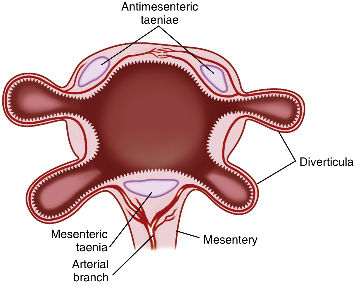
Fig. 121.1 - Sleisenger & Fordtran's Gastrointestinal and Liver Disease: Colonic diverticula and their relationship to the taeniae coli
Distribution:
- Western countries: ~90% left-sided (sigmoid colon predominant); rectum almost never affected (complete muscle coat + wider lumen)
- South East Asia / East Asia: ~75-85% right-sided predominance
- Diverticula found in left colon in ~75% of those over 70 years in the Western world
- Typical size: 3-10 mm diameter; giant diverticula >4 cm (rare, up to 25 cm)
Aetiology
The combination of multiple factors drives diverticulum formation:
| Factor | Role |
|---|---|
| Low-fibre Western diet | Decreased stool bulk -> prolonged transit -> raised intraluminal pressure (Burkitt's hypothesis); classic dogma but relationship is "limited and conflicting" in recent data |
| Ageing | Collagen cross-linking changes - increased elastin deposition in the taeniae -> shortening -> accordion pleating (myochosis or myochoresis); increased wall compliance allows herniation |
| Disordered colonic motility | Segmentation contractions generate high-pressure zones, especially in the narrow sigmoid |
| Raised intraluminal pressure | Most pronounced in the narrow sigmoid; by LaPlace's law (T = Pr), the sigmoid generates the highest wall tension for a given pressure |
| Weakness at vascular penetration sites | Oblique penetration of the vasa recta creates natural points of weakness in the circular muscle |
| Genetics | Familial aggregation well-documented; monozygotic twin concordance OR 7.15; heritable component ~40%; TNFSF15 gene linked to diverticulitis requiring surgery |
| Obesity, smoking, alcohol | Independent risk factors for diverticulosis and its complications |
| Physical inactivity | Associated with higher risk |
| NSAIDs/aspirin | Associated with increased risk of bleeding and perforation |
Why the rectum is spared: The rectum has a complete, continuous muscular coat (no gaps for vascular penetration) and a wider lumen (lower wall tension). - Bailey and Love's Short Practice of Surgery, 28th Edition
Why diverticular disease is rare in Africa/Asia: Traditional diets high in natural fibre; lower prevalence. Japanese migrants to Hawaii show diverticulosis rates approaching Western levels, confirming the dietary/environmental component. - Sleisenger & Fordtran's Gastrointestinal and Liver Disease
Pathogenesis
Diverticulosis Formation
The myochosis model: Increased elastin deposition and collagen cross-linking with ageing causes the taeniae coli to shorten and corrugate the circular muscle into haustral folds. This is associated with circular muscle hypertrophy. The resulting high-pressure segmentation contractions (particularly in the sigmoid) cause mucosa and submucosa to herniate through the weakest points - the sites of vascular penetration.
Wall structure changes: Colons with diverticula show thickening of both longitudinal (taeniae) and circular muscle layers with shortening of the taeniae - a "myochosis" pattern. The thickened wall actually contracts more forcefully and generates higher intraluminal pressure, creating a self-reinforcing cycle.
Diverticulitis - How Inflammation Develops
The process parallels appendicitis pathogenesis:
- The neck of a diverticulum becomes obstructed by inspissated stool (a faecalith)
- The faecalith abrades the mucosal wall of the sac -> low-grade inflammation
- Obstruction -> increased bacterial flora within the sac
- Diminished venous outflow -> localised ischaemia
- Altered mucosal defence mechanisms (defective CD2 pathway-induced apoptosis in lamina propria lymphocytes - similar to IBD)
- Bacteria breach the mucosa and the process extends transmurally
- Microperforation results - contained by pericolic fat/mesentery OR spreading to form:
- Phlegmon (localised inflammatory mass)
- Pericolic abscess
- Pelvic/intra-abdominal abscess
- Generalised purulent or faecal peritonitis
Recent insight: Diverticulitis may be primarily an inflammatory rather than purely infective process in uncomplicated cases - this has shifted antibiotic policy. - Sleisenger & Fordtran's Gastrointestinal and Liver Disease
Clinical Features
Diverticulosis (Asymptomatic)
- Majority (70-80%) are completely asymptomatic
- Found incidentally on colonoscopy, CT, or barium enema
- No symptoms = no treatment beyond dietary advice
Symptomatic Uncomplicated Diverticular Disease (SUDD)
- Intermittent left iliac fossa (LIF) pain
- Bloating, flatulence, sensation of heaviness in the lower abdomen
- Altered bowel habit (constipation or diarrhoea)
- Symptoms overlap significantly with irritable bowel syndrome (IBS) and can be very difficult to distinguish
Acute Diverticulitis
- Persistent LIF pain (left-sided appendicitis" - "appendicitis of the left side")
- Fever, malaise, anorexia
- Nausea, vomiting
- Accompanying diarrhoea or constipation
- Urinary symptoms if bladder is adjacent and inflamed (frequency, dysuria)
- LIF tenderness on palpation; sigmoid colon may be thickened and tender
- Rectal examination may reveal a tender mass (abscess)
- Generalised peritonitis (free perforation): systemic upset, generalised tenderness and rigidity
Diverticular Haemorrhage
- Typically painless and profuse (the most common cause of significant lower GI haemorrhage in older patients)
- Bright red rectal bleeding (sigmoid) or darker blood (right-sided)
- 70-80% stop spontaneously; 10-15% have brisk, significant bleeds
- Often not painful - this distinguishes it from diverticulitis
Fistula Presentation
| Fistula Type | Symptoms |
|---|---|
| Colovesical (most common ~65%) | Pneumaturia (pathognomonic), faecaluria, recurrent UTI; men > women (uterus protects bladder in women) |
| Colovaginal | Faeculent vaginal discharge; more common post-hysterectomy |
| Colocutaneous | Faeculent skin discharge; rare without prior surgery/drainage |
| Colopsoas/coloinguinal | Psoas abscess; groin fistulation; rare |
Investigations
Bloods
- FBC: leucocytosis in diverticulitis (WBC >11×10⁹/L); anaemia if chronic/acute bleeding
- CRP: elevated; useful for monitoring treatment response
- U&E, LFTs
- Blood cultures if septic
Imaging
Plain radiograph (AXR + CXR):
- May show pneumoperitoneum (free perforation) on erect CXR/AXR
- Ileus pattern
- Not diagnostic of diverticulitis itself
CT scan (first-line investigation of choice for suspected diverticulitis):
- Excellent sensitivity and specificity
- Shows: bowel wall thickening, pericolic fat stranding, abscess, extraluminal gas/fluid, fistula
- Identifies Hinchey grade
- Guides CT-guided percutaneous drainage of abscesses
- CT angiography for diverticular haemorrhage (detects bleeding >1 mL/min as contrast "blush")
Ultrasound:
- Useful if CT not available; operator-dependent
- Can show thickened sigmoid wall and pericolic fluid
Colonoscopy:
- NOT during acute attack (risk of perforation - avoid for 6 weeks after acute episode)
- Shows necks of diverticula within lumen; luminal narrowing
- Essential to exclude coexisting carcinoma (key differential diagnosis) - a cancer can mimic diverticular disease
- Right-sided diverticula appear as dark openings at endoscopy
Barium/water-soluble contrast enema:
- Shows diverticula ("saw-tooth" mucosal pattern), luminal narrowing, fistula tract
- Avoided in acute phase; used electively
MRCP/MRI: Useful for fistula delineation and in patients avoiding radiation
Radionuclide scan (99mTc-labelled red cell scan): Localises source of lower GI bleeding if bleeding rate <1 mL/min; more sensitive than CT angiography for slow bleeds
CT angiography then mesenteric angiography: For active haemorrhage - allows therapeutic embolisation
Classification of Complicated Diverticulitis
Hinchey Classification (Original)
| Grade | Description | Mortality |
|---|---|---|
| I | Mesenteric or pericolic abscess | Low (~3%) |
| II | Pelvic abscess | Moderate |
| III | Generalised purulent peritonitis | High (~13%) |
| IV | Generalised faecal peritonitis | Very high (~43%) |
Patients with inflammatory masses/abscess (Grade I-II) have far lower mortality than those with free perforation (Grade III-IV): 3% vs ~33%. - Bailey and Love's Short Practice of Surgery, 28th Edition
Modified Hinchey Classification
| Stage | Description |
|---|---|
| 0 | Mild clinical diverticulitis (no CT changes) |
| Ia | Confined pericolic inflammation/phlegmon |
| Ib | Confined pericolic abscess (<3 cm) |
| II | Pelvic, distant intra-abdominal, or retroperitoneal abscess |
| III | Generalised purulent peritonitis |
| IV | Generalised faecal peritonitis |
Complications Summary
| Complication | Key Features |
|---|---|
| Diverticulitis | Inflammation/micro-perforation of a diverticulum |
| Abscess | Pericolic (Hinchey I) or pelvic (Hinchey II); percutaneous drainage |
| Perforation | Free perforation -> purulent (III) or faecal (IV) peritonitis; surgical emergency |
| Intestinal obstruction | Fibrotic stricture from repeated attacks; may mimic carcinoma |
| Haemorrhage | Painless profuse PR bleeding; vasa recta erosion; usually stops spontaneously |
| Fistula | Colovesical (most common), colovaginal, colocutaneous, coloenteric |
| Colovesical fistula | Pneumaturia, faecaluria, recurrent UTI |
| Smouldering/chronic diverticulitis | Recurrent pain without fever/leucocytosis; SUDD |
Management
Diverticulosis (Asymptomatic)
- High-fibre diet and adequate fluid intake (evidence is "limited and conflicting" but low risk, conditional recommendation by AGA)
- Bulk-forming laxatives
- Avoid NSAIDs (increase bleeding/perforation risk)
- No surgical intervention indicated for asymptomatic disease
- Mesalazine: may reduce symptoms in SUDD; evidence modest
Acute Uncomplicated Diverticulitis (Hinchey 0/Ia)
Conservative (outpatient in mild cases):
- Bowel rest (liquid diet initially; not necessarily strict NBM)
- Antibiotics - increasingly selective use:
- Immunocompetent patients with mild, uncomplicated diverticulitis and no systemic sepsis may not require antibiotics (self-limiting inflammatory process)
- When used: broad-spectrum coverage of Gram-negative and anaerobic organisms
- Options: co-amoxiclav; or ciprofloxacin + metronidazole; or cefuroxime + metronidazole
- IV antibiotics for hospital admission
- Adequate analgesia (avoid opioids if possible; paracetamol/NSAIDs - caution with NSAIDs)
- Exclude perforation by CT before managing as outpatient
Indications for hospital admission:
- Unable to tolerate oral fluids
- Severe pain
- Fever/leucocytosis
- Immunocompromised
- Diagnostic uncertainty
- Failure to improve after 48-72h outpatient treatment
Complicated Diverticulitis
Hinchey I (small pericolic abscess <3-4 cm):
- IV antibiotics + bowel rest
- Abscesses <3-4 cm may resolve with antibiotics alone
- Monitor carefully; if not improving -> escalate to drainage
Hinchey II (pelvic/intra-abdominal abscess):
- IV antibiotics
- CT-guided percutaneous drainage (success rate 74-80%): rapid sepsis control without anaesthesia; avoids multi-stage surgery; allows subsequent single-stage elective resection after 3-4 weeks
- If multiloculated, inaccessible, or not resolving -> emergency surgery
- ~20-25% require urgent surgery despite attempted drainage
Hinchey III/IV (purulent or faecal peritonitis) - Surgical Emergency:
Emergency laparotomy indications:
- Generalised peritonitis
- Failure to respond to maximum medical management
- Haemodynamic instability
Surgical options:
| Procedure | Description | Indication |
|---|---|---|
| Hartmann's procedure | Sigmoid resection + end colostomy (LIF) + closure of rectal stump | Most common emergency procedure; Hinchey III/IV; higher risk patients |
| Sigmoid resection + primary anastomosis | Resection + immediate bowel reconnection ± defunctioning stoma | Selected patients; Hinchey III (purulent) in fit patients; lower contamination |
| Laparoscopic peritoneal lavage | Washout without resection | Hinchey III (purulent peritonitis) only - controversial; NOT for faecal peritonitis |
Emergency laparotomy carries mortality of ~15% overall; ~50% mortality in faecal peritonitis. - Bailey and Love's Short Practice of Surgery, 28th Edition
Note: ~1/3 to 1/2 of Hartmann's patients never undergo stoma reversal due to frailty/co-morbidity.
Laparoscopic peritoneal lavage: A randomised approach for Hinchey III - washes out purulent contamination without resection, allowing elective interval sigmoid resection. Currently under active debate (LADIES and SCANDIV trials showed higher re-intervention rates vs resection).
Diverticular Haemorrhage
- Resuscitation - IV access, crossmatch, transfuse as needed; withhold anticoagulants
- Localise the source:
- CT angiography (first-line if active bleeding >1 mL/min) - shows "blush" of contrast; allows therapeutic embolisation (80-90% success)
- Colonoscopy (after bowel prep/lavage) - directly identifies and allows treatment (coagulation, epinephrine injection, clips)
- 99mTc-labelled red cell scan - for slow/intermittent bleeding
- Interventional radiology embolisation - first-line therapeutic option for active bleeding
- Surgery (sigmoid/subtotal colectomy) - if:
- Bleeding continues despite interventional radiology
- Haemodynamically unstable
- Unable to localise source -> subtotal colectomy (removes ~80% of colonic mucosa but has higher morbidity)
Elective/Interval Surgery for Diverticular Disease
Indications for elective sigmoid resection:
- Recurrent attacks of acute diverticulitis (traditionally after 2 episodes; now individualised)
- After complicated diverticulitis (abscess, perforation) once recovered
- Persistent/chronic symptoms not responding to conservative management
- Inability to exclude carcinoma
- Fistula (requires resection of diseased segment + repair of involved organ)
- Obstruction
Operation of choice: Sigmoid colectomy (laparoscopic preferred where expertise available) with primary anastomosis; should include all of the thickened sigmoid colon down to the rectosigmoid junction.
Fistula surgery: En bloc resection of the fistula, diseased colon, and repair of the involved organ (bladder/vagina); often requires urology/gynaecology involvement.
Key Differentials
| Condition | Distinguishing Features |
|---|---|
| Colorectal carcinoma | Most important differential; CT + colonoscopy; CA 19-9/CEA |
| Irritable bowel syndrome | No fever/leucocytosis; normal CT; symptoms overlap with SUDD |
| Appendicitis | Right iliac fossa; younger patients; USS/CT |
| Crohn's disease | Any age; transmural; fistulae; skip lesions; terminal ileum |
| Ischaemic colitis | Elderly; often splenic flexure/sigmoid; bloody diarrhoea; acute onset |
| Ovarian pathology | Female patients; pelvic USS |
| Pelvic inflammatory disease | Sexually active females; bilateral |
Summary Box
| Feature | Diverticulosis | Diverticulitis |
|---|---|---|
| Inflammation | No | Yes |
| Symptoms | None / IBS-like | LIF pain, fever, leucocytosis |
| Treatment | Diet, observation | Antibiotics ± drainage ± surgery |
| Risk of progression | ~4% develop diverticulitis | Depends on Hinchey grade |
| Bleeding | Can occur (painless) | Not characteristic |
Sources consulted:
- Bailey and Love's Short Practice of Surgery, 28th Edition
- Sleisenger and Fordtran's Gastrointestinal and Liver Disease
- Fischer's Mastery of Surgery, 8th Edition
- Mulholland and Greenfield's Surgery, 7th Edition
This is a shared conversation. Sign in to Orris to start your own chat.