
Cholesterol granuloma
cholesterol granuloma microscopy histology
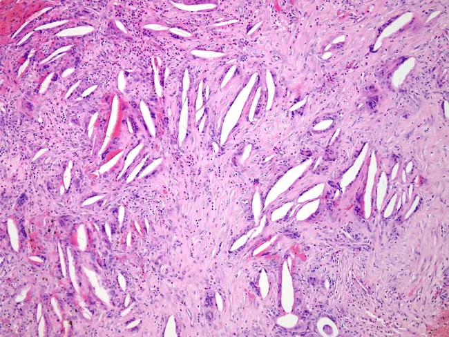
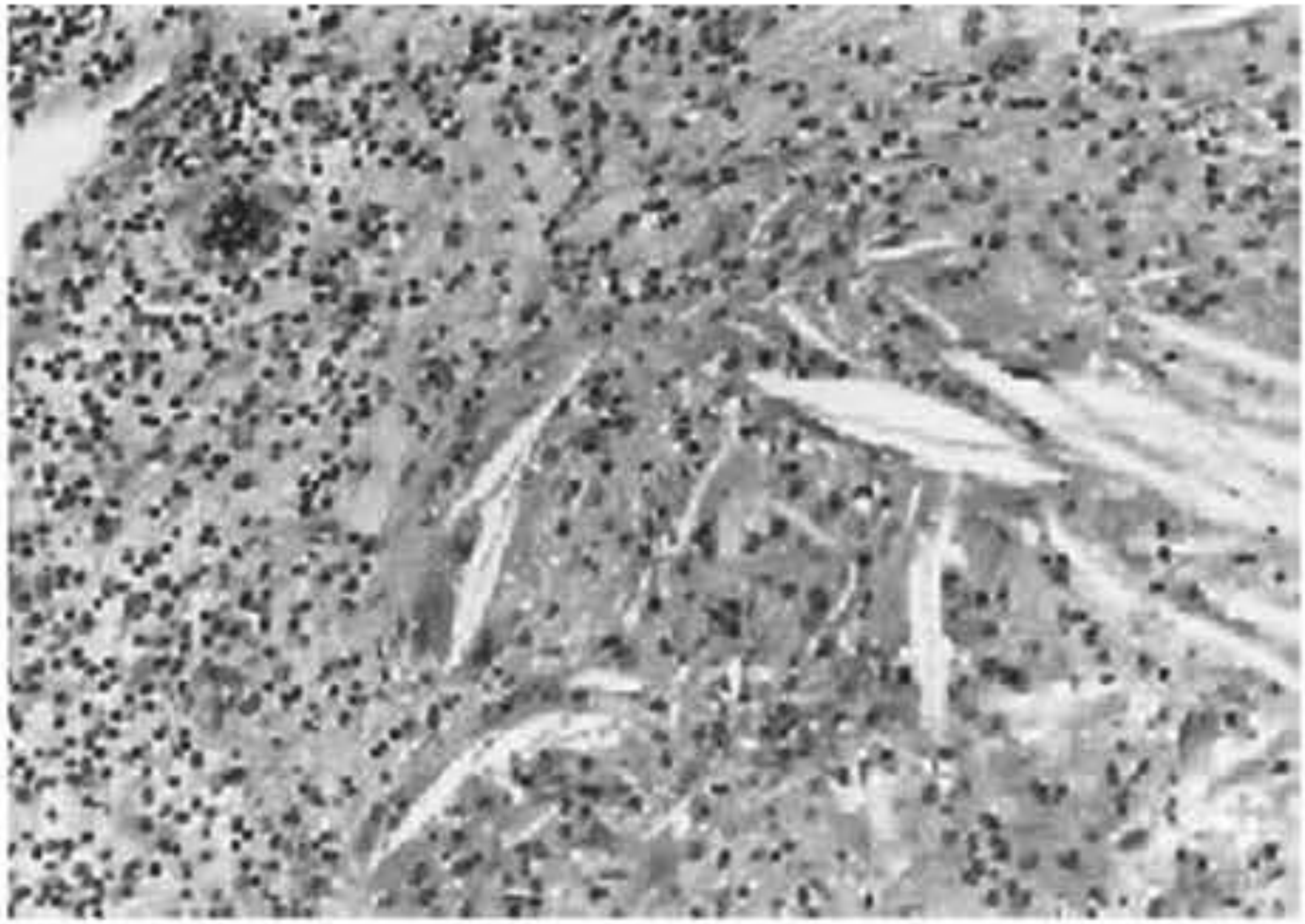
Light microscopy of a thickened hydrocele wall from tunica vaginalis demonstrates cross-sectional histology with dense fibrous stroma, scattered inflammatory cells, and cholesterol clefts. Acute neutrophilic bands and chronic lymphoplasmacytic infiltrates coexist, while multinucleated foreign body giant cells form around cholesterol clefts, reflecting a cholesterol granulomatous reaction. Fibrosis is prominent, with fascicular, collagen-rich architecture and minimal residual lining. The observed histology indicates chronic inflammatory and granulomatous response to leaked or degraded hydrocele contents, superimposed infection, and scarring. Diagnostic significance includes evidence of infectious complication and chronicity; findings support secondary infection in a long-standing hydrocele and help differentiate from neoplastic or purely serous processes. Clinically relevant search terms: hydrocele wall pathology, tunica vaginalis inflammation, cholesterol granuloma, foreign body giant cell reaction, chronic infection, fibrosis, scrotal hydrocele, hydrocelectomy planning, granulomatous scrotal disease.
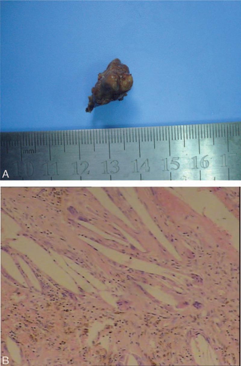
This composite educational image presents the macroscopic and microscopic features of a resected cholesterol granuloma, typically found in the sellar or suprasellar regions. Panel A is a clinical photograph of the gross specimen, showing an irregularly shaped, lobulated tumor that is ash-black in color. A surgical ruler provides scale, indicating dimensions of approximately 2.5 cm by 1.5 cm. Panel B displays a light microscopy image (hematoxylin and eosin staining, 200x magnification) revealing characteristic histopathological features. Prominent needle-like or spindle-shaped empty spaces represent cholesterol clefts where crystals have dissolved during processing. These are surrounded by a chronic inflammatory infiltrate, including multinucleated foreign body giant cells and macrophages. Additionally, areas of hemorrhage and brownish hemosiderin-laden deposits are visible, indicating prior intralesional bleeding. This visual material is used in neurosurgery and pathology to differentiate cholesterol granulomas from other sellar lesions like craniopharyngiomas based on classic pathological landmarks.
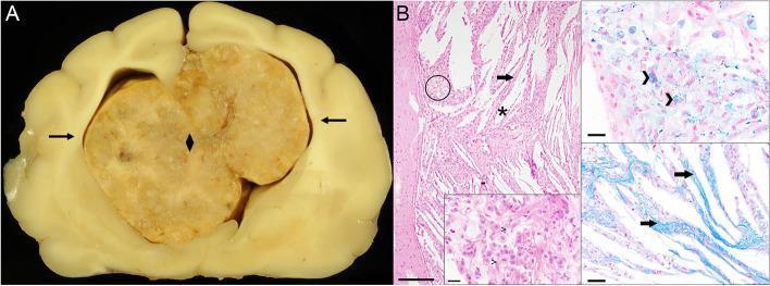
Educational medical composite featuring gross pathology and histopathology of a cerebral cholesterol granuloma. Panel A shows a transverse gross brain section containing a large (2.0 x 2.0 x 2.5 cm), yellowish-grey, lobulated intraventricular mass. The mass almost completely fills the dilated lateral ventricles, causing disruption of the interventricular septum and significant mass effect with compression of the adjacent cerebral parenchyma (indicated by arrows). Panel B displays light microscopy of the mass revealing granulomatous inflammation. Key features include numerous acicular, optically empty 'cholesterol clefts' (*) and prominent fibrous septae (arrows). High-magnification inserts demonstrate a dense population of foamy, hemosiderin-laden macrophages. Specialized stains facilitate diagnosis: Prussian blue (top right) highlights positive blue-stained hemosiderin deposits within macrophages, indicating chronic hemorrhage; Azan stain (bottom right) highlights blue-stained collagen within the fibrous septae. The presentation is characteristic of a cholesterol granuloma originating from the choroid plexus, typically seen in cases of chronic ventricular hemorrhage or obstructive hydrocephalus.
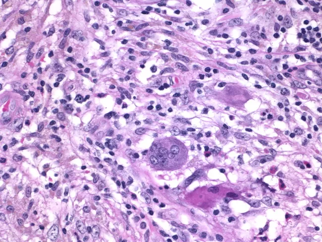
Histology of granulomatous mastitis on bright-field light microscopy. A dense inflammatory infiltrate forms granulomas centered on Langhans-type giant cells surrounded by epithelioid histiocytes, scattered lymphocytes, and occasional eosinophils. The image depicts a high-power view of the center of a granuloma within breast tissue biopsy; multinucleated giant cells with horseshoe-shaped nuclei are evident. The granulomatous architecture includes a peripheral rim of lymphocytes and histiocytes with minimal necrosis described. Staining by Hematoxylin and Eosin highlights basophilic nuclei and eosinophilic cytoplasm; pink collagenous stroma and adipose breast tissue may be visible in the background. Clinically, granulomatous mastitis can mimic infectious etiologies or neoplasia; the presence of Langhans giant cells strongly suggests a granulomatous process and warrants infectious workup for tuberculosis and fungal pathogens, as well as consideration of sarcoidosis or granulomatous reaction to foreign material. This image is diagnostic for granulomatous inflammation in breast tissue when correlated with clinical data and microbiologic studies; useful for education, differential diagnosis, and research on breast granulomatous disease. Additional educational value lies in distinguishing nonnecrotizing granulomas from caseating TB patterns, correlating histology with mammography and ultrasound, and guiding microbiologic testing and patient management in suspected granulomatous breast disease. The image supports teaching of granuloma biology and pathology.
"cholesterol granuloma"
Cholesterol Granuloma
Definition
Sites of Occurrence
- Frontal sinus (most common paranasal sinus site)
- Maxillary sinus
- Middle ear / mastoid
- Middle cranial fossa / cerebellopontine angle
- Orbit (rarely)
Pathogenesis
- Cummings Otolaryngology, p. 1007; Shambaugh Surgery of the Ear
Histopathology
- Foreign body-type multinucleated giant cells
- Lymphocytes and histiocytes
- Hemosiderin-laden macrophages
- Granulation tissue with fibrosis
Clinical Features
- Often asymptomatic and found incidentally on MRI
- When symptomatic: headache (ipsilateral, retroorbital or temporoparietal), hearing loss (conductive if Eustachian tube compressed; sensorineural if cochlea or IAC involved), tinnitus, vertigo, facial numbness or palsy, diplopia (CN VI compression)
- Rarely: pulsatile tinnitus, otalgia, aural fullness/pressure
- Nasal obstruction, headache, discharge
- Diplopia and proptosis (if orbital involvement)
- Cheek swelling (maxillary sinus involvement)
- Bony expansion producing cosmetic deformity
- Otalgia, hearing loss, tinnitus, vertigo
- Ossicular erosion can occur via the same inflammatory mechanism as in chronic otitis media
Radiology
| Modality | Findings |
|---|---|
| CT | Expansile, well-demarcated cystic lesion; smooth bone erosion/remodeling; thin rim of reactive sclerosis (slowly growing lesion); no calcification |
| MRI T1 | High signal intensity (hyperintense) - key feature, due to cholesterol/blood products |
| MRI T2 | High signal intensity (hyperintense) |
| Contrast | No appreciable enhancement (distinguishes from vascular tumors) |
| DWI | No diffusion restriction (distinguishes from cholesteatoma/epidermoid) |
| Fat suppression | High signal on T1 persists (not suppressed by fat-sat sequences) - distinguishes from fatty marrow |
Differential Diagnosis
| Lesion | T1 | T2 | DWI | Enhancement |
|---|---|---|---|---|
| Cholesterol granuloma | High | High | No restriction | None |
| Cholesteatoma (epidermoid) | Low-intermediate | High | Restricted | None |
| Mucocele | Variable | High | No restriction | Rim only |
| Retained effusion | Low | High | No restriction | None |
| Asymmetric fatty marrow | Suppressed on fat-sat | Low-intermediate | - | None |
Treatment
- Endoscopic marsupialization or complete resection via endoscopic or external approach
- Curettage alone carries ~15% recurrence rate; complete removal of granulation tissue is preferred
- Infracochlear or infralabyrinthine drainage - the preferred minimally morbid route when inferior petrous pneumatization allows
- Transpetrous (transcochlear / translabyrinthine) - reserved for large lesions when hearing sacrifice is acceptable
- Middle fossa approach - when superior pneumatization is present and hearing must be preserved
- Endoscopic endonasal transsphenoidal approach - increasingly used, particularly for medially placed lesions; a 2026 systematic review (PMID 41521199) reports long-term efficacy of nasoseptal mucosal flap marsupialization via this route
Key Points
- CG is a benign expansile cyst, not a neoplasm
- Pathogenesis = chronic hemorrhage + impaired drainage → cholesterol crystal deposition → foreign body granulomatous reaction
- Most common primary lesion of the petrous apex; also occurs in frontal/maxillary sinuses and middle ear
- Hallmark histology: acellular cholesterol clefts + multinucleated giant cells + hemosiderin-laden macrophages
- Hallmark MRI: T1 and T2 hyperintense, no enhancement, no DWI restriction
- Treatment = marsupialization/drainage; complete removal of granulation tissue reduces recurrence
- Asymptomatic incidental lesions may be observed conservatively