make a note of bacterial corneal ulcer
Bacterial Corneal Ulcer (Bacterial Keratitis)
Definition
- Tintinalli's Emergency Medicine, p. 1584; Wills Eye Manual, p. 202
Causative Organisms
| Category | Organisms |
|---|---|
| Gram-positive | Staphylococcus aureus, Staphylococcus spp., Streptococcus pneumoniae, S. pyogenes |
| Gram-negative | Pseudomonas aeruginosa (>60% of contact lens-related cases), Moraxella spp., Serratia spp. |
| Penetrates intact epithelium | Neisseria gonorrhoeae, N. meningitidis, Corynebacterium diphtheriae, Haemophilus influenzae |
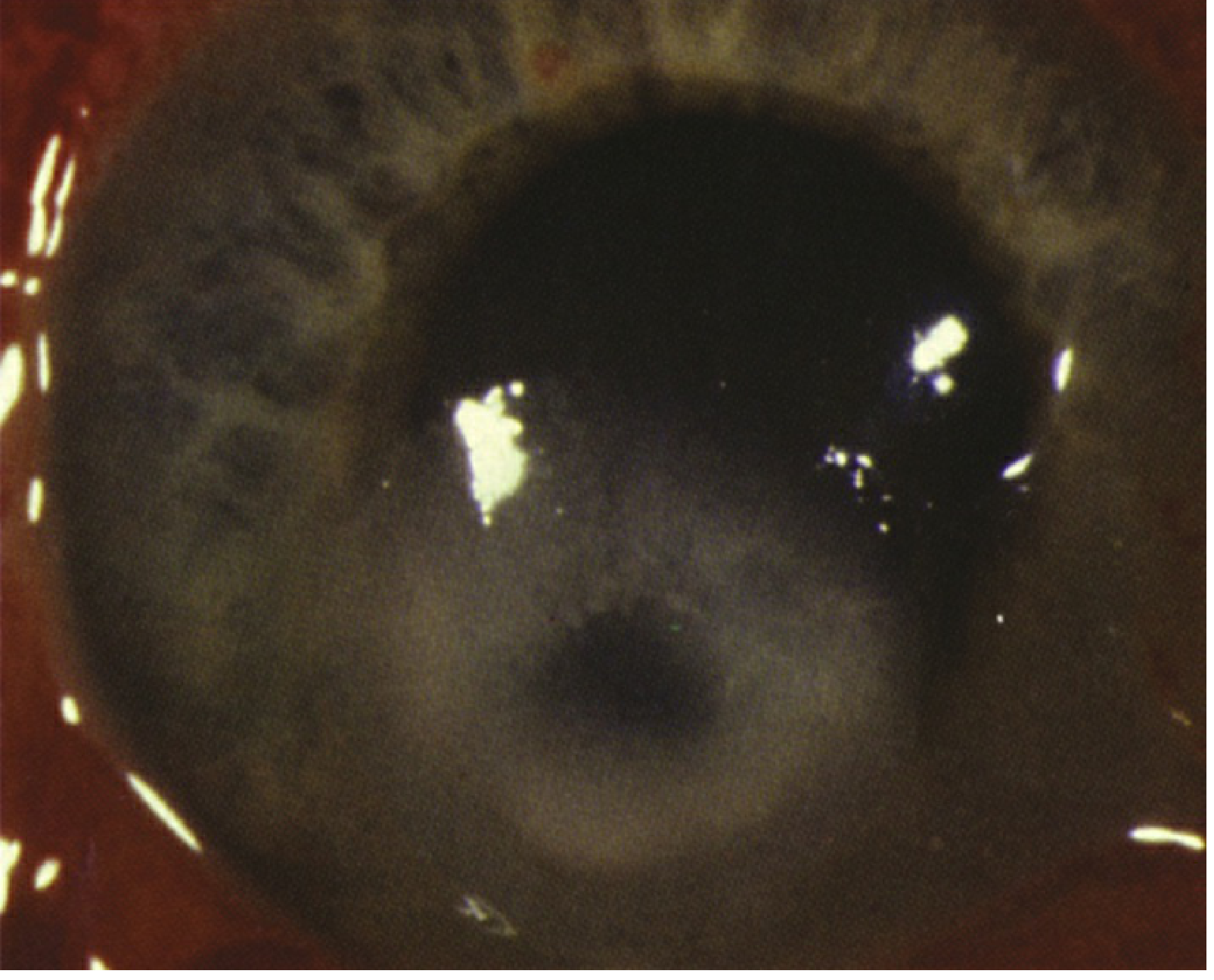
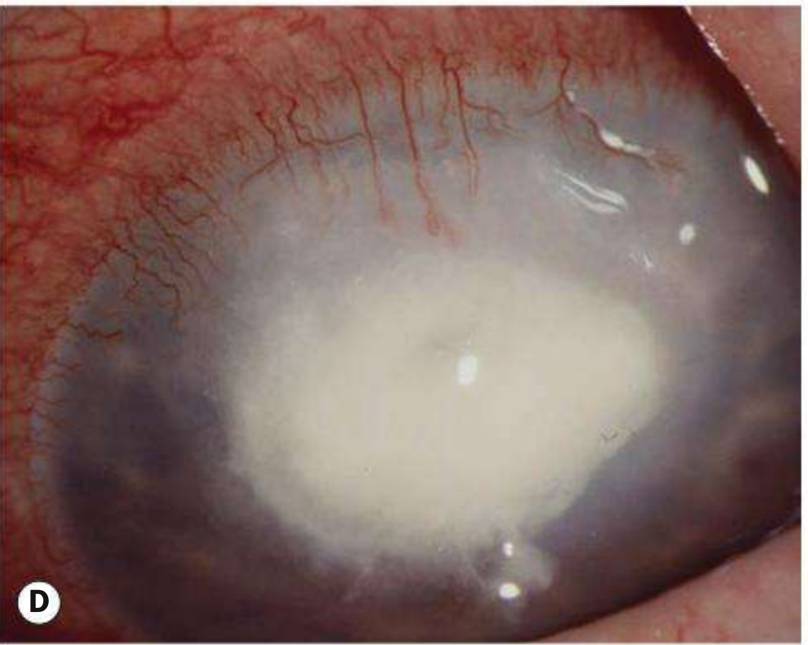
Pseudomonas - most common in soft contact lens wearers, causes rapidly progressive, suppurative, necrotic infiltrate Staph. aureus - focal, well-defined white or yellow-white infiltrate Streptococcus - very purulent or crystalline form; acute fulminant onset with hypopyon Moraxella - indolent, inferior cornea, full-thickness, occurs in immunocompromised patients
- Kanski's Clinical Ophthalmology, p. 226-227; Wills Eye Manual, p. 202-203
Risk Factors
-
Contact lens wear - the single most important risk factor, especially extended-wear soft lenses, sleeping with lenses in, poor lens hygiene, exposure to water (swimming/hot tubbing)
-
Corneal trauma (including vegetable matter, refractive surgery - especially LASIK)
-
Topical or systemic immunosuppressants / steroids
-
Pre-existing ocular surface disease
-
Previous ocular surgery or corneal grafts
-
Bell's palsy (exposure keratitis)
-
Systemic illness / immunocompromise
-
Kanski's Clinical Ophthalmology, p. 227
Clinical Features
Symptoms
- Red eye, moderate-to-severe ocular pain
- Photophobia (including consensual)
- Decreased / blurred vision
- Purulent or mucopurulent discharge
- Acute contact lens intolerance
- Foreign body sensation
Signs (Slit-lamp Findings)
- Mucopurulent discharge
- Stromal edema, folds in Descemet membrane
- Anterior chamber reaction (cells and flare) - iritis
- Hypopyon (sterile inflammatory - does not indicate perforation)
- Conjunctival injection and chemosis; eyelid swelling in moderate-severe cases
- Posterior synechiae, hyphaema, raised IOP in severe cases
- Endothelial fibrin/cell deposition
- Wills Eye Manual, p. 199-200; Kanski's, p. 228
Differential Diagnosis
| Condition | Distinguishing Feature |
|---|---|
| Fungal keratitis | After vegetable matter trauma; feathery borders, satellite lesions; Candida mimics bacterial ulcer |
| Acanthamoeba | Extremely painful; perineural invasion; ring-shaped infiltrate at 3-8 weeks; contact lens wearers with water exposure |
| HSV keratitis | Eyelid vesicles, corneal dendrites; history of recurrent unilateral disease |
| Atypical mycobacteria | Follows ocular surgery (LASIK); indolent course; AFB smear positive |
| Staphylococcal hypersensitivity | Peripheral infiltrates, bilateral, clear space from limbus, minimal pain, associated blepharitis |
| Sterile infiltrates | Multiple small peripheral subepithelial infiltrates, minimal staining, minimal AC reaction |
| Topical anesthetic abuse | Poor response to therapy; large ring opacity |
- Wills Eye Manual, p. 200-201
Investigations / Workup
History
- Contact lens use: type, wear schedule (daily vs. extended), hygiene, solutions, water exposure while wearing lenses
- Trauma, corneal surgery (including refractive surgery)
- Prior corticosteroid/immunosuppressant use
- History of genital herpes or other infections
- Systemic illness
Slit-lamp Examination
- Fluorescein staining to delineate epithelial defect
- Document: size, depth, and location of infiltrate and epithelial defect
- Assess and measure hypopyon
- Measure IOP (Tono-Pen preferred)
Corneal Scraping (Culture Indications)
-
1-2 mm in size (Wills Eye) / >2 mm (Kanski's)
- In the visual axis
- Involving middle to deep stroma
- Unresponsive to initial treatment
- Atypical in appearance, chronic, or suspicious for unusual organism
- Instill non-preserved topical anesthetic (proxymetacaine 0.5%)
- Scrape margins and base with a No. 11 scalpel blade, 20/21-gauge needle, or sterile spatula
- Remove loose mucus/necrotic tissue before scraping
- Thin smear on glass slides + plate onto culture media
| Stain | Organism Detected |
|---|---|
| Gram | Bacteria, fungi, microsporidia |
| Giemsa | Bacteria, fungi, Acanthamoeba |
| Calcofluor white | Acanthamoeba, fungi, microsporidia |
| Acid-fast (ZN / auramine) | Mycobacterium, Nocardia |
| GMS silver | Fungi, Acanthamoeba |
| PAS | Fungi, Acanthamoeba |
- Kanski's, p. 229-230; Wills Eye Manual, p. 203
Treatment
General Measures
- Discontinue contact lens wear (mandatory)
- No eye patching (risk of Pseudomonas rapid ulceration and corneal melting)
- Protective clear plastic eye shield (not a pressure patch) if significant thinning/perforation
- Oral pain medication as needed
Cycloplegic Drops
- Cyclopentolate 1% t.i.d. for comfort and to prevent posterior synechiae
- Atropine 1% b.i.d.-t.i.d. if hypopyon is present
Antibiotic Algorithm (Wills Eye)
- Non-contact lens wearer: Topical fluoroquinolone (moxifloxacin, gatifloxacin, besifloxacin, levofloxacin) or polymyxin B/trimethoprim q1-2h while awake
- Contact lens wearer: Fluoroquinolone q1-2h ± polymyxin B/trimethoprim q1-2h ± tobramycin or ciprofloxacin ointment 1-4x/day
- Fluoroquinolone q1h around the clock ± polymyxin B/trimethoprim q1h ATC
- Loading dose: q5min x 5 doses → q30min until midnight → q1h
- Fortified tobramycin or gentamicin (15 mg/mL) q1h, alternating with fortified cefazolin (50 mg/mL) or vancomycin (25 mg/mL) q1h (= drop every 30 min around the clock)
- Vancomycin reserved for resistant organisms, MRSA risk, or penicillin/cephalosporin allergy
- If Pseudomonas suspected: fortified tobramycin q30min + fortified cefazolin q1h ± fortified ceftazidime q1h or fluoroquinolone q1h
Note: Moxifloxacin and besifloxacin have slightly better gram-positive coverage. Gatifloxacin and ciprofloxacin have slightly better Pseudomonas/Serratia coverage.
Special Situations
| Situation | Treatment |
|---|---|
| Neisseria infection | Ceftriaxone 1 g IV q12-24h (corneal involvement) or 1 g IM single dose (conjunctival only) |
| Haemophilus infection | Amoxicillin/clavulanate orally (20-40 mg/kg/d ÷ 3 doses) |
| Corneal thinning | Doxycycline 100 mg PO b.i.d. (MMP inhibitor) + vitamin C 1-2 g/day (collagen synthesis) |
| Scleral extension / perforation | Oral ciprofloxacin 500 mg PO b.i.d. (preferred for Pseudomonas/Serratia) or moxifloxacin 400 mg PO daily |
| Gonococcal infection | Systemic antibiotics required (fluoroquinolones no longer recommended due to resistance) |
Topical Steroids
- Relatively contraindicated in viral infections; never initiated by the ER physician without ophthalmologist guidance
- May be added once organism and sensitivities are known, infection is controlled, and severe inflammation persists
- Avoid especially in Pseudomonas, fungal, atypical mycobacteria, and Nocardia infections
Indications for Hospital Admission
- Sight-threatening infection or impending perforation
- Patient unable to self-administer drops at required frequency
- High likelihood of non-compliance
- IV antibiotics needed (e.g., Neisseria with corneal involvement)
- Suspected topical anesthetic abuse
- Aggressive disease, particularly involving the only seeing eye
Follow-up
- Daily until significant improvement is demonstrated
- Improvement heralded by: reduced lid edema/chemosis, shrinking epithelial defect, decreasing infiltrate density, reduced AC signs
- Referral to ophthalmologist within 12-24 hours from emergency setting
Complications
- Corneal scarring (may cause irregular astigmatism)
- Corneal perforation
- Descemetocele formation
- Anterior and posterior synechiae
- Secondary glaucoma
- Cataract
- Vascularization
- Endophthalmitis (rare without perforation)
Sources
- The Wills Eye Manual, 4.11 Bacterial Keratitis, pp. 199-208
- Kanski's Clinical Ophthalmology (10th ed.), Bacterial Keratitis, pp. 226-232
- Tintinalli's Emergency Medicine, Corneal Ulcer section, pp. 1584-1585
i am final year ophthalmology resident, make in detail, what are the viva questions that can be asked during long case? what are the organisms involved? how do we identify them? different types of stains. everything from basic to advanced level and also add clinical trials and recent advances to it
bacterial keratitis treatment outcomes clinical trial
bacterial corneal ulcer recent advances 2023 2024 antibiotic resistance moxifloxacin clinical trial
bacterial keratitis steroid adjuvant SCUT trial
bacterial corneal ulcer antibiotic resistance recent advances treatment
SCUT steroids corticosteroid adjuvant bacterial keratitis randomized
SCUT corticosteroids bacterial keratitis moxifloxacin Steroids Corneal Ulcer
corneal cross-linking PACK-CXL infectious keratitis
- Section 1 - How to open and present the long case like a senior resident
- Section 2 - Predicted Q&A organized from basic → advanced (anatomy, organisms, stains, Gram stain technique and mechanism, culture media with C-streak method, pathogenesis + virulence factors, clinical assessment parameters, treatment algorithm with concentrations, systemic antibiotics, anti-collagenase therapy)
- Section 3 - All major clinical trials with PMID numbers: SCUT, SCUT II (2025), CLAIR, Cochrane NMA 2025, meta-analysis 2024
- Section 4 - Recent advances including AMR crisis, bacteriophage therapy, cefiderocol, metagenomic NGS, AI-assisted diagnosis, IVCM
- Section 5 - Examiner trap answers (common wrong answers vs. correct answers)
- Section 6 - Quick-reference summary table
- SCUT II (JAMA Ophthalmology, July 2025) - The latest RCT confirming adjunctive steroids offer no benefit AND CXL actually increased scar size. Most examiners may not know this yet.
- Cochrane NMA 2025 - Moxifloxacin is now the top-ranked monotherapy; vancomycin + ceftazidime best dual therapy by SUCRA ranking.