Watershed area of splenic flexure
Reading File
Finding Sources
Finding Sources
Reading File
Reading File
Loading Image
Here is a comprehensive summary of the watershed area of the splenic flexure:
Watershed Area of the Splenic Flexure — Griffiths' Critical Point
Definition
A watershed area is a region of the colon that lies at the most distal reaches of two separate arterial territories. Because only the smallest-caliber terminal branches supply these zones, they are highly vulnerable to ischemia when perfusion pressure falls.
The splenic flexure harbors the most clinically important watershed zone in the colon: Griffiths' critical point (also written "Griffith's point").
Vascular Anatomy
The splenic flexure sits at the junction of the SMA and IMA territories:
| Artery | Origin | Territory |
|---|---|---|
| Middle colic artery (left branch) | Superior mesenteric artery (SMA) | Distal transverse colon → splenic flexure |
| Ascending branch of the left colic artery | Inferior mesenteric artery (IMA) | Splenic flexure → descending colon |
These two vessels anastomose — often tenuously — at the splenic flexure via the marginal artery of Drummond. This point of communication is Griffiths' critical point.
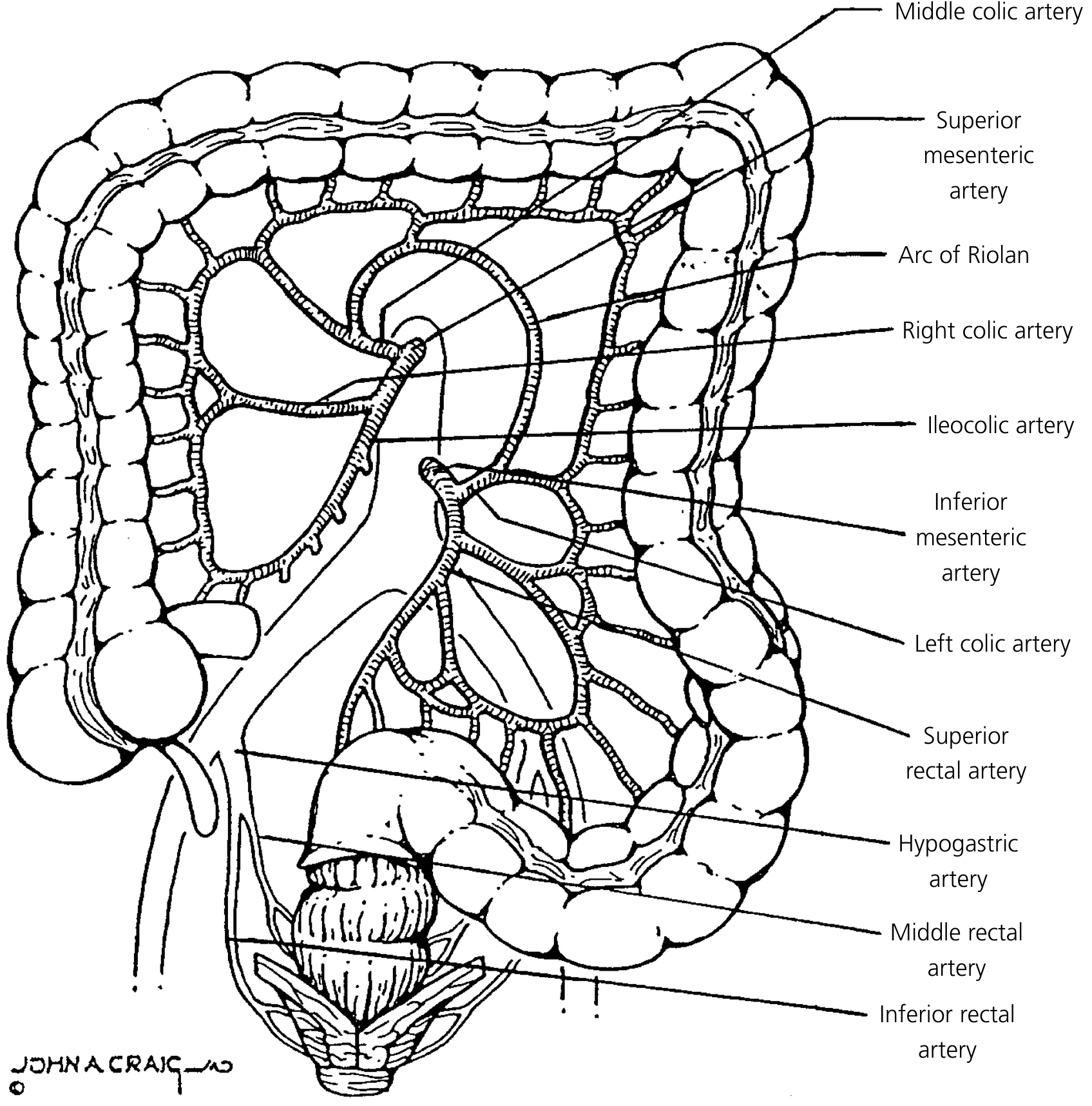
Why Is It Vulnerable?
- The anastomosis at Griffiths' point is formed by the most distal branches of both arterial systems — vessels of the smallest diameter.
- In states of global hypoperfusion (hemorrhagic shock, cardiac failure) or splanchnic vasoconstriction, flow through these terminal branches drops disproportionately.
- The splenic flexure thus becomes ischemic before other colonic segments.
"The watershed zone at the splenic flexure is extremely vulnerable to ischemia." — Gray's Anatomy for Students
"Generalized hypotension or hypoxemia can cause localized injury in watershed zones, and ischemic disease should be considered in the differential diagnosis of focal colitis of the splenic flexure." — Robbins & Cotran Pathologic Basis of Disease
The Two Colonic Watershed Zones
| Watershed Point | Location | Arteries Involved |
|---|---|---|
| Griffiths' critical point | Splenic flexure | Middle colic (SMA) ↔ Ascending left colic (IMA) |
| Sudeck's point | Rectosigmoid junction | Last sigmoid branch (IMA) ↔ Superior rectal artery / Internal iliac |
Together, these two zones account for ~80% of ischemic colitis (IC) cases.
Collateral Pathways
- Marginal artery of Drummond: runs along the mesenteric border of the entire colon; main collateral between SMA and IMA systems.
- Arc of Riolan (meandering artery): a larger, more central communicating arc between the middle colic artery and the ascending branch of the left colic artery. It enlarges and becomes visible when either the SMA or IMA is occluded in atherosclerotic disease, serving as an important compensatory route.
Clinical Relevance
- Ischemic colitis: The splenic flexure is the most common site; presents with left-sided abdominal pain and rectal bleeding after hypotensive episodes.
- Surgical anastomosis: Anastomoses at Griffiths' point should be avoided if possible. If the splenic flexure cannot be avoided, adequate mobilization and tension-free technique are essential — Campbell-Walsh-Wein Urology recommends placing the anastomosis away from these watershed points toward a segment with larger-caliber feeding vessels.
- Aortic surgery: After aortic cross-clamping or IMA ligation (e.g., during aortic aneurysm repair), the left colon and splenic flexure are at risk of ischemia.
- Splenic flexure tumors: Oncologic resection must account for the tenuous blood supply when planning anastomosis.
Sources: Campbell-Walsh-Wein Urology | Yamada's Textbook of Gastroenterology | Robbins & Cotran Pathologic Basis of Disease | Gray's Anatomy for Students | Current Surgical Therapy 14e | Robbins & Kumar Basic Pathology
This is a shared conversation. Sign in to Orris to start your own chat.