
Pampigus Diesese details?? Pom
Reading File
The user is asking about "Pampigus Diesese" — this appears to be Pemphigus (a group of autoimmune blistering diseases). Let me search the library.
Finding Sources
Finding Sources
Good results. Let me read the comprehensive sections from Fitzpatrick's (the gold standard dermatology text) and Robbins Pathology in parallel.
Reading File
Reading File
I have excellent content from both Fitzpatrick's and Robbins. I also noticed image URLs in the content. Let me fetch a couple of the relevant images while synthesizing the answer.
Loading Image
Searching Images
pemphigus vulgaris blisters acantholysis histology
I now have comprehensive information. Let me compile a thorough answer about Pemphigus disease.
Pemphigus — Autoimmune Blistering Disease
Note: "Pampigus Diesese" = Pemphigus (from Greek pemphix = blister/bubble)
What Is Pemphigus?
Pemphigus is a group of chronic autoimmune blistering diseases of the skin and mucous membranes. It is characterized by:
- Intraepidermal blisters due to acantholysis (loss of cell-to-cell adhesion between keratinocytes)
- IgG autoantibodies targeting desmogleins — transmembrane desmosomal adhesion proteins
- The antibodies disrupt intercellular adhesion and may activate intercellular proteases
It was first understood as an autoimmune disease in 1964 when Ernst Beutner and Robert Jordon discovered circulating anti-keratinocyte antibodies in pemphigus vulgaris.
Types / Classification
| Subtype | Autoantibody Target | Level of Blister | Clinical Features |
|---|---|---|---|
| Pemphigus Vulgaris (PV) | DSG1 + DSG3 | Suprabasal | Mucosal + skin erosions; flaccid bullae |
| Pemphigus Foliaceus (PF) | DSG1 only | Subcorneal (stratum granulosum) | Skin only; crusted, scaly lesions; no mucosal involvement |
| Pemphigus Vegetans | DSG1 + DSG3 | Suprabasal | Localized variant of PV; verrucous plaques in flexures |
| Pemphigus Erythematosus | DSG1 | Subcorneal | Localized variant of PF; seborrheic/lupus-like distribution |
| Fogo Selvagem (Endemic PF) | DSG1 | Subcorneal | Endemic in Brazil; triggered by insect bites |
| IgA Pemphigus | Desmocollin 1 | Variable | IgA deposits; less severe |
| Paraneoplastic Pemphigus | Multiple (desmoplakin, etc.) | Suprabasal | Associated with internal malignancy; severe mucosal ulceration |
| Drug-Induced Pemphigus | DSG1 or DSG3 | Variable | ACE inhibitors (captopril), penicillamine, gold |
Pathogenesis
- Autoantibodies (IgG) target desmoglein-1 (DSG1) and/or desmoglein-3 (DSG3) — critical components of desmosomes
- Antibody binding → disruption of intercellular adhesion → acantholysis (cells separate from one another)
- Blister location depends on where desmoglein is expressed:
- DSG3 predominates in mucosa and deeper epidermis → suprabasal blisters in PV
- DSG1 predominates in superficial epidermis → subcorneal blisters in PF
- Associated with specific HLA alleles (genetic predisposition)
- Complement activation also contributes to tissue damage
Clinical Features
Pemphigus Vulgaris (most common)
- Demographics: Older adults; more common in women; higher incidence in Ashkenazi Jews and people of Mediterranean descent
- Sites: Oropharyngeal mucosa (almost always involved first), scalp, face, axillae, groin, trunk, pressure points
- Lesions: Fragile, flaccid bullae that rupture easily → painful, extensive erosions covered with serum crust
- Nikolsky sign: Positive — lateral pressure on normal-looking skin causes epidermal separation
- Lesions are painful and prone to secondary infection
Pemphigus Foliaceus
- Milder form; no mucosal involvement
- Crusted, scaly lesions; blisters are superficial and erythematous
- Widespread in fogo selvagem (endemic variant in Brazil)
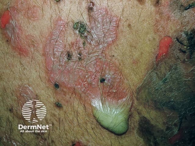
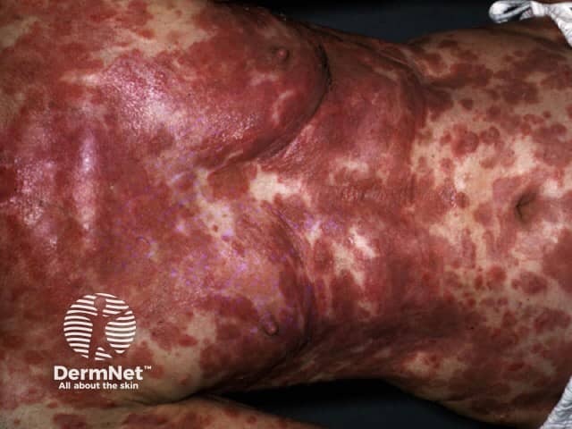
Histology
| Feature | Pemphigus Vulgaris | Pemphigus Foliaceus |
|---|---|---|
| Acantholysis level | Suprabasal | Subcorneal (stratum granulosum) |
| Basal cells | "Tombstone row" — remain attached to basement membrane | Detached superficially |
| Blister type | Intraepidermal, suprabasal | Subcorneal |
Immunofluorescence
- Direct IF (DIF): Fishnet / chicken-wire pattern of intercellular IgG and C3 deposits throughout the epidermis — pathognomonic
- Indirect IF: Circulating anti-desmosomal IgG in serum
- ELISA: Anti-DSG1 and anti-DSG3 antibody titers — useful for diagnosis and monitoring disease activity
Diagnosis
- Clinical — flaccid blisters + mucosal erosions + positive Nikolsky sign
- Biopsy — intraepidermal acantholysis
- Direct immunofluorescence — fishnet IgG intercellular pattern
- ELISA — anti-DSG antibody titers
- Indirect immunofluorescence — circulating antibodies on monkey esophagus substrate
Treatment
| Approach | Agents |
|---|---|
| First-line | Systemic corticosteroids (prednisone) |
| Steroid-sparing | Azathioprine, mycophenolate mofetil, cyclophosphamide |
| Biologic | Rituximab (anti-CD20 monoclonal antibody) — now considered first-line in moderate-severe PV |
| Adjuncts | IV immunoglobulin (IVIG), plasmapheresis |
| Topical | Topical corticosteroids for localized lesions |
| Long-term | Many patients require prolonged immunosuppression |
Rituximab has significantly improved outcomes — it depletes B cells and reduces autoantibody production.
Key Comparisons: Pemphigus vs. Bullous Pemphigoid
| Feature | Pemphigus | Bullous Pemphigoid |
|---|---|---|
| Blister location | Intraepidermal | Subepidermal |
| Blister quality | Flaccid, fragile | Tense, firm |
| Nikolsky sign | Positive | Negative |
| Mucosal involvement | Common (especially PV) | Uncommon |
| Autoantibody target | Desmoglein 1/3 (desmosomes) | BP180/BP230 (hemidesmosomes/BMZ) |
| IF pattern | Fishnet intercellular | Linear at BMZ |
| Age group | Middle-aged to older adults | Elderly |
Sources: Fitzpatrick's Dermatology, Vol. 1–2, p. 939 | Robbins & Kumar Basic Pathology, p. ~22 | Dermatology 2-Volume Set, 5e
This is a shared conversation. Sign in to Orris to start your own chat.