
Nose anatomy clearly with all references
Reading File
Now let me search the medical library for nose anatomy comprehensively, running parallel queries:
Finding Sources
Finding Sources
Searching the Web
nose anatomy external internal nasal cavity structure overview
Excellent - rich sources found. Now let me read the most comprehensive textbook sections in parallel:
Reading File
Reading File
Reading File
Now let me read the KJ Lee ENT book (most detailed nasal cavity section) and the Bailey & Love section further, plus fetch the key diagrams:
Reading File
Reading File
Now let me get the remaining KJ Lee content (nasal cavity details) and fetch the key diagrams in parallel:
Reading File
Loading Image
Loading Image
Loading Image
Now let me get the blood supply/nerve diagrams and more content on nasal blood supply, innervation, and the lateral wall:
Loading Image
Loading Image
Reading File
Excellent data collected. I now have everything needed to compile a thorough, well-illustrated answer. Let me compile the complete nose anatomy reference:
Anatomy of the Nose - Complete Reference
Sources: Bailey & Love's Short Practice of Surgery 28th ed; KJ Lee's Essential Otolaryngology; Cummings Otolaryngology Head and Neck Surgery; Miller's Anesthesia 10th ed
Overview
The nose consists of two anatomically distinct parts:
- External nose - the visible projecting structure on the face
- Internal nose (nasal cavity) - a passage approximately 6-7 cm long running above the roof of the mouth
1. EMBRYOLOGY
Development of the nasal airway begins during the 4th week of gestation (Cummings Otolaryngology, p. 564):
- Neural crest cells proliferate to form nasal placodes
- Medial and lateral nasal processes develop adjacent to the placodes
- Medial nasal processes fuse with the frontal prominence to form: nasal bones, frontal bones, cartilaginous nose, ethmoid bones, central incisors, and hard palate
- Nasobuccal membrane ruptures by week 10, opening communication between nose and nasopharynx
- Nasal septum develops as a downgrowth from the merged medial nasal processes; fuses with the palatine processes anteriorly at week 9, completed posteriorly by week 12
Turbinate embryology (from KJ Lee's Essential Otolaryngology, p. 509):
| Embryonic structure | Adult derivative |
|---|---|
| Maxilloturbinal (from maxillary process) | Inferior turbinate |
| 1st ethmoturbinal (ascending) | Agger nasi |
| 1st ethmoturbinal (descending) | Uncinate process |
| 2nd ethmoturbinal | Middle turbinate |
| 3rd ethmoturbinal | Superior turbinate |
| 4th-5th ethmoturbinals | Supreme turbinate (when present) |
Primary furrows form the meati separating the ethmoturbinals.
2. EXTERNAL NOSE ANATOMY
2a. Surface Landmarks
(KJ Lee's Essential Otolaryngology, p. 509-510)
| Landmark | Description |
|---|---|
| Nasion | Corresponds to the nasofrontal suture |
| Radix | Nasal root; centered at nasion, extends to level of lateral canthus |
| Rhinion | Bony-cartilaginous junction along the nasal dorsum |
| Supratip break | Break in nasal profile, separating dorsum and lobule, just superior to tip-defining point |
| Tip-defining point | Highest, medial, cephalic portion of the lateral crus; corresponds to light reflex externally |
| Columella | The skin-covered midline strut between the nostrils |
| Alar rim | The free margin of the nostril |
| Nares | The external openings (nostrils) |
2b. Nasal Skeleton
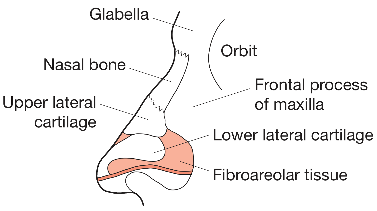
Figure: The nasal skeleton (Bailey & Love's, p. 785)
Bony framework:
- Nasal bones (paired) - form the bony bridge; articulate superiorly with the nasal process of the frontal bone, laterally with the frontal process of the maxilla
- Pyriform aperture - the bony opening into the nasal cavity, bounded superiorly by the caudal margin of nasal bones, inferiorly by the alveolar process of maxilla, laterally by the frontal process of maxilla, medially by the nasal septum
Cartilaginous framework (KJ Lee, p. 510):
-
Upper Lateral Cartilage (ULC)
- Attached superiorly to nasal bones
- Articulates inferiorly with the cephalic margin of the lower lateral cartilage (LLC), often forming an interlocking scroll
- Thickens medially, becoming continuous with the quadrangular (septal) cartilage, forming the cartilaginous dorsum
-
Lower Lateral Cartilage (LLC)
- Provides shape of the nasal tip
- Composed of medial and lateral crura
-
Sesamoid cartilages - small cartilages lateral to the lateral crus
2c. Subcutaneous Layers
Mnemonic "Subcutaneous Fat DeeP" (KJ Lee, p. 509):
| Layer | Content |
|---|---|
| S | Superficial fatty layer (connected to dermis) |
| F | Fibromuscular layer (nasal SMAS) |
| D | Deep fatty layer (contains neurovascular structures) |
| P | Periosteum / perichondrium |
The optimal surgical dissection plane lies between D and P - it is avascular and heals with minimal fibrosis.
2d. Nasal Musculature
Elevators - shorten nose and dilate nostrils:
- Procerus, levator labii superioris alaeque nasi, anomalous nasi
Depressors - lengthen nose and constrict nostrils:
- Alar nasalis, depressor septi, compressor narium minor
Minor muscles - nasalis (transverse head), dilator naris posterior
All nasal muscles are supplied by the facial nerve (CN VII).
3. NASAL SEPTUM
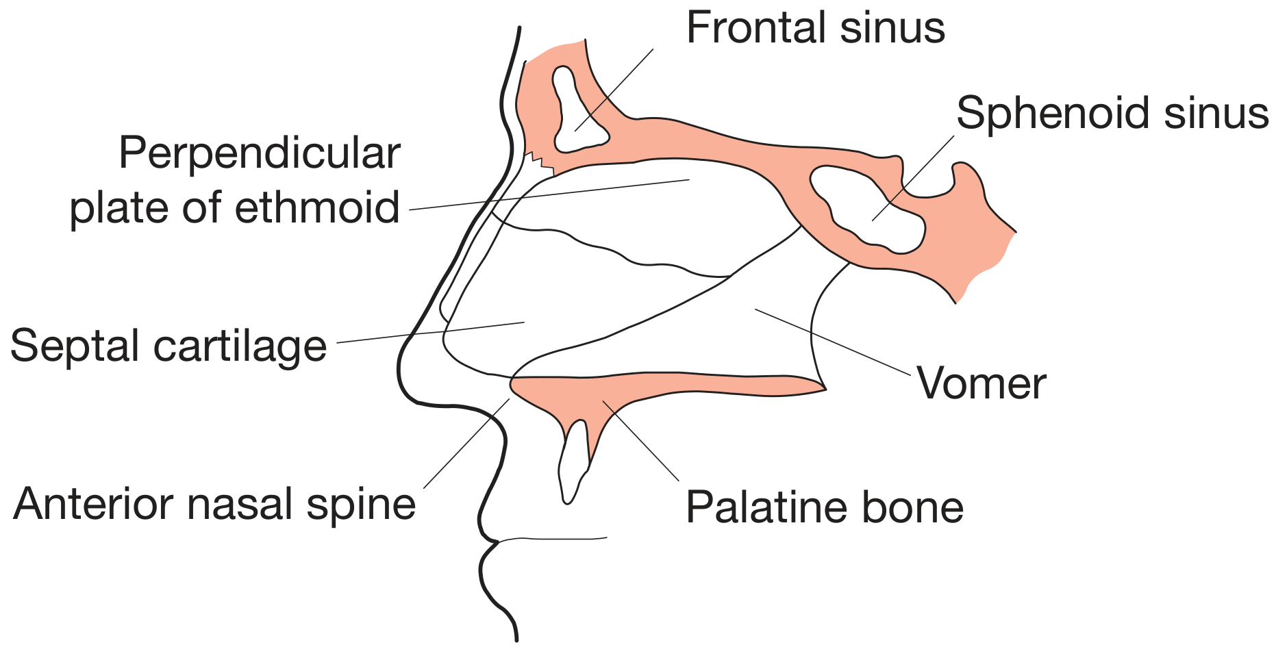
Figure: The left side of the nasal septum (Bailey & Love's, p. 785)
The nasal septum divides the nasal cavity into right and left halves. It is composed of:
Cartilage:
- Quadrangular cartilage - the main component, forms the anterior portion
Bone (Bailey & Love's, p. 785 / KJ Lee, p. 510):
- Perpendicular plate of the ethmoid (posterosuperior)
- Vomer (posteroinferior)
- Crest of the maxillary bone
- Crest of the palatine bone
- Minor contributions: crest of sphenoid bone, nasal spine of frontal bone
Special regions:
- Membranous (mobile) septum - cartilage-deficient membrane adjoining the columella to the caudal septum; site of hemitransfixion/transfixion incision
- Keystone area - convergence of the caudal margin of nasal bone, perpendicular plate of ethmoid, cephalic margin of ULC, and cartilaginous septum; failure to preserve this during surgery causes nasal collapse
- Vomeronasal organ (Jacobsen's organ) - auxiliary olfactory organ (largely regresses in utero); identified as a groove in the anterior-inferior nasal septum
4. NASAL CAVITY
4a. Boundaries
The nasal cavity extends from the vestibule anteriorly to the nasopharynx posteriorly.
| Wall | Structure |
|---|---|
| Medial | Nasal septum |
| Lateral | Turbinates + lateral wall bones |
| Roof | Cribriform plate of ethmoid (central), nasal bones (anterior), body of sphenoid (posterior) |
| Floor | Hard palate (palatine process of maxilla + horizontal plate of palatine bone) |
4b. Divisions of the Nasal Cavity
-
Vestibule - entrance lined by hair-bearing skin, sebaceous and sweat glands
- Boundaries: medially = nasal septum; superior/lateral = LLC; inferior = alveolar process of maxilla; posterior = limen nasi (caudal margin of ULC)
- The limen nasi marks the transition from skin to pseudostratified ciliated columnar epithelium (respiratory mucosa)
-
Olfactory region - superior, adjacent to the cribriform plate; lined with specialized olfactory epithelium
-
Respiratory region - the majority of the cavity; lined with pseudostratified ciliated columnar epithelium (respiratory mucosa) with goblet cells
4c. Lateral Wall and Turbinates
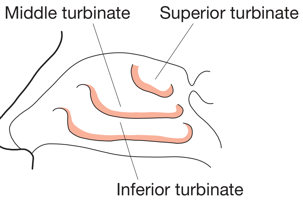
Figure: The right lateral nasal wall - turbinates (Bailey & Love's, p. 785)
Three curved shelves of bone project from the lateral wall - the turbinates (conchae) - which dramatically increase the surface area of the nasal cavity for warming, humidifying, and filtering inspired air.
Osteology of lateral nasal wall (anterior to posterior - KJ Lee, p. 510):
- Frontal process of maxilla
- Lacrimal bone
- Medial maxillary wall inferiorly / lamina papyracea (LP) superiorly
- Perpendicular plate of the palatine bone
- Medial pterygoid plate of the sphenoid bone (the latter two form the sphenopalatine foramen)
Five lamellae of the lateral nasal wall (anterior to posterior):
| Lamella | Structure |
|---|---|
| 1st | Uncinate process (UP) |
| 2nd | Ethmoid bulla (EB) |
| 3rd | Basal lamella of middle turbinate |
| 4th | Basal lamella of superior turbinate |
| 5th | Basal lamella of supreme turbinate (when present) |
4d. Meatuses and Sinus Drainage
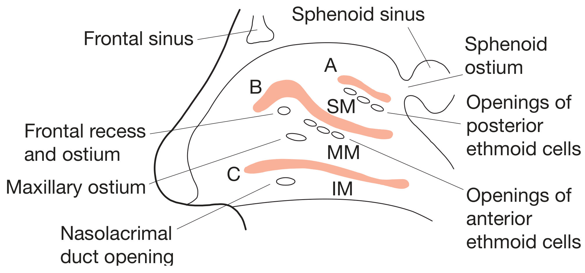
Figure: Right lateral nasal wall with turbinates removed (Bailey & Love's, p. 786)
| Meatus | Location | Structures draining into it |
|---|---|---|
| Superior meatus | Below superior turbinate | Posterior ethmoid sinuses |
| Middle meatus | Below middle turbinate | Frontal sinus, maxillary sinus, anterior ethmoid sinuses |
| Inferior meatus | Below inferior turbinate | Nasolacrimal duct |
| Sphenoethmoidal recess | Between superior turbinate and nasal septum | Sphenoid sinus |
The ostiomeatal complex (OMC) in the middle meatus is the critical drainage pathway for the anterior sinuses.
4e. Nasal Valves
(KJ Lee's Essential Otolaryngology, p. 511)
Internal nasal valve:
- Bounded by: nasal septum, caudal edge of ULC, anterior face of inferior turbinate
- Forms a 10-15 degree nasal valve angle
- Narrowest cross-sectional area of the nasal cavity
- Site of greatest nasal airflow resistance
- Does NOT change dimension during inspiration
External nasal valve:
- Bounded by: nasal ala laterally, nasal septum and columella medially
- Situated caudal to the internal nasal valve
- Normally dilates during inspiration
5. BLOOD SUPPLY
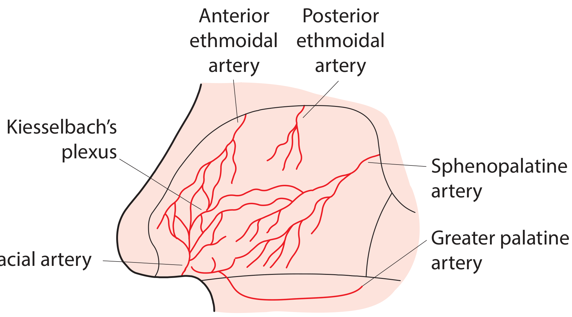
Figure: Arterial blood supply to the nasal septum (Bailey & Love's, p. 786)
The nasal fossae and sinuses receive blood from both the external and internal carotid arteries (Bailey & Love's, p. 785-786):
External Carotid Artery (via maxillary artery):
- Sphenopalatine artery - enters through the sphenopalatine foramen; the dominant supply to the posterior nasal cavity
- Greater palatine artery - supplies the anteroinferior septum via the incisive canal
Internal Carotid Artery (via ophthalmic artery):
- Anterior ethmoidal artery - supplies the anterosuperior nasal cavity
- Posterior ethmoidal artery - supplies the posterosuperior area
Kiesselbach's Plexus (Little's area)
All nasal arteries anastomose on the anterior part of the nasal septum to form Kiesselbach's plexus - the most common site of nosebleeds (epistaxis).
Venous Drainage:
- Via ophthalmic and facial veins and the pterygoid and pharyngeal plexuses
- Intracranial drainage via the cavernous sinus through the ophthalmic vein - clinically important because of potential intracranial spread of nasal sepsis
6. NERVE SUPPLY
(KJ Lee's Essential Otolaryngology; Bailey & Love's)
Sensory Innervation:
| Region | Nerve | Parent nerve |
|---|---|---|
| Skin of dorsum, nasal alae, vestibule | External nasal nerve | Ophthalmic (CN V1) |
| Anterosuperior nasal cavity (septum + lateral wall) | Anterior ethmoidal nerve | Ophthalmic (CN V1) |
| Posteroinferior nasal cavity | Nasopalatine nerve (septum) + posterior nasal nerves (lateral wall) | Maxillary (CN V2), via pterygopalatine ganglion |
| Olfactory mucosa (superior) | Olfactory nerve (CN I) | Filaments through cribriform plate |
Autonomic Innervation:
- Parasympathetic (secretomotor to glands, vasodilatory): postganglionic fibers from the pterygopalatine ganglion (synapse of greater petrosal nerve + deep petrosal nerve = Vidian nerve)
- Sympathetic (vasoconstrictive): postganglionic fibers from the superior cervical ganglion, traveling along the deep petrosal nerve
7. LYMPHATIC DRAINAGE
- Anterior nose: drains to submandibular lymph nodes
- Posterior nose: drains to retropharyngeal and upper deep cervical lymph nodes
8. PARANASAL SINUSES (Summary)
Four paired sinuses communicate with the nasal cavity:
| Sinus | Drainage site | Notes |
|---|---|---|
| Frontal | Middle meatus (via frontal recess) | Anterior group |
| Maxillary | Middle meatus (via ostiomeatal complex) | Largest sinus; floor at level of upper teeth |
| Anterior ethmoid | Middle meatus | Anterior group; multiple small cells |
| Posterior ethmoid | Superior meatus | Posterior group |
| Sphenoid | Sphenoethmoidal recess | Most posterior; related to pituitary gland, cavernous sinus, optic nerve |
9. NASAL FUNCTIONS
- Airway - filters, warms, and humidifies inspired air
- Olfaction - olfactory epithelium in the superior nasal cavity detects odors
- Immunologic - mucociliary clearance removes pathogens and particles
- Resonance - contributes to voice quality and speech
- Nasolacrimal drainage - inferior meatus receives lacrimal secretions
References
| Source | Details |
|---|---|
| Bailey & Love's Short Practice of Surgery, 28th ed. | pp. 785-786 - Basic anatomy of nose and paranasal sinuses |
| KJ Lee's Essential Otolaryngology | pp. 509-511 - Nasal embryology, external anatomy, nasal cavity, valves |
| Cummings Otolaryngology Head & Neck Surgery | p. 564 - Anatomy and embryology of the nose |
| Miller's Anesthesia, 10th ed. | p. 9790 - Synopsis of ENT anatomy for anesthesia |
This is a shared conversation. Sign in to Orris to start your own chat.