
Acute lymphoid leukaemia
I'll read the medical library skill first, then research ALL aspects of ALL in parallel.
Reading File
Now I'll run parallel searches across the library for ALL aspects of ALL:
Finding Sources
Finding Sources
Searching PubMed
"acute lymphoblastic leukemia"[MeSH Terms] AND treatment
Let me try broader searches:
Finding Sources
Finding Sources
Searching PubMed
"acute lymphocytic leukemia" AND treatment
Good - I found relevant sections. Let me now read the key blocks in parallel:
Reading File
Reading File
Finding Sources
Excellent! Now reading the key dedicated sections in parallel:
Reading File
Reading File
Reading File
Excellent content. Now reading treatment, clinical features, and the Robbins pathology content:
Reading File
Reading File
Reading File
Excellent content. Let me now get treatment details and the Harrison's content on treatment:
Reading File
Reading File
I now have rich comprehensive content. Let me get a few more key sections - Harrison's incidence/diagnosis data and the prognosis section:
Reading File
Loading Image
I now have comprehensive, authoritative content from multiple major textbooks. Let me compile the complete answer.
Acute Lymphoid (Lymphoblastic) Leukaemia (ALL)
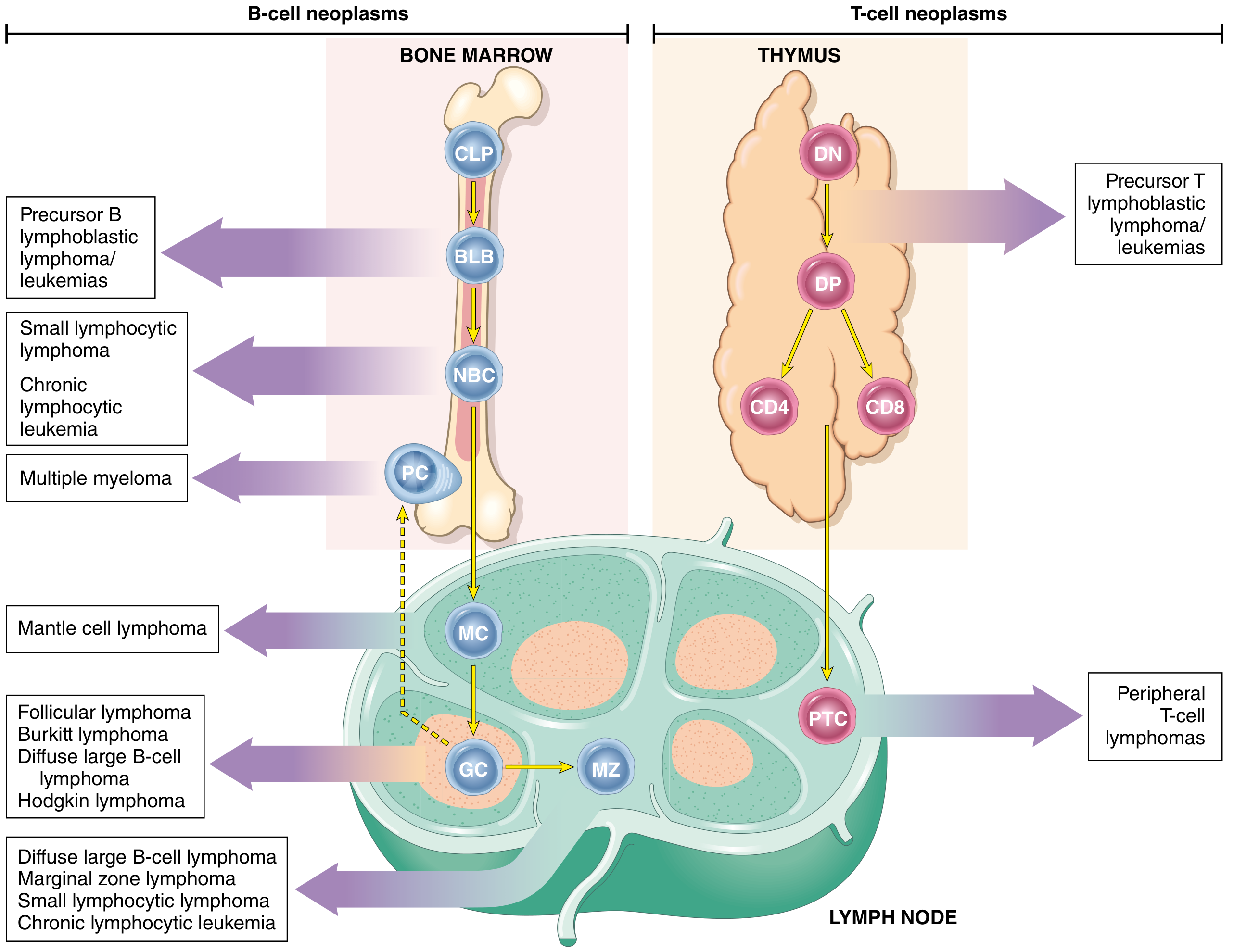
Definition
ALL is a neoplasm of immature B (pre-B) or T (pre-T) lymphocytes called lymphoblasts, arrested at an early stage of differentiation in the bone marrow or lymphatic system, resulting in the accumulation of non-functional leukemic cells that displace normal haematopoiesis.
- ~85% are B-ALLs
- ~15-25% are T-ALLs
Epidemiology
- Most common cancer in children, with a peak incidence at 3-4 years of age
- B-ALL peaks at ~3 years (paralleling the maximum number of normal bone marrow pre-B cells); T-ALL peaks in adolescence (when the thymus reaches maximum size)
- Slightly more frequent in boys than girls
- Hispanic/Latino children have the highest incidence of any ethnic group in the US
- Adult incidence: 0.7-1.8/100,000/year; rises again in the elderly
- Ph+ ALL accounts for ~50% of elderly B-lineage ALL patients
Etiology & Risk Factors
| Factor | Detail |
|---|---|
| Ionizing radiation | Increased risk, more so for AML |
| Prior chemotherapy | Alkylating agents, topoisomerase inhibitors |
| Congenital disorders | Down syndrome (20-fold increased risk), Klinefelter syndrome, Fanconi anaemia, Bloom syndrome, ataxia-telangiectasia, neurofibromatosis |
| Viruses | EBV (Burkitt type); HTLV-I (adult T-cell leukaemia/lymphoma) |
Pathogenesis & Molecular Biology
Most ALLs carry chromosomal aberrations that dysregulate transcription factors needed for normal lymphocyte development. Fewer than 10 driver mutations are generally sufficient.
Key genetic alterations in B-ALL:
| Mutation | Frequency | Significance |
|---|---|---|
| t(9;22) Philadelphia chromosome (BCR-ABL1) | ~5% children, ~25% adults, ~50% elderly | Constitutive ABL1 kinase activation; TKI-targetable |
| t(12;21) ETV6-RUNX1 | ~25% children | Favourable prognosis |
| Hyperdiploidy (>50 chromosomes) | B-ALL only | Better prognosis |
| Hypodiploidy | B-ALL only | Worse prognosis |
| t(4;11) KMT2A-AF4 | ~7% adults | Poor prognosis |
| t(8;14) MYC-IGH | Burkitt variant | Mature B-cell ALL |
| PAX5 mutations | ~30% B-ALL | B-cell maturation arrest |
| IKZF1 mutations | ~25% B-ALL | Poor prognosis |
| CRLF2 + JAK2 mutations | Ph-like ALL (20-25% adults) | Poor prognosis, TKI-targetable |
Key genetic alterations in T-ALL:
- NOTCH1 mutations: 50-70% of T-ALL (essential for T-cell development)
- Translocations at T-cell receptor enhancer regions (chromosomes 7 and 14)
"Ph-like ALL" lacks classic BCR-ABL1 but has kinase-activating mutations in the same signalling pathway - accounting for 20-25% of adult ALLs and carrying a poor prognosis.
Classification
Immunophenotype (B-cell lineage, ~75% of ALL)
| Subtype | Key Markers | Frequency |
|---|---|---|
| Pro-B (null) ALL | HLA-DR, TdT, CD19; no CD10 | ~10-12% adults |
| Common ALL (CALLA+) | CD19, CD22, CD10 (CALLA) | ~50-60% of ALL |
| Pre-B ALL | CD10 + cytoplasmic Ig | ~10% |
| Mature B-ALL (Burkitt) | Surface IgM | ~3-5% adults |
Best therapeutic outcomes among B-cell types: CALLA-positive ALL
T-cell lineage (~25% of ALL)
| Subtype | Key Markers |
|---|---|
| Early T-cell precursor (ETP-ALL) | CD7+, CD1a-, surface CD3- |
| Thymic T-ALL | CD1a+ |
| Mature T-ALL | Surface CD3+ |
The WHO now classifies ALL primarily on cytogenetic and molecular features rather than morphology alone.
FAB Morphology (historical)
- L1: Small blasts (common in children)
- L2: Large heterogeneous blasts (common in adults)
- L3: Vacuolated blasts = Burkitt leukaemia (mature B-ALL); ~5% of adults
Clinical Features
Signs and symptoms result from marrow failure (replacement of normal haematopoiesis) and organ infiltration:
Marrow failure:
- Anaemia - fatigue, pallor, headache, dyspnoea
- Thrombocytopenia - petechiae, ecchymoses, bleeding gums, epistaxis (~1/3 of patients have clinically evident bleeding at diagnosis)
- Granulocytopenia - bacterial infections (~1/3 have significant or life-threatening infections at presentation)
Organ infiltration (more pronounced in ALL than AML):
- Lymphadenopathy, hepatosplenomegaly - common at diagnosis
- Mediastinal mass - characteristic of T-ALL (thymic origin)
- Bone pain - from periosteal infiltration or marrow expansion; especially in children
- CNS involvement (leukemic meningitis) - headache, nausea, cranial nerve palsies
- Leukemia cutis - raised, non-pruritic rash
- Testicular infiltration (especially in relapsed childhood ALL)
Diagnosis
Peripheral Blood
Initial workup includes CBC with differential (Wright-Giemsa stain).
| Lab Parameter | Common finding at diagnosis |
|---|---|
| WBC < 10 × 10⁹/L | 41% |
| WBC 10-50 × 10⁹/L | 31% |
| WBC > 50 × 10⁹/L | 28% |
| WBC > 100 × 10⁹/L | 16% |
| Neutropenia, anaemia, thrombocytopenia | Nearly universal |
Bone Marrow
-
20% blasts required for diagnosis
- Morphology, cytochemistry (TdT+, Sudan black-, MPO- in ALL)
Immunophenotyping (flow cytometry) - essential
- B-lineage: CD19, CD22, CD10, CD20, cytoplasmic/surface Ig
- T-lineage: CD7, CD1a, CD3 (surface/cytoplasmic)
- A marker considered positive if >20% cells stain positive
Cytogenetics / Molecular
- Karyotype, FISH, PCR for BCR-ABL1, KMT2A rearrangements, etc.
- Identifies prognostic subgroups and therapy targets
CSF Examination (Lumbar Puncture)
- Essential in all ALL patients
- CNS involvement defined as ≥5 cells/μL or leukemic blasts on morphology
- Intrathecal methotrexate should be given at the first LP to eliminate any transferred blast cells
- Only in patients with adequate platelets (>20 × 10³/L) and without active haemorrhage
Antigen Targets for Immunotherapy in B-ALL
| Antigen | Expression | Monoclonal Antibody |
|---|---|---|
| CD20 | 86-100% (Burkitt); 30-40% (B-precursor) | Rituximab, Ofatumumab |
| CD22 | 93-98% (B-precursor); ~100% (mature B) | Inotuzumab ozogamicin, Epratuzumab |
| CD19 | 95-100% (B-precursor and mature) | Blinatumomab; CAR-T cells |
(Harrison's Principles of Internal Medicine, 22nd Ed.)
Treatment
Treatment is divided into three phases:
1. Induction (3-4 weeks)
Goal: complete remission (blasts undetectable, normal marrow restored)
Core regimen (all adults): vincristine + prednisone/dexamethasone + L-asparaginase + daunorubicin
- CR achieved in 90% of children and 80-90% of adults
- Dexamethasone preferred over prednisone (better CNS penetration, acts on resting blasts)
- CD20-positive, Ph-negative ALL: add rituximab (375 mg/m²) - improves outcome
- Two widespread adult regimens:
- BFM (Berlin-Frankfurt-Münster) protocol - common in Europe
- Hyper-CVAD (hyperfractionated cyclophosphamide, vincristine, doxorubicin, dexamethasone) - common in North America
2. Post-remission Consolidation (6-8 courses)
Without further therapy, virtually 100% relapse.
- High-dose methotrexate (1-5 g/m²) and/or high-dose cytarabine (1-3 g/m²) - essential for sanctuary site penetration (CNS)
- Ph+ ALL: add a TKI (imatinib, dasatinib, ponatinib)
- Allogeneic stem cell transplantation (SCT) for high-risk patients in first CR
3. Maintenance Therapy (2-2.5 years)
- 6-mercaptopurine (daily) + methotrexate (weekly) + intrathecal therapy
- For Ph+ ALL: add a TKI throughout maintenance; continue 2-2.5 years guided by MRD
- Exception: Burkitt leukaemia requires only 6-12 months maintenance
CNS Prophylaxis and Treatment
- Intrathecal methotrexate is standard
- High-dose systemic methotrexate/cytarabine reaches CNS sanctuary sites
- Cranial irradiation now largely replaced by intrathecal chemotherapy to avoid neurocognitive side effects
Ph+ ALL - Special Considerations
- BCR-ABL1 fusion (190 kD in ALL vs. 210 kD in CML)
- TKI (imatinib, dasatinib, ponatinib) added to chemotherapy - dramatically improves outcomes
- A chemotherapy-free induction with dasatinib + dexamethasone + blinatumomab achieves:
- CR rate 98%
- 2-year OS 95%, DFS 88%
- Blinatumomab eliminates Ph+ cells with TKI-resistant mutations
Immunotherapy & Novel Agents
| Agent | Class | Target | Key Use |
|---|---|---|---|
| Blinatumomab | Bispecific antibody (CD19 × CD3) | CD19 | Relapsed/refractory; Ph+ ALL; MRD+ |
| Inotuzumab ozogamicin | Antibody-drug conjugate | CD22 | Relapsed/refractory B-ALL |
| Tisagenlecleucel / Axicabtagene | CAR-T cells | CD19 | Relapsed/refractory B-ALL |
| Rituximab | Anti-CD20 mAb | CD20 | CD20+ B-ALL front-line; Burkitt |
| TKIs (dasatinib, ponatinib) | Kinase inhibitors | BCR-ABL1 | Ph+ ALL |
Prognosis
Cure rates by age:
| Population | Approximate OS |
|---|---|
| Children (standard risk) | ~90% |
| AYA (15-35 years) - pediatric-inspired | ≥70-80% |
| Adults 45-55 years | 50-60% |
| Adults 55-65 years | 30-40% |
| Elderly/frail patients | <10% |
| Burkitt leukaemia (+ rituximab, no SCT) | 80-90% |
Favourable prognostic features:
- Age 1-10 years, WBC < 50 × 10⁹/L at diagnosis
- Hyperdiploidy (>50 chromosomes)
- t(12;21) (ETV6-RUNX1) in children
- CALLA-positive (CD10+) immunophenotype
- Rapid early response / early achievement of MRD negativity
Unfavourable prognostic features:
- Age <1 or >10 years (in children); all adult ALL
- WBC > 50 × 10⁹/L at diagnosis
- t(9;22) Ph+ without TKI therapy
- t(4;11) KMT2A rearrangement
- Hypodiploidy (<44 chromosomes)
- Ph-like ALL
- T-ALL with early T-cell precursor (ETP-ALL) phenotype
- CNS involvement at diagnosis
- Slow response to induction; failure to achieve MRD negativity
Key Points Summary
- ALL is the most common childhood cancer - peak at 3-4 years, B-cell type predominantly
- T-ALL predominantly affects adolescent males and often presents with a mediastinal mass
- Cytogenetics and molecular biology (not just morphology/immunophenotype) drive modern classification and risk stratification
- The Philadelphia chromosome (BCR-ABL1) is the single most important cytogenetic abnormality to identify in adult ALL - it is found in ~25% and is treatable with TKIs
- Treatment follows induction → consolidation → maintenance, with CNS prophylaxis throughout
- Blinatumomab, inotuzumab, and CAR-T cells have transformed the management of relapsed/refractory ALL
- MRD monitoring (minimal residual disease) guides treatment decisions and predicts relapse
Sources:
- Harrison's Principles of Internal Medicine, 22nd Ed. (2025), Chapter 111: Acute Lymphoid Leukemia
- Goldman-Cecil Medicine, International Ed., Chapter 168: Acute Lymphoblastic Leukemia
- Robbins, Cotran & Kumar Pathologic Basis of Disease, Chapter 13: Lymphoid Neoplasms
Make 20 mcqs of with and explain the wrong one with gab between question and answer
Here are 20 MCQs on Acute Lymphoid Leukaemia (ALL), with answers and explanations of the wrong options clearly separated.
MCQs on Acute Lymphoid Leukaemia (ALL)
Q1. ALL is most commonly seen in which age group?
A) Neonates (< 1 month)
B) Children aged 3-4 years
C) Middle-aged adults (40-50 years)
D) Elderly (> 70 years)
✅ Answer: B) Children aged 3-4 years
❌ A - Neonates: Neonatal ALL is extremely rare. Infants < 1 year actually carry a poor prognosis when ALL does occur (KMT2A rearrangements common), but this is not the peak incidence age.
❌ C - Middle-aged adults: ALL incidence actually decreases through adulthood (30s-50s). It is predominantly a childhood disease.
❌ D - Elderly: While incidence rises again slightly in the elderly (and Ph+ ALL predominates there), the peak incidence is firmly in early childhood at 3-4 years.
Q2. Which of the following is the most common type of ALL?
A) T-cell ALL
B) Mature B-cell ALL (Burkitt type)
C) B-cell precursor ALL
D) Early T-cell precursor (ETP-ALL)
✅ Answer: C) B-cell precursor ALL
❌ A - T-cell ALL: T-ALL accounts for only ~15-25% of all ALL cases. It predominates in adolescent males and often presents as a thymic mass.
❌ B - Mature B-cell (Burkitt) ALL: This is the least common subtype, comprising only 3-5% of adult ALL and characterised by surface IgM expression and t(8;14).
❌ D - ETP-ALL: This is a specific high-risk subtype of T-ALL and represents a minority of ALL overall. It carries a poor prognosis.
Q3. The Philadelphia chromosome (BCR-ABL1) in ALL produces which fusion protein?
A) 210 kD (same as CML)
B) 190 kD (smaller than in CML)
C) 230 kD (larger than in CML)
D) 170 kD
✅ Answer: B) 190 kD (smaller than in CML)
❌ A - 210 kD: This is the fusion protein size characteristic of CML, not ALL. The difference in BCR breakpoint determines the protein size - ALL uses a more upstream breakpoint (minor BCR breakpoint), producing the smaller p190 protein.
❌ C - 230 kD: The p230 protein is associated with chronic neutrophilic leukaemia, an extremely rare CML variant.
❌ D - 170 kD: This is not a recognised BCR-ABL1 fusion protein size in either ALL or CML.
Q4. Which cytogenetic finding in childhood B-ALL carries the BEST prognosis?
A) Hypodiploidy (< 44 chromosomes)
B) t(4;11) KMT2A rearrangement
C) t(12;21) ETV6-RUNX1 fusion
D) t(9;22) Philadelphia chromosome
✅ Answer: C) t(12;21) ETV6-RUNX1 fusion
❌ A - Hypodiploidy: This is associated with a poor prognosis in B-ALL. Fewer chromosomes correlates with worse outcomes, in contrast to hyperdiploidy (>50 chromosomes), which is favourable.
❌ B - t(4;11): KMT2A-AF4 rearrangement is found in ~7% of adult ALL and carries a poor prognosis. It is also the hallmark of infant ALL (< 1 year), which has the worst outcome.
❌ D - t(9;22): The Philadelphia chromosome confers a poor prognosis when treated with chemotherapy alone. It has improved dramatically with TKI addition, but is still considered high-risk.
Q5. Which of the following is the hallmark surface antigen of "common ALL" (CALLA)?
A) CD7
B) CD10
C) CD20
D) CD3
✅ Answer: B) CD10
❌ A - CD7: CD7 is a T-cell antigen. All T-ALL cases express CD7. It is not a marker of common (B-precursor) ALL.
❌ C - CD20: CD20 is expressed in only 30-40% of B-precursor ALL and 86-100% of Burkitt/mature B-ALL. It is not the defining marker of common ALL. Its presence is important because it makes rituximab applicable.
❌ D - CD3: CD3 (surface or cytoplasmic) is the definitive T-cell marker. Surface CD3 defines mature T-ALL. It has no role in identifying B-lineage ALL.
Q6. T-ALL most commonly presents in which demographic?
A) Female infants under 1 year
B) Elderly women over 65 years
C) Adolescent males
D) Middle-aged females
✅ Answer: C) Adolescent males
❌ A - Female infants: Infant ALL is almost exclusively B-ALL with KMT2A rearrangements. T-ALL in infants is rare. There is no female predominance in T-ALL.
❌ B - Elderly women: The elderly ALL surge is predominantly Ph+ B-lineage ALL, not T-ALL. T-ALL incidence is highest in adolescence when the thymus is at maximum size.
❌ D - Middle-aged females: T-ALL has no female predominance and does not peak in middle age. Its peak mirrors the thymus's maximum functional size, which is in adolescence.
Q7. A patient with T-ALL presents with breathlessness and facial oedema. What is the most likely finding on chest imaging?
A) Pleural effusion
B) Anterior mediastinal mass
C) Bilateral hilar lymphadenopathy
D) Consolidation in the right lower lobe
✅ Answer: B) Anterior mediastinal mass
❌ A - Pleural effusion: While pleural effusions can occur secondary to a mediastinal mass in T-ALL (SVC obstruction, lymphatic obstruction), the primary and most characteristic finding is the thymic/mediastinal mass itself.
❌ C - Bilateral hilar lymphadenopathy: This pattern is classic for sarcoidosis or lymphoma (especially Hodgkin disease). T-ALL characteristically produces an anterior mediastinal mass from thymic origin.
❌ D - Right lower lobe consolidation: This is a pattern of infection or aspiration pneumonia - unrelated to ALL infiltration. T-ALL does not preferentially involve pulmonary parenchyma.
Q8. Which mutation is found in 50-70% of T-ALL cases?
A) BCR-ABL1
B) KMT2A rearrangement
C) NOTCH1 mutation
D) MYC translocation
✅ Answer: C) NOTCH1 mutation
❌ A - BCR-ABL1: This is the hallmark of Ph+ ALL, which is overwhelmingly a B-cell ALL finding. BCR-ABL1 is found in T-ALL only very rarely.
❌ B - KMT2A rearrangement: KMT2A (MLL) rearrangements are most common in infant B-ALL and adult ALL with t(4;11). They are not characteristic of T-ALL.
❌ D - MYC translocation t(8;14): MYC translocations are the hallmark of Burkitt lymphoma/leukaemia (mature B-ALL). They are not typical of T-ALL.
Q9. Complete remission (CR) is achieved after induction chemotherapy in what percentage of adults with ALL?
A) 40-50%
B) 60-70%
C) 80-90%
D) 95-100%
✅ Answer: C) 80-90%
❌ A - 40-50%: This figure would represent a very poor induction response, seen only in heavily pre-treated relapsed/refractory disease or highly resistant subtypes. Standard induction achieves far higher CR rates.
❌ B - 60-70%: This underestimates the efficacy of modern induction regimens. Even older regimens achieved >75% CR rates in adults.
❌ D - 95-100%: This is closer to the paediatric CR rate (~90%+) or the remarkable CR rate seen with blinatumomab + dasatinib in Ph+ ALL (98%). For adults overall, 80-90% is the correct figure.
Q10. Which of the following is the standard maintenance regimen in ALL?
A) Cyclophosphamide + doxorubicin for 6 months
B) 6-mercaptopurine (daily) + methotrexate (weekly)
C) High-dose cytarabine for 2 years
D) Imatinib monotherapy for 1 year
✅ Answer: B) 6-mercaptopurine (daily) + methotrexate (weekly)
❌ A - Cyclophosphamide + doxorubicin: These are induction/consolidation agents, not maintenance agents. Their toxicity profile (myelosuppression, cardiotoxicity) makes them unsuitable for prolonged low-dose outpatient use.
❌ C - High-dose cytarabine for 2 years: High-dose cytarabine is used in consolidation cycles. Prolonged high-dose cytarabine is neither safe nor practical as maintenance due to serious neurotoxicity and myelosuppression.
❌ D - Imatinib monotherapy for 1 year: A TKI is added to maintenance in Ph+ ALL specifically, but it is given alongside 6-MP/methotrexate, not as monotherapy. Duration is 2-2.5 years, not 1 year.
Q11. "Ph-like ALL" is important to recognise because:
A) It is cured by standard chemotherapy alone
B) It has kinase-activating mutations targetable by TKIs
C) It exclusively affects children under 5 years
D) It carries a favourable prognosis
✅ Answer: B) It has kinase-activating mutations targetable by TKIs
❌ A - Cured by standard chemotherapy alone: Ph-like ALL actually has a poor response to standard chemotherapy - this is precisely why recognising it matters. These patients need additional targeted therapy.
❌ C - Exclusively affects children under 5: Ph-like ALL accounts for 20-25% of adult ALL and is actually more common with increasing age. It is not confined to young children.
❌ D - Favourable prognosis: Ph-like ALL carries a poor prognosis when treated with chemotherapy alone. It was first identified as an unexplained poor-outcome group among BCR-ABL1-negative ALL.
Q12. Which of the following is the correct definition of CNS involvement in ALL?
A) Any headache in a patient with ALL
B) ≥5 cells/μL or leukemic blasts on CSF morphology
C) Cranial nerve palsy alone
D) Brain MRI showing leptomeningeal enhancement
✅ Answer: B) ≥5 cells/μL or leukemic blasts on CSF morphology
❌ A - Any headache: Headache is a symptom suggestive of CNS involvement but is completely non-specific. It can result from anaemia, infection, or other causes. Diagnosis requires CSF analysis.
❌ C - Cranial nerve palsy alone: While cranial nerve palsies can occur with CNS leukaemia, the formal diagnostic criterion is CSF-based (cell count + morphology), not clinical signs alone.
❌ D - MRI enhancement alone: MRI findings can support the diagnosis but the standard diagnostic definition used in ALL trials is the CSF criterion (≥5 WBC/μL or leukemic blasts on cytospin). MRI is not the defining criterion.
Q13. Blinatumomab works by:
A) Inhibiting BCR-ABL1 kinase activity
B) Linking CD3 on T cells to CD19 on B-ALL blasts
C) Delivering a cytotoxin to CD22+ cells
D) Blocking PD-1 checkpoint signalling
✅ Answer: B) Linking CD3 on T cells to CD19 on B-ALL blasts
❌ A - Inhibiting BCR-ABL1 kinase: This is the mechanism of TKIs (imatinib, dasatinib, ponatinib). Blinatumomab is a bispecific T-cell engager (BiTE) antibody, not a kinase inhibitor.
❌ C - Delivering a cytotoxin to CD22+ cells: This describes inotuzumab ozogamicin, a CD22-targeted antibody-drug conjugate (ADC) that delivers calicheamicin to CD22+ blasts. Blinatumomab does not carry a cytotoxic payload.
❌ D - Blocking PD-1: PD-1 inhibitors (pembrolizumab, nivolumab) are checkpoint inhibitors. Blinatumomab is a bispecific antibody that activates the patient's own T cells to kill CD19+ leukemic blasts - a different mechanism entirely.
Q14. Which congenital syndrome carries a 20-fold increased risk of developing leukaemia?
A) Turner syndrome
B) Marfan syndrome
C) Down syndrome (Trisomy 21)
D) Noonan syndrome
✅ Answer: C) Down syndrome (Trisomy 21)
❌ A - Turner syndrome (45,X): Turner syndrome is associated with cardiovascular malformations and gonadal dysgenesis but does NOT carry a significantly elevated leukaemia risk compared to the general population.
❌ B - Marfan syndrome: Marfan syndrome is a connective tissue disorder caused by FBN1 mutations. It is not associated with increased leukaemia risk. It carries risks of aortic dissection and lens dislocation.
❌ D - Noonan syndrome: Noonan syndrome (RAS pathway mutations) carries a modestly increased risk of juvenile myelomonocytic leukaemia (JMML), but NOT a 20-fold increased ALL risk. The 20-fold figure is specific to Down syndrome.
Q15. Which of the following best describes the duration of maintenance therapy in ALL (non-Burkitt)?
A) 6 months
B) 1 year
C) 2-2.5 years
D) 5 years
✅ Answer: C) 2-2.5 years
❌ A - 6 months: This is specifically the maintenance duration for Burkitt leukaemia (mature B-ALL), which requires only a short intensive course. It is not correct for standard B-ALL or T-ALL.
❌ B - 1 year: One year is insufficient to prevent relapse in standard ALL. Studies consistently show that 2-2.5 years of maintenance significantly reduces relapse rates compared to shorter durations.
❌ D - 5 years: Five-year maintenance was used in historical protocols and is excessive. Modern trials established that 2-2.5 years is optimal - longer duration adds toxicity (growth retardation, hepatotoxicity, infections) without additional benefit.
Q16. Bone pain in a child with ALL is caused by:
A) Pathological fractures through lytic lesions
B) Leukemic infiltration of the periosteum or expansion of the medullary cavity
C) Avascular necrosis from steroid use
D) Vitamin D deficiency from poor nutrition
✅ Answer: B) Leukemic infiltration of the periosteum or expansion of the medullary cavity
❌ A - Pathological fractures: While osteopenia and fractures can complicate ALL (especially with steroid therapy), the initial presenting bone pain is caused by leukemic infiltration and marrow expansion, not pathological fracture. Fractures are a complication, not the primary cause of bone pain.
❌ C - Avascular necrosis (AVN) from steroids: AVN is a recognised treatment complication of high-dose corticosteroids. However, bone pain at initial presentation - before any therapy has begun - cannot be attributed to steroid use.
❌ D - Vitamin D deficiency: ALL-associated bone pain is mechanical/infiltrative, not metabolic. Vitamin D deficiency causes osteomalacia, which has a very different clinical picture (proximal muscle weakness, diffuse bony tenderness, Looser's zones on X-ray).
Q17. In the induction regimen for ALL, what is the role of L-asparaginase?
A) It alkylates DNA to prevent blast cell replication
B) It depletes asparagine, which leukemic blasts cannot synthesise independently
C) It inhibits topoisomerase II, causing DNA strand breaks
D) It crosses the blood-brain barrier to treat CNS disease
✅ Answer: B) It depletes asparagine, which leukemic blasts cannot synthesise independently
❌ A - Alkylation of DNA: DNA alkylation is the mechanism of cyclophosphamide and other alkylating agents. L-asparaginase is an enzyme, not an alkylating agent - it works by substrate depletion, not DNA damage.
❌ C - Topoisomerase II inhibition: This is the mechanism of doxorubicin/daunorubicin (anthracyclines) and etoposide. L-asparaginase does not interact with topoisomerase.
❌ D - CNS penetration: L-asparaginase does NOT cross the blood-brain barrier. This is precisely why dedicated CNS prophylaxis (intrathecal methotrexate, high-dose systemic methotrexate) is needed. Dexamethasone is preferred over prednisone in induction partly because it does cross the blood-brain barrier.
Q18. "Leukemia cutis" in ALL refers to:
A) Painful ulceration of the oral mucosa
B) A raised, non-pruritic rash from leukemic skin infiltration
C) Purpuric rash due to thrombocytopenia
D) Drug-induced maculopapular eruption
✅ Answer: B) A raised, non-pruritic rash from leukemic skin infiltration
❌ A - Oral ulceration: Oral mucositis and ulcers are treatment complications (from methotrexate, cytarabine). They are not leukemia cutis. Gingival hypertrophy/bleeding is more typical of AML (especially M4/M5 monocytic types).
❌ C - Purpuric rash from thrombocytopenia: Thrombocytopenic purpura (petechiae/ecchymoses) is a flat, non-raised lesion caused by blood extravasation, not leukemic cell infiltration. Leukemia cutis is specifically a raised lesion representing actual leukemic cells in the dermis.
❌ D - Drug-induced eruption: Drug rashes are hypersensitivity reactions and are treatment-related, not disease-related. They may be maculopapular, urticarial, or morbilliform - distinct from the specific raised papules/nodules of leukemia cutis.
Q19. Which surface marker is a target of CAR-T cell therapy in B-ALL?
A) CD7
B) CD19
C) CD56
D) CD33
✅ Answer: B) CD19
❌ A - CD7: CD7 is a T-cell marker. While CD7-targeted CAR-T is under investigation for T-ALL, the established, approved CAR-T therapies in ALL (tisagenlecleucel, etc.) target CD19 on B-lineage blasts.
❌ C - CD56: CD56 (NCAM) is expressed on NK cells and plasma cells. It is a target under investigation in multiple myeloma (daratumumab indirectly relevant) but is not the primary CAR-T target in ALL.
❌ D - CD33: CD33 is expressed on myeloid cells. It is the target of gemtuzumab ozogamicin in AML, not ALL. Using CD33-targeted therapy in B-ALL would miss the target entirely, as B-lymphoblasts do not significantly express CD33.
Q20. Which of the following statements about the outcome of ALL is CORRECT?
A) Adults have better outcomes than children due to more aggressive chemotherapy
B) The major risk of relapse peaks at 5 years after remission
C) Burkitt leukaemia treated with intensive chemotherapy + rituximab achieves ~80-90% overall survival
D) Allogeneic SCT is recommended for all ALL patients in first remission
✅ Answer: C) Burkitt leukaemia treated with intensive chemotherapy + rituximab achieves ~80-90% overall survival
❌ A - Adults have better outcomes than children: The opposite is true. Children achieve ~90% cure rates while adult outcomes are strictly worse with age, falling to < 10% in elderly/frail patients. The improved adult outcomes with "pediatric-inspired" protocols actually comes from adopting children's regimens.
❌ B - Major relapse risk peaks at 5 years: The major risk of relapse is in the first 2 years after achieving remission. Relapse is much less likely after 5 years. This is why consolidation and maintenance therapy are most intensive in the first 2-2.5 years.
❌ D - Allogeneic SCT for ALL patients in first remission: SCT is reserved for high-risk patients in first CR (e.g., Ph+, KMT2A rearrangement, poor induction response, MRD+ after consolidation). It is not recommended for all patients - particularly standard-risk paediatric and adolescent patients do very well without SCT and its attendant morbidity/mortality.
Based on: Harrison's Principles of Internal Medicine 22nd Ed., Goldman-Cecil Medicine, Robbins & Cotran Pathologic Basis of Disease
Cml
Reading File
Finding Sources
Finding Sources
Reading File
Reading File
Reading File
Reading File
Reading File
Reading File
Reading File
Reading File
Loading Image
Excellent - rich content from Harrison's 22nd Ed and Robbins/Goldman-Cecil. I now have everything needed for a thorough CML overview.
Chronic Myeloid Leukaemia (CML)
Definition
CML is a clonal hematopoietic stem cell myeloproliferative neoplasm driven by the BCR-ABL1 chimeric oncogene, produced by a reciprocal translocation between chromosomes 9 and 22 - t(9;22)(q34.1;q11.2) - known as the Philadelphia (Ph) chromosome.
- The Ph chromosome = elongated chromosome 9 + shortened chromosome 22
- BCR-ABL1 encodes a constitutively active p210 tyrosine kinase (in CML)
- This kinase drives uncontrolled granulocytic proliferation via RAS, JAK/STAT, and PI3K/AKT signalling pathways
- There is no BCR-ABL1-negative CML - cases without the Ph chromosome by cytogenetics still harbour BCR-ABL1 detectable by FISH or PCR
Epidemiology
| Parameter | Data |
|---|---|
| % of all leukaemias | ~15% |
| Annual incidence | 2/100,000; ~9,000 new cases/year (USA) |
| Median age at diagnosis | 55-65 years |
| Male predominance | Male:Female ratio 1.6:1 |
| Children (<20 years) | Only ~3% of CML |
| Peak incidence | Fifth to sixth decade of life |
With TKI therapy, annual CML mortality has fallen from 10-20% to just ~1-2%, meaning the prevalence of CML is projected to plateau at ~450,000 in the USA by 2040 - making CML an increasingly common oncology diagnosis despite its relatively low incidence.
Etiology
- No familial association - no increased risk in monozygotic twins or relatives
- No benzene, insecticide, fertilizer, or viral associations (unlike AML)
- Ionizing radiation is the only established risk factor:
- Risk peaks 5-10 years after exposure
- Dose-related
- Mean time to CML development after atomic bomb exposure: 6.3 years
Pathophysiology
The t(9;22) translocation juxtaposes ABL1 (chromosome 9q34) to BCR (chromosome 22q11). Two main BCR breakpoints (e13 or e14) produce transcripts e13a2 and e14a2, both encoding the p210BCR-ABL1 oncoprotein.
Signalling consequences:
- Constitutive tyrosine kinase activity → phosphorylation of downstream substrates
- Activation of RAS pathway (proliferation)
- Activation of JAK/STAT pathway (survival, inhibited apoptosis)
- BCR-ABL1 dimerisation (via BCR coiled-coil domain) is required for kinase activation
- Preferentially drives granulocytic and megakaryocytic progenitor expansion
- Causes abnormal release of immature granulocytes from marrow into blood
- Normal stem cells persist and can re-emerge with effective TKI therapy
Compare BCR-ABL1 fusion proteins:
Protein Size Associated condition p210 210 kDa CML (most); rare in Ph+ ALL p190 190 kDa Ph+ ALL (2/3 of cases) p230 230 kDa Rare CML/chronic neutrophilic leukaemia
Phases of CML
CML follows a biphasic or triphasic course:
1. Chronic Phase (CP) - ~90% at diagnosis
- Symptomatic but manageable
- Blasts < 10% (WHO) or < 15% (ELN) in peripheral blood/marrow
- Duration: average 3-6 years without treatment
- >75% of CML in developed world diagnosed in this phase
2. Accelerated Phase (AP)
| Feature | ELN criteria | WHO criteria |
|---|---|---|
| Blast % | 15-30% | 10-20% |
| Additional cytogenetic abnormalities | Clonal evolution (e.g., trisomy 8, isochromosome 17q, duplication of Ph) | Similar |
| Other features | Basophils ≥20%, refractory thrombocytopenia | Similar |
Note: ELN criteria are used in clinical practice; WHO criteria are rarely used in trials.
3. Blast Phase (Blast Crisis) - BC
| Criterion | ELN | WHO |
|---|---|---|
| Blast % | ≥30% | ≥20% |
- 70% myeloid blast crisis (resembles AML)
- 30% lymphoid blast crisis (usually pre-B cell) - evidence CML originates from a pluripotent stem cell with both myeloid and lymphoid potential
- Blast crisis is triggered by acquisition of additional mutations in transcription factor genes (e.g., RUNX1, GATA2, WT1, TP53)
- Prognosis: median survival 5-7 months in blast crisis
Morphology
Peripheral Blood:
- Leukocytosis, often exceeding 100,000 cells/μL
- Full granulocytic spectrum: neutrophils, band forms, metamyelocytes, myelocytes
- Eosinophilia and basophilia (characteristic)
- Blasts usually < 10% in chronic phase
- Thrombocytosis (sometimes marked)
- Low Leukocyte Alkaline Phosphatase (LAP) score - key differentiator from leukaemoid reaction
Bone Marrow:
- Markedly hypercellular - massively increased maturing granulocytic precursors
- Increased megakaryocytes (small, dysplastic forms)
- Sea-blue histiocytes (scattered macrophages with wrinkled green-blue cytoplasm) - characteristic
- Increased reticulin; overt fibrosis rare in chronic phase
Spleen:
- Greatly enlarged due to extensive extramedullary haematopoiesis
- Contains infarcts of varying age
- Mild hepatomegaly and lymphadenopathy also possible
Clinical Features
Onset: insidious
- Fatigue, weakness, weight loss, anorexia (hypermetabolic state)
- Dragging sensation in the left upper abdomen from massive splenomegaly
- Acute left upper quadrant pain from splenic infarction
- Mild-to-moderate anaemia
- Symptoms of hyperviscosity at very high WBC counts
- ~25-50% diagnosed incidentally on routine FBC
Hepatosplenomegaly:
- Splenomegaly is the most prominent finding on examination
- Can be massive - extending to the pelvis in advanced disease
Diagnosis
Step 1 - FBC + Peripheral Blood Film
- Leukocytosis with left shift (entire granulocytic series)
- Basophilia + eosinophilia
- Low/absent LAP score
Step 2 - Confirm BCR-ABL1
| Test | Role |
|---|---|
| Cytogenetics (karyotype) | Detects Ph chromosome in >90%; also detects additional cytogenetic abnormalities (clonal evolution) |
| FISH | Detects BCR-ABL1 in Ph-negative CML; quantitative |
| RT-PCR (qPCR) | Gold standard for monitoring MRD (minimal residual disease); quantifies BCR-ABL1 transcript on International Scale (IS) |
Step 3 - Bone Marrow Biopsy
- Confirms phase, assesses fibrosis
- Cytogenetics for clonal evolution
- Not always required for diagnosis in classical CML
TKI Therapy - The Revolution
Before TKIs (pre-2001): median survival 3-7 years, 10-year survival ≤30%.
After TKIs: 10-year survival >85%, approaching normal age-matched population.
FDA-approved BCR-ABL1 TKIs:
| Generation | Agent | Dose | Key Toxicities |
|---|---|---|---|
| 1st | Imatinib (Gleevec) | 400 mg daily | Oedema (37%), muscle cramps (41%), hypophosphataemia (28%) |
| 2nd | Dasatinib (Sprycel) | 100 mg daily | Pleural/pericardial effusions (28%), neutropenia, pulmonary hypertension |
| 2nd | Nilotinib (Tasigna) | 300-400 mg BID | Rash (38%), QTc prolongation, hyperglycaemia, cardiovascular events |
| 2nd | Bosutinib (Bosulif) | 400 mg daily | Diarrhoea (70%), elevated ALT (23%), thrombocytopenia |
| 3rd | Ponatinib (Iclusig) | Variable | Arterial thrombosis, hypertension (inhibits VEGFR), pancreatitis |
| 3rd/STAMP | Asciminib (Scemblix) | Variable | Unique mechanism - targets ABL myristoyl pocket (STAMP inhibitor) |
Key potency comparisons vs imatinib:
- Nilotinib: ~30× more potent
- Bosutinib: 30-50× more potent
- Dasatinib: ~300× more potent; also inhibits SRC family kinases
Front-line Response Data (12-month milestones):
| Drug | CCyR at 12m | MMR at 12m |
|---|---|---|
| Imatinib | 65-66% | 22-37% |
| Nilotinib | 78-80% | 43-44% |
| Dasatinib | 83% | 46% |
| Bosutinib | 77% | 47% |
(CCyR = complete cytogenetic response; MMR = major molecular response = BCR-ABL1 ≤0.1% IS)
TKI Response Milestones (ELN 2020):
| Timepoint | Optimal | Warning | Failure |
|---|---|---|---|
| 3 months | BCR-ABL1 ≤10% IS | BCR-ABL1 >10% IS | No CHR or Ph+ >95% |
| 6 months | BCR-ABL1 <1% IS (CCyR) | BCR-ABL1 1-10% IS | BCR-ABL1 >10% or Ph+ >35% |
| 12 months | BCR-ABL1 ≤0.1% IS (MMR) | BCR-ABL1 0.1-1% IS | BCR-ABL1 >1% or Ph+ >0% |
Resistance & T315I Mutation
The T315I "gatekeeper" mutation is the most important resistance mutation:
- Threonine → Isoleucine substitution at position 315 of ABL1
- Confers resistance to imatinib, dasatinib, nilotinib, and bosutinib
- Only ponatinib and asciminib retain activity against T315I
- Asciminib targets the ABL myristoyl pocket (allosteric site) rather than the ATP-binding site - unique mechanism among TKIs
Treatment-Free Remission (TFR)
A major modern goal - stopping TKI therapy in patients who achieve deep molecular response:
- Requires ≥4.5-log reduction in BCR-ABL1 (MR4.5) sustained for ≥2 years
- ~50% of carefully selected patients maintain molecular remission after stopping TKI
- The other ~50% relapse (usually within 6 months) but re-respond to TKI re-initiation
- TFR eliminates lifelong drug costs and toxicities
Allogeneic SCT
- Was the only curative option pre-TKI era
- Now reserved for TKI failure (multiple TKIs) or blast crisis
- Risks include transplant-related mortality (10-30%), GvHD, infections
- Accelerated phase with TKIs: median survival now 88 months (vs 28 months pre-TKI)
- Blast crisis remains devastating: median survival only 5-7 months
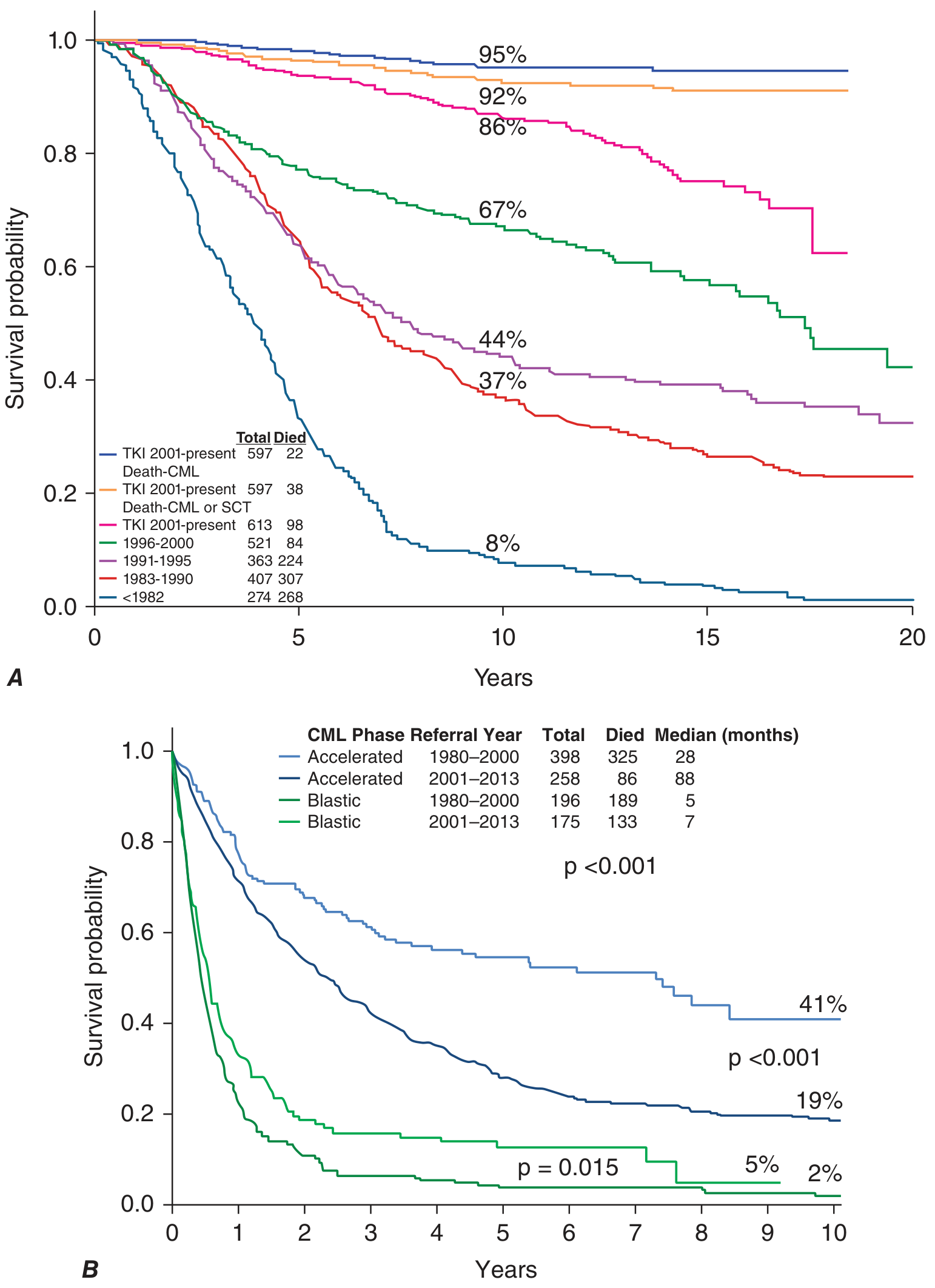
Sokal and ELTS Scoring - Risk Stratification at Diagnosis
Sokal score (older) and ELTS (EUTOS Long-Term Survival) score (preferred now) use:
- Age
- Spleen size
- Platelet count
- % blasts at diagnosis
These stratify patients into low, intermediate, and high risk - influencing choice of front-line TKI.
Key Differentials
| Feature | CML | Leukaemoid Reaction |
|---|---|---|
| LAP score | Low/absent | Very high |
| BCR-ABL1 | Present | Absent |
| Basophilia | Prominent | Absent/mild |
| Cause | Neoplastic | Infection/inflammation |
| Splenomegaly | Massive | Mild/absent |
Summary - Key Points
- CML is caused by t(9;22)(q34;q11) - the Philadelphia chromosome - present in >90% of cases; the rest have cryptic BCR-ABL1 detectable by FISH/PCR
- The cell of origin is a pluripotent HSC - explains why blast crisis can be myeloid OR lymphoid
- BCR-ABL1 produces p210 kDa protein in CML (vs p190 in Ph+ ALL)
- Disease course: chronic → accelerated → blast crisis (the latter carrying very poor prognosis)
- TKIs (especially imatinib and 2nd-gen TKIs) have transformed CML from a uniformly fatal disease to one with near-normal life expectancy
- T315I mutation = pan-resistance to 1st/2nd gen TKIs; only ponatinib/asciminib work
- MRD monitoring by qPCR (BCR-ABL1 on International Scale) drives all treatment decisions
- Treatment-free remission is now an achievable goal in ~50% of patients with deep molecular response
Sources: Harrison's Principles of Internal Medicine 22nd Ed. (2025), Chapter 110; Goldman-Cecil Medicine, Chapter 170; Robbins, Cotran & Kumar Pathologic Basis of Disease, Chapter 13
AML
Finding Sources
Finding Sources
Reading File
Reading File
Reading File
Reading File
Reading File
Reading File
Reading File
Reading File
Reading File
Reading File
Reading File
I now have comprehensive content from Harrison's 22nd Ed and Goldman-Cecil. Let me compile the complete, detailed AML overview.
Acute Myeloid Leukaemia (AML)
Definition
AML is an aggressive clonal neoplasm of immature myeloid lineage blast cells (myeloblasts, monoblasts, erythroblasts, or megakaryoblasts) that replace the bone marrow and suppress normal haematopoiesis. It is characterised by:
- Arrest of myeloid differentiation at an early precursor stage
- Accumulation of non-functional blast cells
- Resultant bone marrow failure (anaemia, thrombocytopenia, neutropenia)
Diagnostic threshold: ≥20% blasts in bone marrow or peripheral blood (exceptions: AML with t(8;21), inv(16), or t(15;17) are diagnosed regardless of blast count given their defining genetic abnormalities)
Epidemiology
| Parameter | Data |
|---|---|
| Most common acute leukaemia in adults | Yes (ALL is more common in children) |
| Median age at diagnosis | ~68 years |
| Annual incidence (USA) | ~20,000 new cases/year |
| Male slight predominance | Yes |
| 5-year overall survival (all ages) | ~29% overall; ~40-50% in younger fit adults with favourable genetics |
Aetiology & Risk Factors
1. Acquired / Environmental
| Exposure | Comment |
|---|---|
| Alkylating agents (chlorambucil, cyclophosphamide, melphalan) | Therapy-related AML; latency 5-10 years; often complex karyotype / del(5q) / del(7q) |
| Topoisomerase II inhibitors (etoposide, doxorubicin) | Therapy-related AML; latency 1-3 years; often KMT2A (11q23) rearrangements |
| Benzene / petroleum products | Chronic occupational exposure |
| Ionizing radiation | Historical (radiologists, atomic bomb survivors) |
| Cigarette smoking | Associated, possibly via benzene metabolites |
| Prior MDS or MPN | Secondary AML - worse prognosis |
2. Hereditary / Germline Predisposition
| Syndrome | Mechanism |
|---|---|
| Down syndrome (Trisomy 21) | GATA1 mutation → megakaryoblastic AML (before age 4); excellent outcomes with dose-reduced chemo |
| Fanconi anaemia | Defective DNA repair → AML |
| Bloom syndrome | Defective DNA repair |
| Ataxia-telangiectasia | Defective DNA repair |
| Kostmann syndrome (congenital neutropenia) | G-CSF receptor + ELANE mutations → AML |
| Shwachman-Diamond syndrome | Ribosome assembly defect |
| Li-Fraumeni syndrome | Germline TP53 mutation |
| Familial AML with DDX41 | Germline DDX41 - increasingly recognised |
Anticancer drugs are the leading cause of therapy-related AML.
Pathogenesis
AML results from two complementary categories of mutations ("two-hit" model):
| Class | Examples | Effect |
|---|---|---|
| Class I - activate proliferation/survival | FLT3-ITD, RAS mutations, KIT mutations | Growth advantage, anti-apoptosis |
| Class II - impair differentiation | RUNX1-RUNX1T1, CBFB-MYH11, PML-RARA, CEBPA, NPM1 | Maturation arrest at blast stage |
AML cells carry on average 10-15 mutations per cell (~5 driver mutations), far fewer than solid tumours.
Key driver mutations and their frequency:
| Gene | Frequency | Prognostic Impact |
|---|---|---|
| FLT3-ITD | 30-35% | Adverse (especially high allelic ratio) |
| NPM1 | ~30% | Favourable (if no FLT3-ITD) |
| DNMT3A | ~20% | Intermediate-adverse |
| IDH1 | ~8% | Targetable (ivosidenib) |
| IDH2 | ~12% | Targetable (enasidenib) |
| CEBPA (bZIP biallelic) | 4-15% | Favourable |
| TP53 | ~8-15% | Strongly adverse |
| RUNX1 | ~10% | Adverse |
| ASXL1 | ~10% | Adverse |
| KIT | ~5-10% | In CBF-AML |
WHO 2022 Classification
Group 1: AML with Defining Genetic Abnormalities
(diagnosed regardless of blast count if genetic lesion is present)
| Subtype | Genetic Lesion | Notes |
|---|---|---|
| APL | PML::RARA fusion; t(15;17) | Medical emergency; ATRA + ATO treatment |
| CBF-AML | RUNX1::RUNX1T1 fusion; t(8;21) | Favourable; slender Auer rods; CD19+ |
| CBF-AML | CBFB::MYH11; inv(16)/t(16;16) | Favourable; abnormal eosinophils in marrow |
| DEK::NUP214; t(6;9) | Adverse | |
| RBM15::MRTFA; t(1;22) | Megakaryoblastic; infants | |
| BCR::ABL1 fusion* | Rare | |
| KMT2A rearrangement | Adverse (most partners); monocytic features | |
| MECOM rearrangement | Adverse | |
| NUP98 rearrangement | Adverse | |
| NPM1 mutation | Most common (~30%); favourable if no FLT3-ITD | |
| CEBPA bZIP mutation | Favourable |
Group 2: AML, Myelodysplasia-Related (AML-MR)
- Prior MDS or MDS/MPN history, OR
- Complex karyotype, specific chromosomal aberrations, OR
- Mutations in: ASXL1, BCOR, EZH2, SF3B1, SRSF2, STAG2, U2AF1, ZRSR2
- Carries adverse prognosis
Group 3: AML, Defined by Differentiation (NOS)
(when no defining genetic abnormality found)
- AML with minimal differentiation (M0)
- AML without maturation (M1)
- AML with maturation (M2)
- Acute myelomonocytic leukaemia (M4)
- Acute monocytic leukaemia (M5)
- Acute erythroid leukaemia (M6)
- Acute megakaryoblastic leukaemia (M7)
- Acute basophilic leukaemia
Note: The 2022 WHO classification superseded the 2016 version. The older FAB (French-American-British) M0-M7 classification is now largely replaced but still used in some contexts.
Morphology & Special Features
Auer Rods
- Pathognomonic of AML (never seen in ALL)
- Needle-shaped pink cytoplasmic inclusions = fused lysosomes
- Multiple Auer rods in a single cell = "faggot cells" - hallmark of APL (M3)
Key Morphologic-Genetic Correlations:
| Morphologic Feature | Associated Genetics |
|---|---|
| Faggot cells (multiple Auer rods) | APL / t(15;17) / PML-RARA |
| Slender Auer rods + CD19 expression + normal eosinophils | t(8;21) / RUNX1-RUNX1T1 |
| Abnormal marrow eosinophils | inv(16) / CBFB-MYH11 |
| Cup-shaped nuclear blasts | NPM1 mutation (especially + FLT3) |
| Monocytic features + gingival hypertrophy | KMT2A rearrangements; AML M5 |
| Dysplastic morphology | Complex karyotype / TP53 mutation |
| DIC at presentation | APL / t(15;17) |
Immunophenotype (Flow Cytometry):
| Marker | Expression Pattern |
|---|---|
| CD34, CD117, HLA-DR | Most immature AML forms |
| CD13, CD33 | More differentiated myeloid AML |
| CD14, CD15, CD11b | Monocytic AML |
| CD36, CD71, CD235a (glycophorin A) | Erythroid leukaemia |
| CD41, CD61 | Megakaryoblastic AML |
Clinical Features
From Marrow Failure:
- Anaemia - fatigue, dyspnoea, decreased exercise tolerance
- Thrombocytopenia - petechiae, ecchymoses, bleeding from unusual sites, DIC (especially APL)
- Neutropenia - recurrent or life-threatening bacterial/fungal infections; fever is the most common presenting symptom
From Organ Infiltration:
- Gingival hypertrophy - characteristic of monocytic AML (M5) / KMT2A rearrangements
- Leukemia cutis (skin nodules/rash) - most common in monocytic subtypes
- Myeloid sarcoma (chloroma) - extramedullary tumour of myeloid blasts; green colour from myeloperoxidase; associated with t(8;21)
- Hepatosplenomegaly, lymphadenopathy (less prominent than in ALL)
- CNS involvement - headache, visual changes, cranial nerve palsies (rare in AML vs ALL)
- Back pain + lower extremity weakness - spinal granulocytic sarcoma; associated with t(8;21)
- DIC - characteristic of APL; ecchymoses, oozing from IV sites
Hyperleukocytosis (WBC > 100,000/μL):
- Occurs in ~5-10% of AML
- Leukostasis: slugging of blasts in capillaries → respiratory failure, stroke, priapism
- Medical emergency - requires emergent leukapheresis
Diagnosis
Initial Workup:
- CBC with differential - anaemia, thrombocytopenia, leukocytosis (or leukopenia); blasts on smear
- Bone marrow aspirate + biopsy (essential):
- Morphology (Auer rods, blast %)
- Flow cytometry (immunophenotype)
- Cytogenetics (karyotype) - standard
- FISH - specific translocations
- Molecular studies (NGS panel): NPM1, FLT3, IDH1/2, CEBPA, TP53, ASXL1, DNMT3A, etc.
- Chemistry panel: LDH, uric acid (tumour lysis risk), electrolytes, renal/hepatic function, DIC screen (PT, aPTT, fibrinogen, d-dimer)
- Cardiac function: echocardiogram or MUGA scan (before anthracyclines)
- HLA typing: for potential allogeneic SCT
- Viral serologies: CMV, HSV, VZV (reactivation risk)
ELN 2022 Risk Stratification (Crucial for Treatment Decisions)
Favourable Risk:
- t(8;21)/RUNX1::RUNX1T1
- inv(16) or t(16;16)/CBFB::MYH11
- Mutated NPM1 without FLT3-ITD
- bZIP in-frame mutated CEBPA
Intermediate Risk:
- Mutated NPM1 with FLT3-ITD
- Wild-type NPM1 with FLT3-ITD (no adverse lesions)
- t(9;11)/MLLT3::KMT2A
- Other cytogenetic/molecular abnormalities
Adverse Risk:
- t(6;9)/DEK::NUP214
- t(v;11)/KMT2A-rearranged (most partners)
- t(9;22)/BCR::ABL1
- inv(3) or t(3;3)/GATA2,MECOM(EVI1)
- -5 or del(5q); -7; -17/abnl(17p)
- Complex karyotype (≥3 abnormalities)
- Monosomal karyotype (≥2 monosomies or 1 monosomy + structural abnormality)
- Wild-type NPM1 + FLT3-ITD high
- Mutated RUNX1, ASXL1, TP53
Treatment
Overall Framework:
Treatment is divided into Induction → Consolidation (Post-remission therapy), selected based on:
- Patient age and fitness
- ELN genetic risk category
- Availability of targeted agents
Induction Chemotherapy
Standard-intensity ("7+3 regimen") - for fit patients:
- Cytarabine 100-200 mg/m² continuous IV infusion × 7 days
- Anthracycline (daunorubicin 60-90 mg/m² or idarubicin 12 mg/m²) × days 1, 2, 3
- CR rate: 60-80% in younger patients; lower in older/unfit
Mechanism of key drugs:
- Cytarabine: S-phase specific antimetabolite → inhibits DNA synthesis (phosphorylated to ara-CTP intracellularly)
- Anthracyclines: DNA intercalators → topoisomerase II inhibition → DNA strand breaks
Addition to 7+3:
- Gemtuzumab ozogamicin (anti-CD33 ADC) added in CBF-AML and CD33+ AML
- Midostaurin added for FLT3-mutated AML (first FDA approval in AML with a targeted agent)
- Gilteritinib (FLT3 inhibitor) for relapsed/refractory FLT3-mutated AML
Low-intensity regimens - for older/unfit patients:
- Venetoclax + azacitidine (or decitabine) - now standard of care for unfit elderly patients
- Venetoclax = BCL-2 inhibitor → overcomes apoptosis resistance
- CR rates ~65-70% in previously untreated elderly patients
- IDH1 inhibitor (ivosidenib) ± azacitidine - for IDH1-mutated AML
- IDH2 inhibitor (enasidenib) - for IDH2-mutated AML
- Low-dose cytarabine (LDAC) ± glasdegib
Post-remission / Consolidation
After achieving CR, further therapy is mandatory (relapse is near-universal without it):
| Risk Category | Preferred Post-remission Strategy |
|---|---|
| Favourable (CBF-AML, NPM1 without FLT3) | High-dose cytarabine (HiDAC) × 3-4 cycles; no SCT needed in first CR |
| Intermediate | HiDAC consolidation or allogeneic SCT - individualised based on MRD |
| Adverse | Allogeneic SCT in first CR is standard |
| Therapy-related / AML-MR | Allogeneic SCT if possible |
High-dose cytarabine (HiDAC): 1-3 g/m² given 4-12 doses per course
- Key toxicity: cerebellar toxicity (ataxia, dysarthria) - monitor carefully
Acute Promyelocytic Leukaemia (APL) - Special Case
APL (AML with PML::RARA / t(15;17)) is a medical emergency but also has the best prognosis of all AML subtypes.
Pathophysiology: PML-RARA fusion protein blocks myeloid differentiation at the promyelocytic stage and releases granule contents → DIC.
Treatment:
- ATRA (all-trans retinoic acid) + ATO (arsenic trioxide) = standard of care for low-risk APL
- ATRA: 45 mg/m²/day orally → induces differentiation of leukemic promyelocytes
- ATO: 0.15 mg/kg/day IV
- CR approaches 100%; long-term survival >90%
- High-risk APL (WBC >10,000/μL): add cytoreductive chemotherapy immediately due to risk of APL syndrome and DIC
APL (Differentiation) Syndrome:
- Occurs within first 3 weeks of ATRA/ATO therapy
- Features: fever, fluid retention, dyspnoea, chest pain, pulmonary infiltrates, pleural/pericardial effusions, hypoxaemia
- Mechanism: adhesion of differentiated neoplastic cells to pulmonary vasculature
- Treatment: dexamethasone ± cytoreduction; temporarily stop ATRA if severe
- Mortality: ~10% if unrecognised
MRD monitoring in APL: PCR for PML-RARA - disappearance of signal = long-term DFS; reemergence = relapse.
Targeted Agents in AML (Summary)
| Drug | Target | Indication |
|---|---|---|
| Midostaurin | FLT3 inhibitor (multi-kinase) | Frontline + 7+3 for FLT3-mutated AML |
| Gilteritinib | FLT3 inhibitor (selective) | Relapsed/refractory FLT3-mutated AML |
| Quizartinib | FLT3 inhibitor | FLT3-ITD AML |
| Ivosidenib | IDH1 inhibitor | IDH1-mutated AML (frontline or R/R) |
| Enasidenib | IDH2 inhibitor | IDH2-mutated AML (R/R) |
| Venetoclax | BCL-2 inhibitor | + azacitidine for unfit elderly AML |
| Gemtuzumab ozogamicin | Anti-CD33 ADC | CBF-AML + 7+3; CD33+ AML |
| ATRA | RAR-α ligand → differentiation | APL (PML-RARA) |
| Arsenic trioxide | Degrades PML-RARA fusion | APL |
| Revumenib | Menin inhibitor | KMT2A-rearranged or NPM1-mutated AML (R/R) |
Allogeneic SCT in AML
- Gold standard for adverse-risk AML in first CR
- Only potentially curative approach for many high-risk patients
- Requires HLA-matched sibling or unrelated donor (or haploidentical if no match)
- Main benefit: Graft-vs-Leukaemia (GvL) effect in addition to high-dose conditioning
- Main risks: transplant-related mortality, graft-vs-host disease (GvHD), infections
- Decision guided by ELN risk + MRD status after induction
MRD (Minimal Residual Disease) Monitoring
MRD assessment after induction and consolidation is increasingly standard:
- Flow cytometry MRD (leukemia-associated immunophenotype)
- Molecular MRD (NPM1, RUNX1-RUNX1T1, CBFB-MYH11 by PCR)
- MRD negativity after first consolidation = strong predictor of long-term remission
- MRD persistence → escalate therapy / proceed to SCT
Prognosis
| Risk Group | 5-year OS (approximate) |
|---|---|
| Favourable (CBF-AML, NPM1+/FLT3-) | ~50-70% |
| Intermediate | ~25-40% |
| Adverse (complex karyotype, TP53, monosomal) | <10-15% |
| APL (low-risk, ATRA + ATO) | >90% |
| Therapy-related AML | ~10-20% |
| Elderly patients overall | ~10-20% |
Key Points Summary
- AML is diagnosed by ≥20% blasts in marrow/blood (except APL, t(8;21), inv(16) - diagnosed by genetics)
- Auer rods are pathognomonic of AML; faggot cells (multiple Auer rods) = APL
- The two-hit model: Class I mutations (FLT3, RAS) drive proliferation; Class II mutations (NPM1, RUNX1 fusions) block differentiation
- APL is a haematological emergency - DIC is the key early threat; ATRA + ATO achieves ~100% CR and >90% long-term survival
- FLT3-ITD (~30%) = most common adverse mutation; NPM1 (~30%) = most common favourable mutation
- Treatment: 7+3 induction → HiDAC consolidation for fit patients; venetoclax + azacitidine for unfit elderly patients
- ELN risk stratification determines whether allogeneic SCT is needed in first CR
- Targeted therapies (midostaurin, gilteritinib, ivosidenib, venetoclax) have significantly improved outcomes across AML subtypes
Sources: Harrison's Principles of Internal Medicine 22nd Ed. (2025), Chapter 109; Goldman-Cecil Medicine, Chapter 168; Robbins, Cotran & Kumar Pathologic Basis of Disease, Chapter 13
Make 40 mcqs pf aml clinical based scenarios along with explanation of answers and wrong ones covering every mahor aspect of topic at final year mbbs level with gap between question and answer
Make 40 clinical based mcqs for aml woth explaning answers and the wrong ones with gap between question and answer Mcqs should be final year med student level
Here are 40 clinical scenario-based MCQs on AML at final year MBBS level, with full explanations of correct and wrong answers.
40 Clinical MCQs — Acute Myeloid Leukaemia (AML)
Q1. A 58-year-old man presents with 3 weeks of fatigue, gum bleeding, and easy bruising. Examination reveals pale conjunctivae, petechiae on the lower limbs, and gingival hypertrophy. FBC shows Hb 7.2 g/dL, platelets 28 × 10⁹/L, WBC 64 × 10⁹/L. Blood film shows numerous large blasts with reniform nuclei and abundant cytoplasm. Which AML subtype is most likely?
A) APL (M3)
B) Acute myelomonocytic leukaemia (M4)
C) Acute monocytic leukaemia (M5)
D) AML with minimal differentiation (M0)
✅ Answer: C) Acute monocytic leukaemia (M5)
Gingival hypertrophy is the hallmark extramedullary feature of monocytic leukaemia (M5) and, to a lesser extent, M4. Monoblasts are large cells with reniform (kidney-shaped) or folded nuclei and abundant cytoplasm. Skin infiltration is also more common in monocytic subtypes.
❌ A - APL (M3): APL blasts are heavily granulated promyelocytes - they do NOT cause gingival hypertrophy. The hallmark of APL is DIC and faggot cells (multiple Auer rods). Gum hypertrophy is not a feature.
❌ B - M4 (myelomonocytic): M4 can cause gingival hypertrophy when monocytic differentiation predominates, but the purely monocytic morphology described (reniform nuclei, abundant cytoplasm, NO granulation) and prominent gingival hypertrophy points more specifically to M5.
❌ D - AML M0: M0 blasts are completely undifferentiated - minimal cytoplasm, no granules, no monocytic features. It does NOT cause gingival hypertrophy and is essentially a diagnosis of exclusion.
Q2. A 32-year-old woman is admitted with a 1-week history of spontaneous bruising and heavy menstrual bleeding. She is found to have petechiae all over her body and oozing from her venepuncture site. Labs show Hb 9.1 g/dL, WBC 4.2 × 10⁹/L (with 78% blasts), platelets 18 × 10⁹/L, PT prolonged, fibrinogen 0.8 g/L (low), D-dimer markedly elevated. Bone marrow shows numerous hypergranular blasts with multiple Auer rods in single cells ("faggot cells"). What is the IMMEDIATE priority in management?
A) Start 7+3 induction chemotherapy urgently
B) Administer ATRA immediately
C) Perform allogeneic stem cell transplantation
D) Give platelet transfusion and wait for cytogenetics
✅ Answer: B) Administer ATRA immediately
This is classic APL (M3) - faggot cells, DIC, young woman, hypergranular blasts. ATRA must be started immediately on clinical suspicion, without waiting for cytogenetic confirmation of t(15;17). Delaying ATRA while awaiting results is dangerous - APL carries a high early mortality from haemorrhage if untreated. ATRA induces differentiation of promyelocytes and reduces DIC.
❌ A - 7+3 chemotherapy immediately: Standard 7+3 is NOT the treatment for APL. Anthracycline alone without ATRA in APL would worsen DIC by causing rapid cell lysis and release of procoagulant granule contents - historically this led to fatal haemorrhage. ATRA + ATO is the correct regimen.
❌ C - Allogeneic SCT: SCT has no role in the initial management of newly diagnosed APL. It is reserved for relapsed or refractory disease. APL is one of the most curable leukaemias with ATRA + ATO.
❌ D - Wait for cytogenetics: This is the most dangerous option. APL patients die from haemorrhage within hours to days if ATRA is not started promptly. ATRA must be started on clinical/morphologic suspicion alone - cytogenetics confirms but should not delay treatment.
Q3. A 45-year-old man with APL is started on ATRA. On day 10 of treatment he develops fever (38.9°C), progressive dyspnoea, bilateral pleural effusions on chest X-ray, and a rising WBC count. His oxygen saturation is 88% on room air. What is this complication and what is the first-line treatment?
A) Bacterial pneumonia; start broad-spectrum antibiotics
B) ATRA-induced hepatotoxicity; stop ATRA permanently
C) APL differentiation syndrome; start dexamethasone
D) Pulmonary embolism; start anticoagulation
✅ Answer: C) APL differentiation syndrome; start dexamethasone
This is the APL (differentiation) syndrome - a complication of ATRA (and less commonly ATO) occurring in the first 3 weeks of treatment. Features: fever, fluid retention, dyspnoea, pleural/pericardial effusions, pulmonary infiltrates, hypoxaemia, often with a rising WBC. It is caused by differentiated neoplastic cells adhering to the pulmonary vasculature. Treatment: dexamethasone (10 mg IV BID), ± temporary hold of ATRA in severe cases (respiratory failure, renal failure requiring ICU). Mortality is ~10% if unrecognised.
❌ A - Bacterial pneumonia: While pneumonia can occur, the combination of fever + fluid retention + pleural effusions + rising WBC on day 10 of ATRA is the classic presentation of differentiation syndrome. Antibiotics alone would miss the diagnosis.
❌ B - Hepatotoxicity; stop ATRA permanently: ATRA can cause hepatotoxicity but does not cause bilateral pleural effusions, fluid retention, and progressive hypoxaemia. Stopping ATRA permanently would be wrong - the patient may need ATRA re-started after differentiation syndrome is controlled.
❌ D - Pulmonary embolism: PE would not cause fever, bilateral pleural effusions, and a rising WBC. APL patients do have coagulopathy and bleeding risk, but the complete clinical picture here is differentiation syndrome, not PE.
Q4. A 67-year-old woman presents with fatigue and recurrent infections. She has a history of breast cancer treated 4 years ago with cyclophosphamide and radiation. FBC shows pancytopenia with 35% blasts. Karyotype reveals deletion of chromosome 7 and complex cytogenetics. Which AML category does she have, and what is its prognosis?
A) De novo AML with favourable risk; good prognosis
B) Core binding factor AML; intermediate prognosis
C) Therapy-related AML with adverse risk; poor prognosis
D) APL; excellent prognosis with ATRA
✅ Answer: C) Therapy-related AML with adverse risk; poor prognosis
This is classic therapy-related AML (t-AML) following alkylating agent chemotherapy (cyclophosphamide) and radiation for breast cancer. Latency is typically 5-10 years for alkylating agents. The presence of del(7) and complex karyotype are hallmark adverse cytogenetic features of t-AML. This category carries a very poor prognosis (~10-20% 5-year OS), partly because these cells are inherently resistant to chemotherapy and often harbour TP53 mutations.
❌ A - De novo AML with favourable risk: Favourable risk AML includes t(8;21), inv(16), and NPM1 mutation without FLT3-ITD. Del(7) and complex karyotype are in the adverse-risk category. This patient's history of prior chemotherapy also immediately places her in the therapy-related category.
❌ B - CBF-AML with intermediate prognosis: CBF-AML (t(8;21) or inv(16)) is a favourable-risk genetic subgroup found predominantly in younger adults. This patient's karyotype shows del(7) and complex cytogenetics, not CBF translocations.
❌ D - APL with excellent prognosis: APL requires t(15;17)/PML-RARA. There is no morphologic or cytogenetic feature of APL here. Faggot cells, DIC, and t(15;17) are not mentioned.
Q5. A 52-year-old man is newly diagnosed with AML. Karyotype shows t(8;21)(q22;q22) and molecular testing confirms RUNX1-RUNX1T1 fusion. His blast percentage is 18%. He is otherwise fit with good performance status. What is the appropriate management?
A) Watchful waiting since blasts are < 20%
B) Allogeneic SCT in first complete remission
C) Intensive 7+3 induction chemotherapy + gemtuzumab ozogamicin
D) Venetoclax + azacitidine
✅ Answer: C) Intensive 7+3 induction chemotherapy + gemtuzumab ozogamicin
AML with t(8;21)/RUNX1-RUNX1T1 is a defining genetic AML - diagnosis is made regardless of blast count (even < 20%). This is core binding factor (CBF) AML, which is ELN favourable risk. The correct approach is intensive induction (7+3) with the addition of gemtuzumab ozogamicin (anti-CD33 antibody-drug conjugate), which has been shown to improve outcomes in CBF-AML specifically.
❌ A - Watchful waiting because blasts < 20%: This is WRONG. AML with defined genetic abnormalities (including t(8;21)) is diagnosed and treated regardless of blast count. The 20% threshold does not apply when a defining genetic abnormality is present.
❌ B - Allogeneic SCT in first CR: SCT in first CR is reserved for adverse-risk AML. CBF-AML is favourable risk - patients do well with intensive chemotherapy alone and do NOT need SCT in first remission. Adding SCT adds toxicity without survival benefit in favourable-risk disease.
❌ D - Venetoclax + azacitidine: This low-intensity regimen is for older/unfit patients who cannot tolerate intensive chemotherapy. A 52-year-old with good performance status should receive intensive chemotherapy to maximise cure potential.
Q6. A 71-year-old man with no prior medical history presents with fatigue, weight loss, and night sweats. Bone marrow biopsy shows 28% blasts. He has an ECOG performance status of 3 due to severe COPD, with poor exercise tolerance. Molecular testing shows IDH2 mutation. What is the most appropriate treatment?
A) 7+3 intensive induction chemotherapy
B) Observation alone
C) Venetoclax + azacitidine
D) Immediate allogeneic SCT
✅ Answer: C) Venetoclax + azacitidine
This elderly, unfit patient (ECOG 3, severe COPD) cannot tolerate intensive 7+3 chemotherapy. The standard of care for unfit elderly AML patients is now venetoclax (BCL-2 inhibitor) + azacitidine (hypomethylating agent), which achieves CR rates of ~65-70% with acceptable toxicity. IDH2 mutation may additionally support consideration of enasidenib (IDH2 inhibitor), but venetoclax + azacitidine remains the primary standard.
❌ A - 7+3 intensive induction: Intensive chemotherapy in a frail ECOG 3 patient with severe COPD carries very high treatment-related mortality. The benefit of intensive therapy is controversial in unfit older patients and should not be applied to ECOG 3 patients with significant comorbidities.
❌ B - Observation alone: Studies consistently show that treatment is better than supportive care for all AML candidates who can tolerate any therapy. Even unfit patients benefit from low-intensity therapy - observing AML in a 71-year-old leads to certain rapid progression.
❌ D - Immediate allogeneic SCT: SCT is only offered after achieving CR and is typically reserved for patients who are fit enough to tolerate transplant-related morbidity. An ECOG 3 patient with severe COPD is not a SCT candidate.
Q7. A 38-year-old woman is diagnosed with AML following molecular testing which reveals FLT3-ITD mutation with high allelic ratio, alongside NPM1 mutation. Cytogenetics are otherwise normal. Per ELN 2022, what is her risk category?
A) Favourable
B) Intermediate
C) Adverse
D) Cannot be determined without additional markers
✅ Answer: B) Intermediate
Per ELN 2022, NPM1 mutation + FLT3-ITD is classified as intermediate risk (regardless of FLT3-ITD allelic ratio). Note: NPM1 mutation without FLT3-ITD = favourable. The presence of FLT3-ITD alongside NPM1 mutation negates the favourable NPM1 effect and moves the patient to intermediate risk.
❌ A - Favourable: NPM1 mutation is favourable ONLY when FLT3-ITD is absent. The co-occurrence of high-allelic-ratio FLT3-ITD negates this benefit. This is a classic exam trap.
❌ C - Adverse: Adverse risk requires specific lesions such as monosomal karyotype, complex karyotype, del(5q), del(7), t(6;9), inv(3), or TP53/RUNX1/ASXL1 mutations. NPM1+FLT3-ITD does not reach the adverse-risk threshold.
❌ D - Cannot be determined: The ELN 2022 classification gives clear guidance for this specific combination. It is intermediate risk - this is a well-defined and important clinical scenario, not an undetermined one.
Q8. A 50-year-old man with AML (FLT3-ITD mutated, intermediate risk) achieves complete remission after 7+3 + midostaurin induction. He has an HLA-matched sibling donor. What is the recommended post-remission strategy?
A) High-dose cytarabine (HiDAC) consolidation × 4 cycles, no SCT
B) Allogeneic SCT in first CR
C) Maintenance midostaurin only
D) Observation; treat at relapse
✅ Answer: B) Allogeneic SCT in first CR
For intermediate-risk AML, allogeneic SCT in first CR is recommended especially when FLT3-ITD is present (a strong relapse risk factor) and an HLA-matched donor is available. The graft-vs-leukaemia (GvL) effect provides protection against relapse that chemotherapy alone cannot. With a matched sibling donor available, SCT is the preferred post-remission approach.
❌ A - HiDAC consolidation alone: HiDAC alone is preferred in favourable-risk AML (CBF-AML, NPM1+/FLT3-). In FLT3-ITD positive intermediate-risk disease, relapse rates with HiDAC alone are high, making SCT the preferred option when a donor is available.
❌ C - Maintenance midostaurin only: While FLT3 inhibitors are used in induction (midostaurin) and maintenance post-SCT (gilteritinib/midostaurin), maintenance alone without consolidation or SCT is not adequate post-remission therapy for this risk category.
❌ D - Observation: Without consolidation therapy, virtually 100% of AML patients relapse after induction CR. There is no role for observation in AML after achieving remission.
Q9. A 42-year-old woman is undergoing induction chemotherapy for AML. On day 15 of the 7+3 regimen, she develops fever (39.2°C), rigors, hypotension, and tachycardia. Her absolute neutrophil count is 0.1 × 10⁹/L. What is the most urgent initial management?
A) Wait for blood culture results before starting antibiotics
B) Start broad-spectrum IV antibiotics immediately (within 1 hour)
C) Start antifungal therapy with voriconazole
D) Administer G-CSF to stimulate neutrophil recovery
✅ Answer: B) Start broad-spectrum IV antibiotics immediately (within 1 hour)
This is febrile neutropenia - a haematological emergency. ANC < 0.5 × 10⁹/L + fever ≥38.3°C = febrile neutropenia. Broad-spectrum IV antibiotics (e.g., piperacillin-tazobactam or ceftazidime/cefepime) must be started within 1 hour of fever onset. Every hour's delay increases mortality. Blood cultures should be drawn BEFORE antibiotics but must not delay their initiation.
❌ A - Wait for culture results: Waiting for culture results before starting antibiotics in febrile neutropenia is potentially fatal. The classic teaching: "Draw cultures, START antibiotics immediately." Culture results take 24-72 hours; a neutropenic patient can deteriorate to septic shock within hours.
❌ C - Start antifungals immediately: Antifungal therapy (voriconazole, caspofungin) is added if fever persists after 4-7 days of broad-spectrum antibiotics despite negative bacterial cultures - this is empirical antifungal escalation. It is NOT the first-line treatment for a new febrile episode.
❌ D - G-CSF first: G-CSF (filgrastim) is not routinely given during active AML induction (where the marrow is intentionally ablated) and is not the immediate priority in a septic patient. The patient needs antibiotics now - not stimulation of marrow that is currently empty of neutrophils.
Q10. A 28-year-old man presents with a 2-week history of fatigue and a sudden onset of severe headache and right-sided hemiplegia. His WBC is 185 × 10⁹/L with 80% blasts. CT head shows no haemorrhage. What is the most likely cause of his neurological deficit and immediate management?
A) CNS leukaemia; start intrathecal methotrexate
B) Leukostasis; emergent leukapheresis
C) Haemorrhagic stroke due to thrombocytopenia; platelet transfusion
D) Bacterial meningitis; lumbar puncture and IV antibiotics
✅ Answer: B) Leukostasis; emergent leukapheresis
WBC 185 × 10⁹/L = hyperleukocytosis. At such extreme counts, blasts aggregate and plug microvascular capillaries causing leukostasis - manifesting as respiratory failure, stroke, priapism, and altered consciousness. CT showing no haemorrhage rules out haemorrhagic stroke. The immediate treatment is leukapheresis (mechanical removal of circulating blasts) to rapidly reduce the blast burden, alongside urgent initiation of cytoreductive chemotherapy.
❌ A - CNS leukaemia; intrathecal methotrexate: CNS leukaemia presents with headache, nausea, cranial nerve palsies, and papilloedema - diagnosed by CSF examination (≥5 cells/μL with blasts). With a WBC of 185 × 10⁹/L, leukostasis is the immediate danger. Intrathecal chemotherapy takes time and does not address the systemic leukostasis.
❌ C - Haemorrhagic stroke; platelet transfusion: CT head shows NO haemorrhage. Haemorrhagic stroke due to thrombocytopenia would show blood on CT. Leukostasis causes ischaemic, not haemorrhagic, neurological events.
❌ D - Bacterial meningitis: Bacterial meningitis can cause headache and hemiplegia but does not explain a WBC of 185 × 10⁹/L with 80% blasts. The clinical picture is overwhelmingly consistent with AML + leukostasis, not an infection.
Q11. A 19-year-old boy with Down syndrome is found to have 35% megakaryoblasts on bone marrow examination. He presents with pallor and bruising. What unique consideration applies to his chemotherapy?
A) He should not receive chemotherapy; supportive care only
B) Standard-dose chemotherapy is given as usual
C) Dose-reduced chemotherapy due to high sensitivity and treatment-related toxicity
D) Allogeneic SCT is the only curative option
✅ Answer: C) Dose-reduced chemotherapy due to high sensitivity and treatment-related toxicity
Children with Down syndrome-associated AML (typically megakaryoblastic, with GATA1 mutation, occurring before age 4) have excellent outcomes but are highly sensitive to cytarabine due to increased drug uptake and reduced cytarabine catabolism. Standard doses cause excessive toxicity. Therefore, dose-reduced regimens are used, yet cure rates are very high (~80-90%).
❌ A - Supportive care only: Down syndrome AML is one of the most curable subtypes of paediatric AML. Withholding chemotherapy would deny a potentially curable disease. Supportive care alone would be inappropriate.
❌ B - Standard dose chemotherapy: Standard doses lead to prohibitive toxicity (mucositis, infections, organ damage) in Down syndrome AML patients. Protocols specifically designed for this population use reduced doses while maintaining efficacy.
❌ D - SCT is the only curative option: Down syndrome AML is highly chemosensitive. SCT is NOT routinely required for first-line treatment. Standard chemotherapy alone (dose-modified) achieves excellent cure rates.
Q12. A 60-year-old man is diagnosed with AML. Cytogenetics show inversion of chromosome 16: inv(16)(p13.1q22), confirmed as CBFB-MYH11 by FISH. Bone marrow shows increased abnormal eosinophils. After induction, he achieves complete remission. What is the most appropriate post-remission therapy?
A) Allogeneic SCT in first CR
B) High-dose cytarabine (HiDAC) × 3-4 cycles
C) Maintenance azacitidine
D) Observation and treat at relapse
✅ Answer: B) High-dose cytarabine (HiDAC) × 3-4 cycles
Inv(16)/CBFB-MYH11 is CBF-AML = ELN favourable risk. For favourable-risk AML in first CR, multiple cycles of high-dose cytarabine (HiDAC) consolidation are the standard - allogeneic SCT is NOT required in first CR. The 5-year OS with HiDAC consolidation in CBF-AML exceeds 50-60%. Abnormal marrow eosinophils are a characteristic morphologic feature of inv(16).
❌ A - SCT in first CR: SCT is reserved for intermediate and adverse risk AML in first CR. Adding SCT in favourable-risk disease (CBF-AML) increases treatment-related mortality without improving outcome compared to HiDAC consolidation.
❌ C - Maintenance azacitidine: There is no standard maintenance therapy in favourable-risk AML outside of clinical trials. Azacitidine maintenance is used in some AML subgroups after intensive therapy, but this patient has excellent prognosis and achieves cure with HiDAC consolidation alone.
❌ D - Observation: Observation after CR is never appropriate in AML. Without post-remission therapy, virtually all patients relapse within months.
Q13. A 55-year-old woman with AML has the following molecular profile: NPM1 mutated, FLT3-ITD absent, normal karyotype. After 7+3 induction she achieves CR. PCR for NPM1 after 2 cycles of HiDAC consolidation shows NPM1 transcript has disappeared (MRD negative). What does this imply?
A) She requires immediate SCT as MRD negativity means nothing
B) She has an excellent prognosis and may not need SCT in first CR
C) She should receive maintenance ATRA
D) She should proceed to investigational therapy as standard treatments have failed
✅ Answer: B) She has an excellent prognosis and may not need SCT in first CR
This patient has NPM1-mutated, FLT3-ITD-negative AML = ELN favourable risk. Achievement of MRD negativity (disappearance of NPM1 PCR signal) after consolidation is associated with an excellent long-term prognosis and strongly supports continuing with HiDAC consolidation alone, without SCT. MRD negativity is one of the strongest predictors of durable remission in AML.
❌ A - MRD negativity means nothing: MRD is one of the most powerful prognostic tools in AML. MRD negativity consistently predicts lower relapse rates and longer overall survival across multiple studies. Ignoring MRD is clinically inappropriate.
❌ C - Maintenance ATRA: ATRA is only used for APL (PML-RARA). There is no role for ATRA in non-APL AML with NPM1 mutation.
❌ D - Investigational therapy: There is no indication for investigational therapy in a patient with favourable-risk AML who has achieved MRD negativity after standard treatment. She is in a best-case scenario, not a treatment failure.
Q14. A 44-year-old man presents with AML. A blood film is shown to the examiner with blasts containing needle-shaped pink cytoplasmic inclusions. Some blasts contain numerous such inclusions arranged in a bundle pattern. What are these inclusions called, and what is their diagnostic significance?
A) Döhle bodies - indicate bacterial infection
B) Auer rods / "faggot cells" - pathognomonic of AML
C) Azurophilic granules - normal myeloid precursors
D) Heinz bodies - indicate G6PD deficiency
✅ Answer: B) Auer rods / "faggot cells" - pathognomonic of AML
Auer rods are needle-shaped pink (azurophilic) cytoplasmic inclusions formed from fused lysosomes. They are pathognomonic of AML - they are never seen in ALL or normal cells. When a single blast contains multiple Auer rods in a bundle arrangement, these are called "faggot cells", which are the hallmark morphology of APL (M3).
❌ A - Döhle bodies: Döhle bodies are small pale blue cytoplasmic inclusions seen in mature neutrophils during severe infections, burns, or pregnancy. They are NOT in blasts and have nothing to do with AML.
❌ C - Azurophilic granules: Primary (azurophilic) granules are normal components of promyelocytes and myelocytes. They can be seen in normal myeloid cells. Auer rods are specifically abnormal, fused, crystalline lysosomes - not normal granules.
❌ D - Heinz bodies: Heinz bodies are precipitates of denatured haemoglobin seen in red cells in G6PD deficiency or oxidative haemolysis. They are in RBCs, not in WBC blasts, and are visualised with supravital stains like crystal violet, not on a standard Wright-Giemsa smear.
Q15. A bone marrow biopsy in a 48-year-old man with AML shows blasts with strong positivity for myeloperoxidase (MPO) on cytochemistry, and Sudan black B staining is also positive. CD33 and CD13 are expressed on flow cytometry. These findings are consistent with which lineage?
A) B-lymphoid lineage
B) T-lymphoid lineage
C) Myeloid lineage
D) Erythroid lineage
✅ Answer: C) Myeloid lineage
Myeloperoxidase (MPO) and Sudan black B positivity on cytochemistry are the definitive markers of myeloid differentiation. MPO positivity in ≥3% of blasts is sufficient to assign myeloid lineage. CD33 and CD13 are the characteristic myeloid surface antigens in AML. MPO is negative in ALL, which is the key cytochemical distinction.
❌ A - B-lymphoid: B-lymphoid blasts are MPO negative and Sudan black negative. They express CD19, CD22, CD10 (in common ALL). B-cell lineage is confirmed by cytoplasmic or surface immunoglobulin, TdT positivity, and B-cell surface markers.
❌ B - T-lymphoid: T-cell blasts are also MPO negative. They express CD7, CD3 (cytoplasmic), CD2. Sudan black negativity is expected in ALL. The presence of MPO and Sudan black positivity definitively excludes lymphoid lineage.
❌ D - Erythroid: Erythroid leukaemia is diagnosed by CD36, CD71, CD235a (glycophorin A) positivity and characteristic erythroblast morphology. Erythroid precursors may be PAS positive (periodic acid-Schiff), not MPO positive.
Q16. A 63-year-old man presents with AML. Karyotype shows: -5, -7, del(5q), and two additional structural abnormalities (5 total cytogenetic abnormalities). What is his risk category per ELN 2022, and what is the implication?
A) Favourable risk; HiDAC consolidation alone
B) Intermediate risk; consider SCT based on MRD
C) Adverse risk; allogeneic SCT in first CR if fit
D) Favourable risk; ATRA + ATO treatment
✅ Answer: C) Adverse risk; allogeneic SCT in first CR if fit
This patient has a monosomal karyotype (monosomy 5 and 7 = loss of chromosomes) and complex karyotype (≥3 cytogenetic abnormalities) - both independently adverse features per ELN 2022. Del(5q) and del(7q) are also specifically listed adverse markers. This places him firmly in adverse risk category. For adverse-risk AML patients who achieve CR and are fit, allogeneic SCT in first CR is the standard recommendation.
❌ A - Favourable risk; HiDAC alone: Favourable risk requires t(8;21), inv(16), NPM1+/FLT3-, or CEBPA bZIP mutations. Monosomal and complex karyotypes are at the opposite end of the spectrum.
❌ B - Intermediate risk; SCT based on MRD: While MRD guides decisions in intermediate risk, this patient clearly has adverse-risk markers (-5, -7, complex karyotype). The adverse risk category mandates SCT recommendation in CR1 regardless of MRD, not just consideration.
❌ D - Favourable risk; ATRA + ATO: ATRA + ATO is the treatment for APL (PML-RARA). This has nothing to do with the cytogenetic findings described.
Q17. During post-remission consolidation with high-dose cytarabine (3 g/m²), a 55-year-old woman with AML develops dysarthria, ataxia, and confusion 48 hours after the first dose. What is the most likely complication?
A) CNS leukaemic relapse
B) Cerebral fungal abscess
C) HiDAC-induced cerebellar neurotoxicity
D) Wernicke's encephalopathy
✅ Answer: C) HiDAC-induced cerebellar neurotoxicity
High-dose cytarabine (HiDAC) at doses ≥1 g/m² can cause cerebellar toxicity - a well-recognised, dose-limiting complication. Features: nystagmus, dysarthria, ataxia, cerebellar tremor. It is more common in patients over 60 years and those with renal impairment (cytarabine metabolites accumulate). Neurological assessment before each dose of HiDAC is mandatory. If cerebellar signs are found, HiDAC must be stopped immediately to prevent irreversible damage.
❌ A - CNS relapse: CNS relapse is possible in AML but would not present acutely within 48 hours of chemotherapy in a patient in remission. CNS relapse typically causes progressive headache, cranial nerve palsies, and CSF pleocytosis - not acute onset ataxia.
❌ B - Cerebral fungal abscess: Fungal CNS infections (Aspergillus, Candida) develop over days-weeks during prolonged neutropenia. They produce focal neurological deficits and are seen on CT/MRI as ring-enhancing lesions. Acute-onset cerebellar signs within 48 hours of a dose of cytarabine is not consistent with this.
❌ D - Wernicke's encephalopathy: Wernicke's is caused by thiamine (B1) deficiency, classically presenting with the triad of confusion, ophthalmoplegia, and ataxia. It develops over weeks in malnourished patients, not acutely within 48 hours of chemotherapy.
Q18. A 53-year-old woman is diagnosed with AML. Molecular testing shows an IDH1 mutation (R132H). She is unfit for intensive chemotherapy. Which novel targeted therapy is most appropriate to combine with a hypomethylating agent?
A) Midostaurin
B) Gilteritinib
C) Ivosidenib
D) Venetoclax
✅ Answer: C) Ivosidenib
Ivosidenib is a selective oral IDH1 inhibitor approved for IDH1-mutated AML. It can be given as a single agent or in combination with azacitidine for unfit older patients with IDH1-mutated AML - achieving deep, durable remissions. IDH1 mutation leads to production of 2-hydroxyglutarate (an oncometabolite) that blocks differentiation; ivosidenib reverses this.
❌ A - Midostaurin: Midostaurin is a FLT3 inhibitor (multi-kinase) approved in combination with 7+3 induction for FLT3-mutated AML. It has no activity against IDH1-mutated disease.
❌ B - Gilteritinib: Gilteritinib is a selective FLT3 inhibitor for relapsed/refractory FLT3-ITD or FLT3-TKD mutated AML. It targets FLT3, not IDH1.
❌ D - Venetoclax: Venetoclax (BCL-2 inhibitor) + azacitidine is a valid option for elderly unfit AML patients regardless of IDH status, but the question specifically asks about a targeted agent for IDH1 mutation. Ivosidenib is the IDH1-specific answer. Venetoclax + azacitidine could be added, but ivosidenib is the direct IDH1-targeted therapy.
Q19. A 47-year-old man presents with AML showing a t(9;22)/BCR-ABL1 fusion (a rare AML subtype). What is the primary treatment implication of this finding?
A) Treat the same as standard AML with 7+3 alone
B) This finding excludes AML - he actually has CML in blast crisis
C) Add a BCR-ABL1 TKI (e.g., dasatinib) to intensive chemotherapy and plan SCT
D) Use ATRA + ATO as for APL
✅ Answer: C) Add a BCR-ABL1 TKI (e.g., dasatinib) to intensive chemotherapy and plan SCT
AML with BCR-ABL1 fusion (t(9;22)) is a rare but recognised AML entity per WHO 2022. It is an adverse-risk finding. Management involves adding a BCR-ABL1 TKI (dasatinib or imatinib) to standard intensive chemotherapy, analogous to its use in Ph+ ALL, followed by allogeneic SCT in first CR due to its poor prognosis.
❌ B - This actually means CML blast crisis: The distinction between de novo AML with BCR-ABL1 vs. CML blast crisis can be difficult, but clinically, CML blast crisis is preceded by chronic phase CML with splenomegaly and leukocytosis, while de novo AML with BCR-ABL1 presents acutely without this history. Both require TKI, but the diagnostic classification matters for management planning.
❌ A - 7+3 alone: Given the adverse risk of BCR-ABL1 in AML, 7+3 alone is insufficient. A TKI must be added to target the constitutively active ABL kinase.
❌ D - ATRA + ATO: These are specifically for PML-RARA / APL. BCR-ABL1 is a completely different molecular target and does not respond to retinoic acid or arsenic.
Q20. A 39-year-old woman with AML is started on intensive induction chemotherapy. On day 3, she develops severe nausea, oliguria, muscle cramps, and ECG changes (peaked T-waves). Labs show K⁺ 6.8 mmol/L, phosphate 2.9 mmol/L, urate 820 μmol/L, Ca²⁺ 1.7 mmol/L, creatinine 280 μmol/L. What is the diagnosis and the key prophylactic agent that should have been started beforehand?
A) Septic shock; prophylactic antibiotics
B) Tumour lysis syndrome; rasburicase (or allopurinol)
C) DIC; fresh frozen plasma
D) ATRA syndrome; dexamethasone
✅ Answer: B) Tumour lysis syndrome; rasburicase (or allopurinol)
This is tumour lysis syndrome (TLS) - the classic triad of hyperuricaemia, hyperphosphataemia, hyperkalaemia + hypocalcaemia (from calcium-phosphate binding), developing with renal failure after cytotoxic therapy in high-burden AML. ECG changes (peaked T-waves from hyperkalaemia) can precipitate fatal arrhythmia. Prophylaxis with rasburicase (recombinant urate oxidase - rapidly reduces uric acid) ± allopurinol and aggressive IV hydration should be started before chemotherapy in high-risk patients.
❌ A - Septic shock: Septic shock causes hypotension, fever, and increased WBC. The metabolic profile (hyperuricaemia, hyperphosphataemia, hyperkalaemia, hypocalcaemia) is specifically TLS - not infection.
❌ C - DIC: DIC presents with bleeding, prolonged PT/aPTT, low fibrinogen, and elevated D-dimer. The metabolic picture described (electrolyte and uric acid derangements) is TLS, not DIC.
❌ D - ATRA syndrome: ATRA/differentiation syndrome occurs in APL patients on ATRA and presents with fever, dyspnoea, and pleural effusions - not with the metabolic derangements of TLS. This patient's drug is not specified as ATRA.
Q21. A 55-year-old man with AML achieves complete remission after induction. He undergoes allogeneic SCT. Three months post-transplant, he develops an erythematous maculopapular rash on the palms and soles, jaundice, and severe diarrhoea with abdominal cramping. What is the most likely diagnosis?
A) Viral haemorrhagic fever
B) Acute graft-versus-host disease (aGvHD)
C) Leukaemic skin relapse
D) Drug-induced skin reaction from tacrolimus
✅ Answer: B) Acute graft-versus-host disease (aGvHD)
Acute GvHD classically affects three target organs: skin (maculopapular rash starting on palms/soles, spreading centripetally), liver (jaundice from biliary epithelial damage), and gut (profuse watery/bloody diarrhoea, abdominal cramping). It occurs within 100 days of SCT (acute GvHD) due to donor T cells attacking host tissues. Skin involvement first, then gut and liver, is the classic staging progression.
❌ A - Viral haemorrhagic fever: VHF is exceedingly rare and would require specific exposure history. The clinical picture post-SCT is characteristic GvHD - this is a common and expected complication of allogeneic transplant.
❌ C - Leukaemic skin relapse: Leukemia cutis from relapse would present as violaceous nodules or indurated plaques, not a diffuse maculopapular rash. It also wouldn't cause jaundice and diarrhoea simultaneously - these are GvHD target organs.
❌ D - Tacrolimus drug reaction: While tacrolimus can cause renal impairment and neurotoxicity, it does not typically cause the specific triad of skin rash + jaundice + bloody diarrhoea that characterises acute GvHD. The clinical context post-SCT makes GvHD by far the most likely diagnosis.
Q22. A 64-year-old woman with AML is found to have RUNX1 mutation and ASXL1 mutation on molecular testing, with normal karyotype. Per ELN 2022, what is her risk category?
A) Favourable
B) Intermediate
C) Adverse
D) Intermediate-high
✅ Answer: C) Adverse
Per ELN 2022, both RUNX1 mutation and ASXL1 mutation are independently listed as adverse-risk molecular markers. These mutations are also frequently associated with AML-MR (myelodysplasia-related AML) and secondary AML. Even with a normal karyotype, these molecular markers drive adverse-risk classification and management decisions (recommending SCT in CR1).
❌ A - Favourable: Favourable molecular markers are: NPM1 without FLT3-ITD, and CEBPA bZIP. RUNX1 and ASXL1 mutations are the exact opposite.
❌ B - Intermediate: Intermediate-risk requires the absence of both favourable and adverse markers. RUNX1 and ASXL1 are specifically listed in the adverse category - they move the patient out of intermediate.
❌ D - Intermediate-high: This is not a recognised ELN 2022 risk category. ELN uses three categories: Favourable, Intermediate, and Adverse.
Q23. A 70-year-old man is started on venetoclax + azacitidine for newly diagnosed AML. After 28 days of venetoclax, he develops profound neutropenia (ANC 0.05 × 10⁹/L) and febrile neutropenia. Which management strategy is most appropriate going forward?
A) Permanently discontinue venetoclax
B) Continue venetoclax at full dose; the neutropenia is expected
C) Dose reduction or cycle interruption of venetoclax + azacitidine; restart when ANC recovers
D) Switch to 7+3 intensive induction
✅ Answer: C) Dose reduction or cycle interruption of venetoclax + azacitidine; restart when ANC recovers
Myelosuppression (neutropenia, thrombocytopenia) is the most common dose-limiting toxicity of venetoclax + azacitidine. Protocols typically involve dose adjustments, cycle delays, or venetoclax interruption during severe cytopenias, followed by restart when ANC recovers above a threshold (typically >0.5-1 × 10⁹/L). This is a manageable toxicity; the regimen remains effective for elderly AML patients.
❌ A - Permanently discontinue venetoclax: Permanent discontinuation for the first episode of neutropenia would deprive the patient of an effective therapy. Venetoclax-associated cytopenias are managed with dose adjustment/interruption, not permanent cessation.
❌ B - Continue full dose through neutropenia: Continuing venetoclax at full dose during profound neutropenia with febrile neutropenia would risk fatal sepsis. The regimen must be interrupted/adjusted - treatment guidelines specifically recommend this.
❌ D - Switch to 7+3: This 70-year-old was started on venetoclax + azacitidine precisely because he is not fit for 7+3 intensive chemotherapy. Switching to 7+3 in response to venetoclax-related cytopenias would expose him to far greater toxicity.
Q24. A medical student examines a 57-year-old man admitted with AML. He has massive hepatosplenomegaly and the WBC is 210 × 10⁹/L. The student asks the attending why the spleen is so enlarged in AML. What is the correct explanation?
A) Leukemic infiltration of the spleen only
B) Extramedullary haematopoiesis (marrow function displaced to spleen)
C) Autoimmune splenomegaly from leukaemia-related immune dysregulation
D) Portal hypertension from hepatic leukaemic infiltration
✅ Answer: B) Extramedullary haematopoiesis (marrow function displaced to spleen)
In AML (and especially in CML and myeloproliferative neoplasms), as the bone marrow is replaced by leukaemic blasts, the spleen and liver resume haematopoietic function - a process called extramedullary haematopoiesis (EMH). This causes massive splenomegaly. The spleen fills with haematopoietic precursors and may contain infarcts. While leukaemic infiltration also contributes, EMH is the primary mechanism.
❌ A - Leukaemic infiltration only: Leukaemic infiltration does contribute to organomegaly in AML, but EMH is the more complete mechanistic answer. In CML particularly, the spleen can be massively enlarged primarily because of EMH.
❌ C - Autoimmune splenomegaly: Autoimmune mechanisms causing splenomegaly occur in conditions like SLE, autoimmune haemolytic anaemia, or ITP. Leukaemic splenomegaly is not primarily autoimmune in mechanism.
❌ D - Portal hypertension: Portal hypertension from hepatic infiltration can theoretically cause splenomegaly, but this is not the primary mechanism in AML. Portal hypertension is characteristically associated with cirrhosis, Budd-Chiari syndrome, or portal vein thrombosis.
Q25. A 34-year-old woman in the 2nd trimester of pregnancy is newly diagnosed with AML (non-APL). She is anxious about starting chemotherapy. What is the most appropriate approach?
A) Delay all treatment until delivery to protect the foetus
B) Initiate intensive chemotherapy after delivery by emergency caesarean section now
C) Initiate intensive AML chemotherapy; cytarabine and anthracyclines can be used in 2nd/3rd trimester
D) Supportive transfusions only throughout the pregnancy
✅ Answer: C) Initiate intensive AML chemotherapy; cytarabine and anthracyclines can be used in 2nd/3rd trimester
AML is rapidly fatal without treatment. Delaying therapy risks the mother's life. Cytarabine and anthracyclines are relatively safer in the 2nd and 3rd trimester (organogenesis is complete after the 1st trimester), with risks to the foetus being substantially lower. The general approach is to treat the mother with standard induction chemotherapy while monitoring the foetus closely. Delivery is planned as soon as foetal maturity is adequate.
❌ A - Delay all treatment until delivery: AML has a median survival of weeks without treatment. Delaying therapy until delivery (potentially months away) would almost certainly result in the mother's death. This is not an acceptable approach.
❌ B - Emergency caesarean section now then chemotherapy: Delivering an extremely premature 2nd-trimester foetus by emergency caesarean to then start chemotherapy would result in foetal death. The foetus is not yet viable in the early-mid 2nd trimester.
❌ D - Supportive transfusions only: Supportive care alone cannot prevent AML progression. AML is rapidly fatal, and transfusions only address anaemia/thrombocytopenia without attacking the underlying disease.
Q26. A 61-year-old man is 18 months post-allogeneic SCT for adverse-risk AML in first CR. He presents with fatigue. Bone marrow biopsy shows 30% blasts and donor chimerism studies show 95% recipient cells. His molecular MRD was turning positive over the past 3 months. What has occurred and what is the management?
A) Delayed engraftment; watchful waiting
B) Post-transplant lymphoproliferative disorder; anti-CD20 therapy
C) AML relapse post-SCT; consider donor lymphocyte infusion (DLI) ± salvage chemotherapy
D) Graft failure; second allogeneic SCT immediately
✅ Answer: C) AML relapse post-SCT; consider donor lymphocyte infusion (DLI) ± salvage chemotherapy
This is AML relapse post-allogeneic SCT - evidenced by rising MRD, increasing blasts, and shift from donor to recipient chimerism (95% recipient cells indicates graft loss/relapse). Management options include: donor lymphocyte infusion (DLI) to boost graft-vs-leukaemia effect, salvage chemotherapy to achieve second CR, second SCT (if feasible), and targeted therapies (e.g., gilteritinib if FLT3+ relapse). Prognosis is very poor but not uniformly hopeless.
❌ A - Delayed engraftment: Engraftment occurs within 2-4 weeks post-SCT. At 18 months, engraftment delay is not possible. Rising blasts + recipient chimerism = relapse.
❌ B - Post-transplant lymphoproliferative disorder (PTLD): PTLD is a complication of T-cell depleted transplants, caused by EBV-driven B-cell proliferation. It presents as lymphadenopathy/lymphoma, not marrow blast percentage increases. Donor chimerism in PTLD would not show this pattern.
❌ D - Graft failure; second SCT immediately: Primary graft failure occurs early (within weeks) and presents with failure to engraft (persistent pancytopenia, recipient chimerism). At 18 months with initially successful engraftment, this is relapse, not graft failure. A second SCT may eventually be considered but only after achieving second remission.
Q27. A 49-year-old woman presents with back pain and bilateral leg weakness that progressed over 2 days. She has a history of AML diagnosed 3 months ago. MRI spine shows an epidural soft tissue mass at T5-T8 compressing the spinal cord. Biopsy reveals a collection of myeloid blasts. What is this lesion called?
A) Multiple myeloma plasmacytoma
B) Myeloid sarcoma (chloroma / granulocytic sarcoma)
C) Epidural abscess
D) Metastatic carcinoma
✅ Answer: B) Myeloid sarcoma (chloroma / granulocytic sarcoma)
A myeloid sarcoma (also called chloroma or granulocytic sarcoma) is an extramedullary tumour mass composed of myeloid blasts. The green colour (giving the name "chloroma") comes from myeloperoxidase in the cells. It is associated with AML, particularly t(8;21). Spinal cord compression from epidural myeloid sarcoma is an oncological emergency requiring urgent radiotherapy ± local treatment to prevent permanent paralysis.
❌ A - Plasmacytoma: Plasmacytomas are extramedullary collections of plasma cells, seen in multiple myeloma. They are typically associated with a monoclonal protein, punched-out lytic bone lesions, and CD138+ plasma cells on biopsy - not myeloid blasts.
❌ C - Epidural abscess: Epidural abscess is caused by bacterial infection (Staph aureus most commonly), presents with fever + back pain + neurological deficits, and shows pus/inflammatory cells on biopsy - not myeloid blasts. Immunocompromised AML patients can develop abscesses, but biopsy showing blasts definitively identifies this as myeloid sarcoma.
❌ D - Metastatic carcinoma: Metastatic carcinoma typically involves vertebral body bone with cord compression from vertebral collapse. It would show epithelial malignant cells (cytokeratin positive), not myeloid blasts (MPO positive, CD33/CD13 positive).
Q28. A 72-year-old man has AML with 40% blasts. Molecular testing shows TP53 mutation. His geriatric assessment reveals multiple comorbidities including heart failure (EF 35%). Which statement about his prognosis is correct?
A) TP53-mutated AML has an excellent response to venetoclax + azacitidine
B) TP53 mutation confers favourable prognosis in AML
C) TP53-mutated AML is one of the most treatment-resistant subtypes with very poor outcomes
D) Allogenic SCT should be performed immediately before any chemotherapy
✅ Answer: C) TP53-mutated AML is one of the most treatment-resistant subtypes with very poor outcomes
TP53 mutation is one of the strongest adverse prognostic markers in AML (ELN 2022 adverse category). It confers resistance to most chemotherapy regimens and is associated with complex karyotype. Median OS is typically < 6-12 months even with treatment. Novel approaches including decitabine, eprenetapopt (APR-246), and clinical trials are being explored but responses are generally short-lived.
❌ A - Excellent response to venetoclax + azacitidine: Early studies were promising, but TP53-mutated AML shows particularly poor durability with venetoclax + azacitidine, with high relapse rates despite initial responses.
❌ B - Favourable prognosis: TP53 is specifically listed in the adverse category by ELN 2022. It is associated with complex karyotype, chemotherapy resistance, and dismal outcomes - essentially the opposite of favourable.
❌ D - SCT immediately before chemotherapy: SCT is only performed after achieving CR (or at least CR with incomplete haematologic recovery). A patient with 40% blasts needs chemotherapy to achieve remission first. Furthermore, with severe heart failure (EF 35%), this patient may not even be a SCT candidate.
Q29. A 44-year-old man undergoes bone marrow biopsy for newly diagnosed AML. The biopsy report notes: "Increased myeloid precursors with morphological dysplasia in ≥2 lineages. No prior chemotherapy exposure. SRSF2 and STAG2 mutations identified." This AML is classified as:
A) De novo AML with favourable genetics
B) Therapy-related AML
C) AML, myelodysplasia-related (AML-MR)
D) AML with NPM1 mutation
✅ Answer: C) AML, myelodysplasia-related (AML-MR)
AML-MR (myelodysplasia-related) per WHO 2022 is defined by one or more of: (1) prior history of MDS/MPN, (2) specific chromosome abnormalities typical of MDS, or (3) mutation in specific MDS-associated genes - which include SRSF2, SF3B1, U2AF1, ZRSR2, ASXL1, EZH2, BCOR, STAG2. SRSF2 and STAG2 are listed AML-MR defining mutations. This category carries adverse prognosis and overlaps with therapy-related AML in terms of poor outcomes.
❌ A - De novo AML with favourable genetics: SRSF2 and STAG2 mutations are adverse, MDS-associated markers. De novo favourable-risk AML would have t(8;21), inv(16), NPM1 without FLT3-ITD, or CEBPA bZIP mutations.
❌ B - Therapy-related AML: Therapy-related AML requires a history of prior chemotherapy, radiation, or immunosuppressive therapy. The question explicitly states "no prior chemotherapy exposure."
❌ D - AML with NPM1 mutation: NPM1 mutation is not mentioned in this case. NPM1 mutation is a favourable-risk marker (without FLT3-ITD) - completely different from the MDS-associated mutations described here.
Q30. A 29-year-old man presents with AML. Bone marrow shows 65% blasts. Flow cytometry shows the blasts are CD13+, CD33+, CD34+, but also CD7+ and CD19+. Which of the following is the correct interpretation?
A) This is mixed phenotype acute leukaemia (MPAL), not AML
B) The CD7 and CD19 expression changes the diagnosis to ALL
C) Co-expression of lymphoid antigens (CD7, CD19) on AML blasts is possible and does not change the AML diagnosis or treatment
D) A diagnosis cannot be made until all lymphoid markers are negative
✅ Answer: C) Co-expression of lymphoid antigens on AML blasts is possible and does not change the AML diagnosis or treatment
In 10-20% of AML cases, blasts co-express antigens typically restricted to B- or T-cell lineages (e.g., CD7, CD19, CD2, CD56). This is called aberrant antigen expression and does NOT change either the AML classification or the response to standard AML therapy. A diagnosis of MPAL requires specific criteria (expression of definitive lineage markers for two lineages), not just any lymphoid antigen positivity. Myeloid lineage is confirmed by MPO positivity and CD13/CD33.
❌ A - MPAL: MPAL has strict WHO criteria. It requires blasts that express definitive markers of more than one lineage (e.g., strong MPO AND cytoplasmic CD3, or MPO AND cytoplasmic CD79a). Mere co-expression of CD7 or CD19 on CD33+ blasts does not meet MPAL criteria.
❌ B - Diagnosis changes to ALL: This is wrong. The strong myeloid markers (CD13, CD33) and the clinical/morphologic picture define this as AML. A few lymphoid antigens expressed "illegitimately" do not convert AML to ALL.
❌ D - Wait until all lymphoid markers negative: This approach would delay essential treatment in an AML patient. Immunophenotyping supports AML diagnosis here. Waiting for all lymphoid markers to disappear before diagnosing AML is not a standard approach.
Q31. A 56-year-old woman is diagnosed with AML. Molecular testing reveals an IDH2 mutation. She achieves CR with 7+3 induction. She is intermediate risk. Which novel approach is being used for maintenance therapy in this setting?
A) Oral azacitidine maintenance
B) Enasidenib (IDH2 inhibitor) in maintenance
C) Continued high-dose cytarabine cycles indefinitely
D) Thalidomide maintenance
✅ Answer: A) Oral azacitidine maintenance
Oral azacitidine (CC-486) is FDA-approved for maintenance therapy in AML patients aged ≥55 years who are in first CR/CRi after intensive chemotherapy and are not candidates for SCT. The QUAZAR AML-001 trial showed it significantly improved overall and relapse-free survival vs placebo. This is currently the only approved AML maintenance therapy after intensive induction.
❌ B - Enasidenib maintenance: Enasidenib (IDH2 inhibitor) is used in relapsed/refractory IDH2-mutated AML. While IDH inhibitor maintenance is an area of active research, oral azacitidine currently holds FDA approval for AML maintenance in this post-induction setting.
❌ C - Indefinite HiDAC cycles: Prolonged indefinite HiDAC consolidation beyond the standard 3-4 cycles adds cumulative toxicity (cerebellar toxicity, myelosuppression) without additional benefit. Standard consolidation is finite.
❌ D - Thalidomide: Thalidomide has no role in AML maintenance. It is used in multiple myeloma and some MDS contexts. Using it in AML post-induction has no evidence base.
Q32. A 24-year-old man is found to have AML with a WBC of 2.1 × 10⁹/L (low) and 65% blasts in the marrow. He has no fever. His bone marrow biopsy shows hypocellular marrow with blasts. Which differential diagnosis is important to exclude before starting AML induction chemotherapy?
A) Myeloma
B) Aplastic anaemia
C) Myelofibrosis
D) Chronic lymphocytic leukaemia
✅ Answer: B) Aplastic anaemia
Aplastic anaemia (AA) can occasionally be difficult to distinguish from hypocellular AML - both can present with pancytopenia and a hypocellular marrow. Key distinguishing features: in AA the marrow shows fatty replacement with few haematopoietic cells but NO blasts; in hypocellular AML, blasts are identified even in a hypocellular marrow. This distinction is critical - AA is treated with immunosuppression (ATG + cyclosporine) or SCT, NOT AML chemotherapy. Misdiagnosing AA as AML and giving 7+3 would be catastrophic.
❌ A - Myeloma: Multiple myeloma presents with bone pain, hypercalcaemia, renal failure, and a monoclonal protein. Marrow shows plasma cell infiltration. The clinical picture here - young man with blasts - is not myeloma.
❌ C - Myelofibrosis: Myelofibrosis presents with massive splenomegaly, leukoerythroblastic blood film, and a dry tap on bone marrow aspiration due to fibrosis. It can transform to AML but does not typically present in a 24-year-old as hypocellular marrow with 65% blasts.
❌ D - CLL: Chronic lymphocytic leukaemia is a disease of the elderly (median age ~70), presents with lymphocytosis (small mature lymphocytes), and is not associated with blast-predominant hypocellular marrow in a 24-year-old.
Q33. A 58-year-old man is diagnosed with AML. His oncologist mentions he has "Cup-shaped nuclear blasts" on marrow morphology. Which molecular mutation is most associated with this morphologic finding?
A) PML-RARA
B) RUNX1-RUNX1T1
C) NPM1 mutation (especially co-occurring with FLT3-ITD)
D) CBFB-MYH11
✅ Answer: C) NPM1 mutation (especially co-occurring with FLT3-ITD)
Cup-shaped nuclear morphology - blasts with a nuclear invagination forming a cup or crescent shape - is a characteristic morphologic feature of NPM1-mutated AML, particularly when FLT3-ITD co-occurs. NPM1 mutations cause the nucleophosmin protein to be abnormally exported from the nucleus, and this nuclear instability is reflected in the cup-shaped appearance. This morphology can serve as a morphologic clue to suspect NPM1 mutation pending molecular results.
❌ A - PML-RARA: APL with PML-RARA is characterised by faggot cells (multiple Auer rods per blast), not cup-shaped nuclei. The blasts are hypergranular promyelocytes.
❌ B - RUNX1-RUNX1T1 (t(8;21)): t(8;21) AML blasts typically have slender Auer rods, CD19 expression, and normal eosinophils in the marrow. Cup-shaped nuclei are not the characteristic morphology.
❌ D - CBFB-MYH11 (inv(16)): inv(16) is characterised by abnormal marrow eosinophils (eosinophil precursors with mixed basophilic and eosinophilic granules). Cup-shaped nuclei are not the morphologic hallmark of this subtype.
Q34. A 50-year-old man with AML is undergoing induction with 7+3. He has a known severe penicillin allergy. On day 12 of chemotherapy, his temperature rises to 38.8°C and his ANC is 0.08 × 10⁹/L. Blood cultures are drawn. He remains febrile after 5 days on meropenem + vancomycin. His chest CT shows a 2 cm pulmonary nodule with surrounding halo sign. What organism and treatment should be started?
A) Pneumocystis jirovecii; start co-trimoxazole
B) Invasive pulmonary aspergillosis; start voriconazole
C) CMV pneumonitis; start ganciclovir
D) Legionella pneumophila; add azithromycin
✅ Answer: B) Invasive pulmonary aspergillosis; start voriconazole
The halo sign on CT (pulmonary nodule surrounded by a ground-glass opacity representing haemorrhagic infarction around a fungal nodule) is the classic radiologic sign of invasive pulmonary aspergillosis (IPA). It occurs during prolonged neutropenia in AML patients. Persistent fever despite broad-spectrum antibiotics for >4-7 days should trigger empirical/targeted antifungal therapy. Voriconazole is the first-line treatment for IPA.
❌ A - Pneumocystis jirovecii (PCP): PCP presents with diffuse bilateral interstitial infiltrates ("ground glass" bilaterally), not a focal nodule with halo sign. It is more common in patients on prolonged steroids. Treatment is co-trimoxazole (but contraindicated in severe sulfa allergy - check this separately).
❌ C - CMV pneumonitis: CMV pneumonitis causes bilateral interstitial infiltrates and occurs in the context of immunosuppression post-SCT. A focal pulmonary nodule with halo sign is not the typical presentation of CMV.
❌ D - Legionella: Legionella causes an atypical pneumonia with lobar infiltrates, not a nodular lesion with halo sign. In a neutropenic AML patient with this CT finding, aspergillosis is the definitive concern.
Q35. A 46-year-old man is about to start high-dose cytarabine (HiDAC) consolidation. His creatinine is mildly elevated at 130 μmol/L (normal up to 100). The attending orders neurological checks before each dose. Why is cerebellar toxicity assessment mandatory and when must HiDAC be stopped?
A) Check for peripheral neuropathy only; stop if tingling occurs
B) Neurological assessment before each dose; stop HiDAC immediately if any cerebellar signs (nystagmus, dysarthria, ataxia) develop
C) MRI brain before each dose; only stop if lesions are visible
D) No neurological monitoring needed if creatinine is only mildly elevated
✅ Answer: B) Neurological assessment before each dose; stop HiDAC immediately if any cerebellar signs develop
HiDAC cerebellar toxicity is dose-limiting and potentially irreversible if the drug is not stopped promptly. Risk factors include: age >60, creatinine elevation (reduced cytarabine clearance), high cumulative dose. Clinical neurological assessment (checking for nystagmus, dysarthria, ataxia, tremor) before EVERY dose is mandatory. If any cerebellar signs are detected, HiDAC must be stopped immediately - continuing risks permanent cerebellar damage. This is one of the most important safety protocols in AML consolidation.
❌ A - Peripheral neuropathy only: HiDAC causes cerebellar (central) toxicity, not primarily peripheral neuropathy. The monitoring target is cerebellar function: nystagmus, slurred speech, finger-nose testing, heel-shin testing, gait.
❌ C - MRI brain before each dose: Clinical neurological examination is the standard required check, not MRI before each dose. MRI may be used to confirm cerebellar changes if the clinical picture is uncertain, but clinical assessment is the mandated protocol.
❌ D - No monitoring if only mild creatinine elevation: Mild renal impairment is actually a risk factor that increases cerebellar toxicity risk (reduced clearance of ara-U metabolite). This patient needs even more careful monitoring, not less.
Q36. A 66-year-old woman is diagnosed with APL. Her WBC is 22 × 10⁹/L (high risk by APL criteria). Which adjustment is required compared to standard low-risk APL treatment?
A) ATRA + ATO alone is sufficient as for low-risk APL
B) High-risk APL requires the addition of cytoreductive chemotherapy (e.g., idarubicin/gemtuzumab) to ATRA + ATO
C) High-risk APL should be treated with 7+3 alone without ATRA
D) High-risk APL is treated only with ATO; ATRA is omitted
✅ Answer: B) High-risk APL requires the addition of cytoreductive chemotherapy to ATRA + ATO
High-risk APL is defined as WBC >10,000/μL at presentation. These patients have a higher risk of:
- Fatal APL (differentiation) syndrome (rapidly rising WBC after ATRA)
- Life-threatening DIC/haemorrhage
Therefore, they require immediate cytoreduction with chemotherapy (idarubicin or gemtuzumab ozogamicin) in addition to ATRA + ATO, to rapidly reduce the blast burden and prevent the dangerous rise in WBC that follows ATRA-induced differentiation.
❌ A - ATRA + ATO alone as for low-risk: ATRA + ATO alone (chemotherapy-free) is the regimen for low-risk APL (WBC ≤10,000/μL). In high-risk APL, ATRA alone causes a life-threatening rise in WBC and differentiation syndrome - cytoreduction is essential.
❌ C - 7+3 without ATRA: 7+3 chemotherapy without ATRA in APL was the historical treatment approach that led to fatal haemorrhage from chemotherapy-induced promyelocyte lysis and DIC. ATRA must always be included in APL treatment.
❌ D - ATO only; omit ATRA: ATO monotherapy has been studied but is not standard. The combination of ATRA + ATO is superior to either alone for APL. There is no clinical rationale for omitting ATRA specifically in high-risk disease.
Q37. A 68-year-old man with AML has his bone marrow tested after 2 cycles of HiDAC consolidation. PCR for an NPM1 mutation shows BCR-ABL1 equivalent log reduction. The NPM1 transcript is still detectable at 0.1% (MRD positive). What is the clinical implication?
A) MRD positivity at this level is meaningless and can be ignored
B) He has achieved satisfactory MRD negativity; proceed without intervention
C) Persistent MRD positivity after consolidation predicts higher relapse risk; escalation (e.g., SCT) should be considered
D) MRD positivity means the diagnosis was wrong
✅ Answer: C) Persistent MRD positivity after consolidation predicts higher relapse risk; escalation should be considered
Molecular MRD monitoring by PCR (NPM1, CBF fusions) after consolidation is a powerful predictor of outcome. Persistent or rising MRD positivity after consolidation cycles is strongly associated with haematological relapse and worse OS. In intermediate-risk patients with persistent MRD after consolidation, this finding supports escalation to allogeneic SCT if the patient is fit and a donor is available.
❌ A - MRD positivity is meaningless: This is incorrect. Multiple large studies (e.g., NEJM 2018, Jongen-Lavrenic et al.) have validated molecular MRD as a key predictor of AML relapse. It is now incorporated into treatment algorithms.
❌ B - Satisfactory MRD negativity: 0.1% is still MRD positive by PCR. MRD negativity requires undetectable transcript. A detectable signal after consolidation, even low-level, indicates residual disease.
❌ D - Wrong diagnosis: MRD positivity for NPM1 after consolidation does not suggest a wrong diagnosis - it confirms ongoing disease from the same NPM1-mutated clone. A wrong diagnosis would mean different cell populations on re-biopsy.
Q38. A 77-year-old man with newly diagnosed AML and a performance status of 4 (completely bedridden) is referred for haematology opinion. His family asks if he should receive any treatment. Which statement best reflects current guidelines?
A) All AML patients, regardless of performance status, must receive 7+3 induction
B) Performance status 4 patients should receive observation alone; chemotherapy is contraindicated
C) Even very unfit patients should be considered for some therapy (hypomethylating agent ± venetoclax) as treatment is better than supportive care for most candidates
D) Allogeneic SCT is the only appropriate treatment at any age
✅ Answer: C) Even very unfit patients should be considered for some therapy as treatment is better than supportive care for most candidates
Harrison's states: "treatment is better than supportive care for all candidates." Even frail, elderly patients may benefit from hypomethylating agents (azacitidine alone), best supportive care plus low-dose cytarabine, or venetoclax + azacitidine (if tolerable). The decision requires a careful patient-centred discussion weighing goals of care, but categorical refusal of any treatment for all PS4 patients is not appropriate. For some very frail patients with very short life expectancy, best supportive care alone is a reasonable palliative choice if desired by the patient.
❌ A - All patients must receive 7+3: This is wrong. PS4 patients cannot tolerate intensive 7+3 chemotherapy. Age and performance status significantly influence treatment choice. Intensive therapy in PS4 would cause death, not cure.
❌ B - PS4: observation only, chemotherapy contraindicated: While PS4 is a poor prognostic indicator, "chemotherapy contraindicated" is too absolute. Very low-intensity options (HMA monotherapy) may still extend meaningful life in selected patients. Patient goals of care determine whether to treat.
❌ D - SCT is the only appropriate treatment at any age: SCT in a 77-year-old, PS4 patient is not appropriate. SCT requires a fit patient who can tolerate conditioning regimens, prolonged hospitalisation, and GvHD risk.
Q39. A 35-year-old woman is 6 weeks post-induction for AML with 7+3. Her day 28 bone marrow shows 3% residual blasts. She has no evidence of normal marrow recovery (platelets 22 × 10⁹/L, ANC 0.15 × 10⁹/L). What response category has she achieved?
A) Complete remission (CR)
B) Complete remission with incomplete haematologic recovery (CRi)
C) Morphologic leukaemia-free state (MLFS)
D) Partial remission
✅ Answer: C) Morphologic leukaemia-free state (MLFS)
Response definitions in AML:
- CR: <5% blasts + ANC >1.0 × 10⁹/L + platelets >100 × 10⁹/L
- CRi (incomplete haematologic recovery): <5% blasts BUT ANC <1.0 × 10⁹/L or platelets <100 × 10⁹/L
- MLFS: <5% blasts with no haematologic recovery of ANC or platelets (deep cytopenia)
With 3% blasts but ANC 0.15 × 10⁹/L and platelets 22 × 10⁹/L - the blast count meets criteria but the blood counts are profoundly inadequate. This is best described as MLFS (or some guidelines classify this as CRi depending on the criteria set used - examiners often accept CRi in this scenario as well).
❌ A - CR: Complete remission requires <5% blasts AND ANC >1.0 × 10⁹/L AND platelets >100 × 10⁹/L. Both blood count criteria are unmet here.
❌ B - CRi: CRi is defined as <5% blasts with incomplete recovery of EITHER ANC or platelets. Here both are very low, which some classify as MLFS. However, CRi is often used broadly for this scenario - examiners may accept it depending on the reference used.
❌ D - Partial remission: Partial remission is not a standard AML response category. AML response is defined as CR, CRi, MLFS, or treatment failure. "Partial remission" is an ALL and lymphoma term.
Q40. A 61-year-old man with AML relapses 8 months after achieving first CR with 7+3 induction and HiDAC consolidation. Repeat molecular testing at relapse shows a new FLT3-ITD mutation (not present at diagnosis). He is fit with good performance status. What is the most appropriate next step?
A) Repeat 7+3 induction - it worked the first time
B) Salvage chemotherapy (e.g., FLAG-Ida) + gilteritinib, then allogeneic SCT in second CR
C) Venetoclax + azacitidine as for unfit elderly patients
D) Palliative care only, as relapsed AML is uniformly fatal
✅ Answer: B) Salvage chemotherapy (FLAG-Ida) + gilteritinib, then allogeneic SCT in second CR
This patient has relapsed AML with a newly acquired FLT3-ITD mutation at relapse - a common finding (clonal evolution). Management of fit, relapsed AML:
- Salvage chemotherapy (FLAG-Ida = fludarabine + cytarabine + idarubicin; or MEC/HAM) to achieve second CR
- Add gilteritinib (selective FLT3 inhibitor) to target the new FLT3-ITD mutation
- Allogeneic SCT in second CR - the only potentially curative option for relapsed AML in a fit patient
The ADMIRAL trial established gilteritinib as superior to chemotherapy in relapsed/refractory FLT3-mutated AML.
❌ A - Repeat 7+3: Standard 7+3 is unlikely to achieve second CR in a patient who relapsed within 8-12 months (poor-risk relapse). Salvage regimens with different drug combinations are used to overcome drug resistance. Also, 7+3 does not target the newly acquired FLT3-ITD.
❌ C - Venetoclax + azacitidine: This low-intensity regimen is for older/unfit patients as frontline therapy. In a fit 61-year-old with relapsed AML, a more intensive salvage approach aiming for second CR and SCT is appropriate.
❌ D - Palliative care only: Palliative care is NOT the default for relapsed AML in a fit patient. Roughly 30-50% of patients can achieve second CR with salvage regimens, and some can be cured with subsequent allogeneic SCT. A fit patient deserves an attempt at salvage therapy.
Based on: Harrison's Principles of Internal Medicine 22nd Ed. (2025), Goldman-Cecil Medicine, Robbins & Cotran Pathologic Basis of Disease, and ELN 2022 AML Guidelines
This is a shared conversation. Sign in to Orris to start your own chat.