
Diabetes mellitus full in detail as I am a doctor I need to treat pt
Reading File
Now let me run a comprehensive parallel search across the medical library for all major DM topics.
Finding Sources
Finding Sources
Searching PubMed
"diabetes mellitus" AND "management guidelines"
Good - I have key file paths. Now let me read the comprehensive DM sections in parallel from the most relevant textbooks.
Reading File
Reading File
Finding Sources
Finding Sources
Good. Now let me read the comprehensive DM content from key textbooks in parallel - Harrison's, Goodman & Gilman, and Tintinalli's for emergency complications.
Reading File
Reading File
Finding Sources
Finding Sources
Reading File
Reading File
Reading File
Reading File
Reading File
Reading File
Reading File
Finding Sources
Reading File
Finding Sources
Reading File
Reading File
Reading File
Reading File
Reading File
Reading File
I now have comprehensive data from multiple authoritative textbooks. Let me compile the full clinical reference for the treating physician.
Diabetes Mellitus - Complete Clinical Reference
Source textbooks: Goldman-Cecil Medicine 22E, Goodman & Gilman's Pharmacology, Washington Manual of Medical Therapeutics, Mulholland & Greenfield's Surgery, Creasy & Resnik's Maternal-Fetal Medicine, Katzung Pharmacology
1. CLASSIFICATION (ADA Framework)
-
Type 1 DM (T1DM): Autoimmune destruction of pancreatic beta cells → absolute insulin deficiency. Accounts for 5-10% of all DM. HLA-associated (DR3, DR4 most common).
-
Type 2 DM (T2DM): Progressive loss of beta-cell insulin secretion, frequently on a background of insulin resistance. >90% of all DM cases.
-
Gestational DM (GDM): Diagnosed in the 2nd-3rd trimester; resolves postpartum in most but increases lifetime T2DM risk.
-
Other specific types:
- MODY (maturity-onset diabetes of the young): autosomal dominant; at least 11 gene mutations; MODY2 = glucokinase mutation; MODY3 = HNF-1α mutation
- Pancreatic disease: pancreatitis, cystic fibrosis, hemochromatosis (Type 3c DM)
- Drug-induced: glucocorticoids, antipsychotics, checkpoint inhibitors, pentamidine, diazoxide, interferons
2. EPIDEMIOLOGY
- ~34 million Americans affected (10.5% of population)
- Prevalence highest in American Indians/Alaska Natives (14.5%), Hispanic (12.5%), non-Hispanic Black (11.7%)
- T1DM: 1 in 400-500 children; mean onset early teenage years, but possible at any age
- T2DM: lifetime risk 40% with one affected parent, 70% if both parents affected; monozygotic twin concordance 70% vs dizygotic 20-30%
- GDM: ~6% of pregnancies in the US (range 1-25% by ethnicity/criteria)
3. PATHOPHYSIOLOGY
Type 1 DM
- Primary autoimmune destruction of beta cells; proinsulin is the most likely autoantigen
- Genetic susceptibility: HLA region (DR3-DQ2, DR4-DQ8 haplotypes) plus >60 associated loci
- Environmental triggers: enteroviruses strongly implicated
- Autoantibodies: anti-GAD65, anti-IA-2, anti-insulin, anti-ZnT8 (zinc transporter 8)
- Insulitis (lymphocytic infiltration of islets) is the pathological hallmark
- At clinical presentation, only 10-20% of original beta-cell mass remains
- Concordance in identical twins <100%, indicating environmental contribution
Type 2 DM
- Initial abnormality: peripheral insulin resistance (predominantly in muscle, fat, and liver)
- Compensatory hyperinsulinemia maintains normoglycemia initially
- Progressive beta-cell exhaustion → hyperglycemia
- Risk factors (Table 210-5, Goldman-Cecil):
- Age ≥45, obesity (BMI ≥30 or >23 in Asians), sedentary lifestyle
- First-degree relative with DM
- Prior GDM or baby >4 kg
- HDL <35 mg/dL or TG >250 mg/dL
- PCOS, acanthosis nigricans, NAFLD
- Hypertension, antipsychotic therapy, sleep disorders (OSA, shift work)
- Long-term glucocorticoid use
4. DIAGNOSTIC CRITERIA
| Test | Diabetes | Prediabetes |
|---|---|---|
| Fasting plasma glucose | ≥126 mg/dL (7.0 mmol/L) | 100-125 mg/dL (IFG) |
| 2-h glucose (75g OGTT) | ≥200 mg/dL (11.1 mmol/L) | 140-199 mg/dL (IGT) |
| HbA1c | ≥6.5% (48 mmol/mol) | 5.7-6.4% |
| Random plasma glucose + symptoms | ≥200 mg/dL | - |
Note: Requires confirmation on a second occasion unless unequivocal hyperglycemia with symptoms or random glucose ≥200.
Screening recommendations:
- All adults with BMI ≥30 (or >23 in Asians) + any risk factor
- Start age 45, repeat every 3 years minimum
- Prediabetes (HbA1c ≥5.7%, IGT, or IFG): test annually
- Post-GDM: test at least every 3 years
5. CLINICAL FEATURES
Classic presentation: Polyuria, polydipsia, polyphagia, unexplained weight loss, fatigue, blurred vision
T1DM: Lean patient, abrupt onset, possible DKA at presentation (13-80% globally, more common in age <5 and in low-resource settings)
T2DM: Typically obese, insidious onset, often discovered incidentally on screening; may present with complications already established
Honeymoon phase (T1DM): Brief period post-diagnosis where residual beta cells may reduce exogenous insulin requirement; does not persist
6. PHARMACOLOGIC TREATMENT
Type 1 DM - Insulin is Mandatory
Insulin dosing:
- Usual dose: 0.4-0.7 units/kg/day for most T1DM
- Obese/pubescent/T2DM with insulin resistance: 1-2 units/kg/day
- Basal dose: 40-50% of total daily dose
- Prandial dose: remainder, adjusted per carbohydrate intake + correction dose
Insulin types:
| Type | Examples | Onset | Peak | Duration |
|---|---|---|---|---|
| Rapid-acting | Lispro, Aspart, Glulisine | 5-15 min | 1-2 h | 3-5 h |
| Short-acting | Regular | 30-60 min | 2-4 h | 6-8 h |
| Intermediate | NPH | 1-3 h | 6-8 h | 12-16 h |
| Long-acting | Glargine, Detemir | 1-2 h | Flat | 20-24 h |
| Ultra-long | Degludec | 30-90 min | Flat | >42 h |
Preferred regimens:
- Basal-bolus: once-daily glargine/degludec + rapid-acting before each meal (gold standard)
- Pump (CSII): continuous subcutaneous rapid-acting insulin - most physiological
- Twice-daily NPH + Regular: less intensive, option where cost is a concern
Adverse effects of insulin: Hypoglycemia (most important), weight gain, lipohypertrophy at injection sites (rotate sites!), allergic reactions (rare)
Type 2 DM - Stepwise Pharmacotherapy
Step 1: Metformin + lifestyle (at diagnosis)
Metformin (Biguanide) - First-line for nearly all T2DM:
- Mechanism: reduces hepatic gluconeogenesis, enhances insulin sensitivity, possible AMPK activation/mTOR inhibition
- HbA1c reduction: 1.0-1.5%
- Dose: Start low (500 mg OD/BD with meals) → titrate over weeks to 1-2g BD (max 2.55 g/day)
- Extended-release: fewer GI side effects
- Does not cause weight gain; may cause mild weight reduction
- No hypoglycemia as monotherapy
- UKPDS: reduced CV disease in obese T2DM (beyond glucose effect)
- Safe with GFR >30 mL/min/1.73m²; use caution 30-45; withhold if GFR <30
- Monitor: renal function before starting, annually thereafter; B12 levels (20-30% reduction)
- Hold: before IV contrast, during acute illness or surgery
- Avoid: severe pulmonary disease, decompensated HF, severe liver disease, chronic alcohol abuse
- GI side effects (10-25%): nausea, diarrhea, bloating - diminish with slow titration and food
- Lactic acidosis: rare; mostly in context of tissue hypoperfusion (sepsis, MI, CHF)
Step 2: Add second agent if HbA1c target not met in 2-3 months
(Choose based on CV/renal comorbidities, side-effect profile, cost):
| Drug Class | Examples | HbA1c ↓ | Key Features |
|---|---|---|---|
| GLP-1 RA | Liraglutide, Semaglutide, Dulaglutide | 1.0-1.5% | Weight loss, CV benefit (ASCVD preferred), GI SE, weekly/daily injection |
| SGLT2 inhibitors | Empagliflozin, Dapagliflozin, Canagliflozin | 0.7-1.0% | HF benefit, nephroprotective, weight loss, UTI/genital mycosis risk; euglycemic DKA risk |
| DPP-4 inhibitors | Sitagliptin, Saxagliptin, Alogliptin | 0.5-0.8% | Weight neutral, safe in renal impairment, well tolerated |
| Sulfonylureas | Glibenclamide, Glipizide, Glimepiride | 1.0-1.5% | Cheap, effective; hypoglycemia + weight gain; avoid in elderly |
| Thiazolidinediones | Pioglitazone | 0.8-1.0% | Insulin sensitizer; fluid retention, HF risk, fractures; avoid if HF NYHA III/IV |
| Alpha-glucosidase inhibitors | Acarbose | 0.5-0.8% | Delays GI glucose absorption; GI flatulence; taken with first bite of meal |
| Insulin | NPH, Glargine, etc. | Most potent | When HbA1c very high (>10%), symptomatic, or other agents inadequate |
Current evidence-based preferences (ADA 2024):
- Established ASCVD: Add GLP-1 RA or SGLT2 inhibitor regardless of HbA1c
- Heart failure: SGLT2 inhibitor preferred
- Diabetic nephropathy/CKD: SGLT2 inhibitor (eGFR-dependent; initiate if eGFR ≥20)
- Obesity: GLP-1 RA preferred (semaglutide has greatest weight loss effect)
- Cost constraint: Sulfonylurea or NPH insulin
Step 3: Triple therapy or insulin intensification if target not met on dual therapy after 3 months
Glycemic targets (ADA):
- HbA1c <7% for most adults (individualise)
- HbA1c <8% for elderly, limited life expectancy, hypoglycemia unawareness
- Fasting glucose: 80-130 mg/dL; Postprandial <180 mg/dL
- In-hospital (critical care): 140-180 mg/dL
- In-hospital (non-critical): fasting <140, postprandial <180 mg/dL
7. ACUTE COMPLICATIONS
Diabetic Ketoacidosis (DKA)
Definition:
- Plasma glucose >250 mg/dL (may be lower in euglycemic DKA)
- Arterial pH <7.30
- Serum bicarbonate <15 mEq/L
- Moderate ketonemia/ketonuria
Euglycemic DKA: Blood glucose <200 mg/dL; occurs in pregnancy, SGLT2 inhibitor use, starvation
Precipitants (5 I's):
- Infection (most common)
- Inadequate insulin
- Infarction (MI, stroke)
- Inflammation (pancreatitis)
- Initial presentation of T1DM
Clinical features:
- Polyuria, polydipsia, weight loss, nausea, vomiting, abdominal pain
- Kussmaul respiration (deep, labored breathing)
- Fruity/acetone breath
- Tachycardia, dehydration, hypotension in severe cases
- Confusion/coma (more with HHS than DKA)
Labs:
- Anion gap metabolic acidosis (AG >12)
- Elevated BHB (beta-hydroxybutyrate - more reliable than nitroprusside ketone test)
- Pseudohyponatremia: correct Na⁺ = measured Na + 1.6 × [(glucose - 100)/100]
- Hyperkalemia at presentation (but total body K depleted - will drop with insulin)
- WBC elevated (may be metabolic, not just infection)
- Amylase elevated (usually non-pancreatic origin)
- Serum ketone testing may underestimate severity (does not detect BHB)
DKA Treatment Protocol (Washington Manual):
1. Fluid replacement:
- 0.9% normal saline: 1 L in first hour (15-20 mL/kg), then 500 mL/h x 2h, then 250 mL/h
- Switch to 0.45% saline once hemodynamically stable if Na corrected or high-normal
- Add D5W when blood glucose <250 mg/dL
2. Insulin:
- Regular insulin IV infusion: 0.1 units/kg/hour (no bolus needed if K⁺ ≥3.5 mEq/L)
- Target glucose fall: 50-75 mg/dL/hour (avoid >100 mg/dL/h - risk of cerebral edema)
- Continue until: HCO₃ >15, anion gap closed, clinical improvement
- Switch to SC basal insulin 2 hours before stopping IV infusion
3. Potassium:
- K⁺ <3.5 mEq/L: Hold insulin; replace K⁺ aggressively (40 mEq/h IV) before starting insulin
- K⁺ 3.5-5.5: Add 20-40 mEq/L to IV fluids
- K⁺ >5.5: Hold K⁺ replacement until <5.5 with good urine output
- Monitor K⁺ every 2-4 hours
4. Bicarbonate: Not routinely recommended. Consider only if:
- pH <6.9, OR HCO₃ <5 mEq/L
- Hemodynamic compromise/shock or coma
- Severe hyperkalemia with cardiac effects
- Dose: 50-100 mEq NaHCO₃ in 1L 0.45% saline over 30-60 min; add 10 mEq KCl
5. Phosphate/Magnesium: Not routinely given; consider if not eating or ventricular arrhythmia (Mg)
6. Monitoring: BG hourly; electrolytes every 2-4h; ABG as needed
Hyperosmolar Hyperglycemic State (HHS)
Definition:
- Blood glucose ≥600 mg/dL (33.3 mmol/L)
- Serum osmolality >320 mOsm/kg
- Absent or mild ketosis (enough residual insulin to suppress lipolysis)
- Often severe mental status change
Presentation: Typically older T2DM patients; gradual onset over days; profound dehydration; neurologic symptoms (stupor, seizures, coma) proportional to hyperosmolality; up to 10% present in frank coma
Treatment: Similar to DKA but:
- More aggressive fluid resuscitation (up to 8-10L deficit)
- Lower insulin rates initially (0.05 units/kg/h)
- Correct osmolality slowly (target <3 mOsm/kg/h fall)
- Search for precipitant (infection, MI, stroke)
- DVT prophylaxis (hypercoagulable state)
Hypoglycemia
Classification (Goldman-Cecil):
| Type | Definition |
|---|---|
| Severe | Neurocognitive impairment requiring third-party assistance |
| Documented symptomatic | Glucose ≤70 mg/dL + symptoms; self-managed |
| Asymptomatic | Glucose ≤70 mg/dL without symptoms (hypoglycemia unawareness) |
| Pseudo-hypoglycemia | Symptoms with glucose >70 mg/dL |
Treatment:
- Conscious patient: 15-20g fast-acting carbohydrate (glucose tablets, juice); repeat if glucose still <70 after 15 min
- Confused/uncooperative: 40% dextrose gel squeezed between gum and cheek
- Unconscious/seizure: IM glucagon (1 mg) OR IV dextrose (50 mL of 50% dextrose)
- Sulfonylurea-induced: hospitalise (prolonged effect)
8. CHRONIC COMPLICATIONS
A. Microvascular Complications
All directly related to duration and degree of hyperglycemia; tight glycemic control (DCCT/UKPDS evidence) significantly reduces their development and progression.
Diabetic Retinopathy:
- Classification: Non-proliferative (microaneurysms, hard exudates, cotton-wool spots, intraretinal hemorrhage) → Proliferative (neovascularization - risk of vitreous hemorrhage and traction retinal detachment)
- Macular edema can occur at any stage
- Screening: Annual dilated fundus exam (T1DM: from puberty or 3-5 years after diagnosis; T2DM: at diagnosis)
- Treatment: Glycemic + BP control (ACEi/ARB), laser photocoagulation (proliferative DR/macular edema), intravitreal anti-VEGF (ranibizumab, bevacizumab), vitrectomy
- Fenofibrate may slow DR progression (FIELD, ACCORD-Eye trials)
Diabetic Nephropathy:
- Most common cause of ESRD in Western societies
- 30-40% of T2DM patients develop nephropathy
- Stages: hyperfiltration → microalbuminuria (30-300 mg/day) → macroalbuminuria (>300 mg/day) → declining GFR → ESRD
- Pathology: Kimmelstiel-Wilson nodules (nodular glomerulosclerosis), diffuse glomerulosclerosis, afferent/efferent arteriolar hyalinosis
- Treatment: ACEi or ARB (first-line for albuminuria), SGLT2 inhibitors (empagliflozin/dapagliflozin - nephroprotective even at reduced GFR), tight glycemia, BP <130/80, protein restriction (0.8 g/kg/day), avoid nephrotoxins
Diabetic Neuropathy:
- Lifetime prevalence ~50%
- Distal symmetric polyneuropathy (DPN): Most common; stocking-and-glove sensory loss; responsible for 50-75% of non-traumatic amputations
- Screening: 10g monofilament + 128 Hz tuning fork + pinprick + temperature annually
- Treatment of painful DPN: Pregabalin (150-300 mg/day), Gabapentin (900-3600 mg/day), Duloxetine (preferred by FDA for painful DPN), Amitriptyline (10-150 mg at bedtime), Topical capsaicin 0.075%
- Autonomic neuropathy: Orthostatic hypotension (fludrocortisone 0.1-0.3 mg/day + compression stockings), gastroparesis (metoclopramide, erythromycin 125-500 mg QID short-term), neurogenic bladder (bethanechol, self-catheterisation), erectile dysfunction (PDE5 inhibitors), diabetic diarrhea
- Radiculoplexopathy (Bruns-Garland syndrome): Severe proximal leg pain with weakness; usually self-limiting
- Cranial neuropathies: CN III most common (with pupillary sparing); recovers spontaneously
B. Macrovascular Complications (Accelerated Atherosclerosis)
Diabetes is an independent CVD risk factor; risk equivalent to prior MI in some patients (Haffner data).
Cardiovascular disease:
- 2-4x increased risk of MI and stroke
- Silent MI more common (due to autonomic neuropathy)
- Management: Aggressive risk factor control - statin (target LDL <70 mg/dL in high-risk), ACEi/ARB, antiplatelet therapy (aspirin if established CVD), BP <130/80
- SGLT2 inhibitors and GLP-1 RAs have proven CV mortality benefit (EMPA-REG, LEADER, SUSTAIN-6 trials)
Peripheral arterial disease (PAD):
- Strongly associated with DM; ankle-brachial index (ABI) <0.9 is diagnostic
- Risk of critical limb ischemia and amputation
- Management: smoking cessation, antiplatelet, statin, revascularization when indicated
Diabetic Foot:
- Combination of sensory neuropathy + PAD + infection
- Typical location: plantar surface, under metatarsal heads
- Wagner classification (Grade 0-5) guides management
- Treatment: debridement, offloading (total contact cast), systemic antibiotics if infected, revascularization if PAD present, amputation if bone involved or non-healing
- Prevention: foot exam at every visit, patient education, proper footwear
9. CARDIOVASCULAR RISK MANAGEMENT (Critical for Reducing DM Mortality)
| Parameter | Target |
|---|---|
| HbA1c | <7% (individualise) |
| BP | <130/80 mmHg |
| LDL | <70 mg/dL (high CV risk); <55 mg/dL (very high risk) |
| TG | <150 mg/dL |
| Aspirin | Low-dose if established CVD; consider for primary prevention in high CV risk |
| Smoking | Cessation mandatory |
| Weight | 5-10% loss significantly improves glycemia, BP, and lipids |
| Exercise | ≥150 min/week moderate-intensity aerobic; resistance training 2-3x/week |
10. DIABETES IN SPECIAL POPULATIONS
Gestational DM:
- Screen at 24-28 weeks (or earlier if high risk) with 50g GCT or 75g OGTT (IADPSG criteria)
- Treatment: Medical nutrition therapy first; insulin if targets not met (metformin/glyburide used off-label)
- Glycemic targets in pregnancy: fasting <95, 1h post-meal <140, 2h post-meal <120 mg/dL
- Postpartum: OGTT at 4-12 weeks to screen for persistent T2DM
Elderly:
- Relaxed HbA1c targets (7.5-8.5%) to minimize hypoglycemia
- Avoid sulfonylureas with long half-lives; prefer DPP-4 inhibitors, GLP-1 RA
- Falls risk from hypoglycemia is significant
Chronic Kidney Disease:
- Metformin: safe if eGFR >30; avoid if <30
- SGLT2 inhibitors: empagliflozin/dapagliflozin - initiate if eGFR ≥20 for renal protection
- Sulfonylureas: avoid glibenclamide (long-acting); glipizide safer
- Dose-adjust insulin (reduced clearance increases hypoglycemia risk)
Inpatient/Perioperative:
- ICU target: 140-180 mg/dL
- Non-ICU target: fasting <140, random <180 mg/dL
- Hold SGLT2 inhibitors, metformin (contrast/NPO), GLP-1 RA in most situations
- Insulin (basal-bolus or insulin infusion) is preferred in hospital
- Resume outpatient regimen at discharge with education
11. PREVENTION
Lifestyle intervention (DPP trial): 5-7% weight loss + 150 min/week exercise → 58% reduction in progression from prediabetes to T2DM
Metformin (500-1000 mg BD): 31% reduction in DM progression (recommended by ADA for prediabetes, especially BMI >35, age <60, or prior GDM)
Acarbose: 25% risk reduction in prediabetes
Liraglutide 3 mg/day: ~20% reduction in incident T2DM in obese patients with prediabetes
Bariatric surgery: Most effective intervention; T2DM remission in 60-80% of obese patients
12. MONITORING TOOLS
| Test | Frequency | Notes |
|---|---|---|
| HbA1c | Every 3 months (unstable/changing therapy); every 6 months (stable) | Unreliable with hemoglobinopathy, hemolysis, recent transfusion |
| Self-monitoring BG (SMBG) | T1DM: ≥4 times/day; T2DM: as indicated | |
| CGM (continuous glucose monitoring) | T1DM standard of care; T2DM on insulin | Time-in-range (70-180 mg/dL) >70% target |
| Urine/serum albumin | Annually | Spot urine albumin:creatinine ratio |
| eGFR | Annually | |
| Fasting lipids | At diagnosis, then annually | |
| Dilated eye exam | As above | |
| Foot exam | Each visit (10g monofilament, pulses, ABI if PAD suspected) | |
| BP | Each visit | |
| Dental exam | Every 6 months | |
| Thyroid function (T1DM) | Periodically (associated autoimmune) |
13. QUICK DIAGNOSTIC WORKUP AT FIRST PRESENTATION
- Fasting BG, random BG, HbA1c, OGTT if needed
- Urine ACR (albumin:creatinine ratio), eGFR/creatinine
- Fasting lipid profile
- LFTs (NAFLD screening)
- BP measurement
- Dilated fundus exam (refer ophthalmology)
- Foot exam (monofilament, pulses, ABI if warranted)
- BMI, waist circumference
- In T1DM: anti-GAD65, anti-IA-2, anti-insulin antibodies, C-peptide; thyroid function; coeliac screen (anti-tTG)
- ECG in T2DM >45 or with cardiac symptoms
Recent evidence note: A 2024 systematic review on diabetic foot infections (PMID 39165660) emphasises culture-guided antibiotics and multidisciplinary teams for DFI management. A 2026 systematic review on hyperosmolar hyperglycemic state (PMID 41587208) highlights significant variability in HHS management guidelines, recommending a more uniform evidence-based protocol.
Key References:
- Goldman-Cecil Medicine International Edition (2-Volume Set) - Chapter 210
- Goodman & Gilman's Pharmacological Basis of Therapeutics - Chapter 51
- The Washington Manual of Medical Therapeutics - Chapter 23
- Creasy & Resnik's Maternal-Fetal Medicine - Chapter 59
- Mulholland & Greenfield's Surgery - Chapter (Metabolic)
How to decide the addition of drug with metformin
Great clinical question. The decision is driven by patient-specific comorbidities, not just HbA1c.
Searching sources...
Finding Sources
Finding Sources
Searching PubMed
"type 2 diabetes" AND "add-on therapy" AND metformin AND "cardiovascular outcomes"
Reading File
Reading File
Finding Sources
Searching PubMed
"type 2 diabetes" AND "glucose lowering" AND "drug selection" AND "comorbidities"
Reading File
Reading File
Finding Sources
Searching PubMed
"type 2 diabetes" AND "second agent" AND "metformin" AND treatment
Reading File
Loading Image
Loading Image
I now have the two authoritative treatment algorithm flowcharts and the full textbook content. Here is the complete, clinically-oriented answer.
Adding a Drug to Metformin: The Decision Framework
The core principle from current guidelines is comorbidity-driven, not just HbA1c-driven. Two parallel triggers exist for adding a second agent.
The Two Triggers for Adding a Drug
Trigger 1 - Comorbidity-Based (Act REGARDLESS of HbA1c or even HbA1c at goal)
If the patient has ASCVD, Heart Failure, or CKD - add a second agent at diagnosis or as soon as identified, independent of glycemic control. These drugs reduce mortality and organ damage beyond their glucose-lowering effect.
Trigger 2 - Glycemia-Based (HbA1c above target at 3 months)
If there is no major comorbidity but HbA1c remains above target after 3 months of metformin + lifestyle, add a second agent chosen based on patient-specific factors.
The Flowchart (from Lippincott & Goldman-Cecil)
Here are the two authoritative decision-tree diagrams:
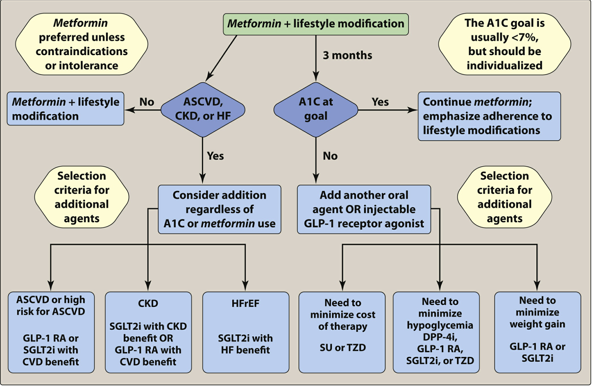

Decision Matrix: Which Drug to Add?
Scenario 1: Patient has ASCVD or high ASCVD risk
Established ASCVD (prior MI, stroke, symptomatic PAD):
- GLP-1 RA (liraglutide, semaglutide, dulaglutide) - proven MACE reduction (LEADER, SUSTAIN-6, REWIND trials)
- SGLT2 inhibitor (empagliflozin, dapagliflozin, canagliflozin) - also reduce MACE (EMPA-REG, CANVAS, DECLARE trials)
- Either class is appropriate; many guidelines prefer GLP-1 RA for ASCVD and SGLT2i for HF/CKD
- If HbA1c still above target after one of these, consider adding the other (both together)
Key: Glucose lowering alone does NOT reduce CV events in T2DM. Only these specific classes do.
Scenario 2: Patient has Heart Failure (especially HFrEF)
- SGLT2 inhibitor is the preferred second agent - empagliflozin and dapagliflozin have dedicated HF trials (EMPEROR-Reduced, DAPA-HF)
- SGLT2i reduce HF hospitalisations via natriuresis, preload/afterload reduction, and improved myocardial energy metabolism
- If SGLT2i contraindicated or not tolerated → GLP-1 RA (though HF benefit less robust)
- Avoid TZDs (pioglitazone) - cause fluid retention, worsen HF
Scenario 3: Patient has CKD (diabetic nephropathy)
- SGLT2 inhibitor is the preferred second agent (reduce albuminuria, slow eGFR decline, prevent ESRD)
- Empagliflozin (EMPA-KIDNEY) and dapagliflozin (DAPA-CKD) - can initiate if eGFR ≥20 mL/min for renal protection even if glucose lowering effect is attenuated below eGFR 45
- GLP-1 RA (semaglutide) also shows renal benefit - second choice or add-on
- Linagliptin (DPP-4i) - no renal dose adjustment needed; safe alternative if above not tolerated
- Avoid sulfonylureas (hypoglycaemia risk increased with reduced renal clearance) - if needed, prefer glipizide
Scenario 4: No ASCVD, HF, or CKD - choose by patient characteristic
| Patient Characteristic | Preferred Add-on | Avoid |
|---|---|---|
| Needs to minimise hypoglycaemia | DPP-4i, GLP-1 RA, SGLT2i, TZD | Sulfonylurea, Insulin |
| Obese, needs weight loss | GLP-1 RA (most effective - semaglutide>liraglutide>dulaglutide), SGLT2i | SU (weight gain), TZD (weight gain) |
| HbA1c very high (>9-10%) or symptomatic | Insulin (basal) ± GLP-1 RA | Any oral agent alone (insufficient potency) |
| Cost constraint / resource-limited setting | Sulfonylurea (glipizide/glimepiride) or TZD (pioglitazone) | GLP-1 RA, SGLT2i (expensive) |
| Most effective glucose lowering needed | GLP-1 RA, Basal insulin, TZD, SU | DPP-4i, SGLT2i (modest HbA1c effect) |
| Elderly (≥65, fall risk, polypharmacy) | DPP-4i (sitagliptin, linagliptin), GLP-1 RA | Glibenclamide (long-acting SU), TZD (fracture risk) |
| PCOS / insulin resistance predominant | TZD (pioglitazone), GLP-1 RA | - |
| Postprandial hyperglycaemia dominant | Alpha-glucosidase inhibitor (acarbose), Meglitinide (repaglinide), DPP-4i | Basal insulin only |
| Fatty liver (NAFLD/NASH) | Pioglitazone (evidence for NASH improvement), GLP-1 RA (semaglutide) | - |
When to Add Insulin Directly to Metformin
Consider metformin + basal insulin (not replacing metformin) when:
- HbA1c >10% or severely symptomatic hyperglycaemia
- Beta-cell reserve is severely depleted (low fasting C-peptide)
- Other oral agents have failed
- Hospitalisation, acute illness, surgery (switch to insulin temporarily)
- Pregnancy (insulin preferred over most oral agents)
Basal insulin dose: Start 10 units at bedtime (or 0.1-0.2 units/kg), titrate by 2 units every 3 days until fasting BG 80-130 mg/dL
Combinations to AVOID
| Combination | Reason |
|---|---|
| DPP-4i + GLP-1 RA | Overlapping mechanism (both work via incretin pathway); no added benefit, increased toxicity |
| SGLT2i during hospitalisation | Risk of euglycaemic DKA, prolonged fasting, acute illness |
| TZD + Insulin together | Fluid retention synergy, increased HF risk |
| TZD in HF (NYHA III/IV) | Worsens fluid overload |
| Glibenclamide in elderly/CKD | Prolonged hypoglycaemia |
| Sulfonylurea + high alcohol intake | Flushing reaction, severe hypoglycaemia |
Practical Monitoring After Adding a Second Drug
- Recheck HbA1c at 3 months after any drug addition
- If target still not met → add a third agent (triple therapy) before considering insulin
- If HbA1c >10% on triple therapy → add or switch to insulin
- Check eGFR before adding SGLT2i; monitor for UTI/genital infections
- Monitor weight, BP when adding GLP-1 RA (both improve)
- Monitor LFTs with TZD; check for oedema
- Reinforce lifestyle at every visit - this is non-negotiable
Quick Decision Summary
Patient on Metformin → 3-month review

├── Has ASCVD? → Add GLP-1 RA or SGLT2i (regardless of HbA1c)
├── Has Heart Failure? → Add SGLT2i (regardless of HbA1c)
├── Has CKD/nephropathy? → Add SGLT2i (regardless of HbA1c)
│
└── No above comorbidities:
    ├── HbA1c at goal? → Continue + reinforce lifestyle
    └── HbA1c not at goal?
        ├── Obese → GLP-1 RA or SGLT2i
        ├── Hypoglycaemia risk → DPP-4i or GLP-1 RA or SGLT2i
        ├── Cost constraint → SU or TZD
        ├── Very high HbA1c (>10%) → Basal insulin or GLP-1 RA
        └── Postprandial hyperglycaemia → Acarbose or meglitinide
Recent evidence note: A 2026 meta-analysis (PMID 42165377) in Diabetic Medicine evaluated dual vs single oral therapy at T2DM diagnosis, supporting early combination in patients with higher baseline HbA1c to achieve targets faster and maintain them longer.
Sources: Goldman-Cecil Medicine (Chapter 210), Lippincott Illustrated Reviews Pharmacology (Chapter 24), Goodman & Gilman's Pharmacological Basis of Therapeutics (Chapter 51), Comprehensive Clinical Nephrology 7e, National Kidney Foundation Primer on Kidney Diseases 8e
This is a shared conversation. Sign in to Orris to start your own chat.