
Ctev
Reading File
CTEV (Congenital Talipes Equinovarus) is clubfoot - a common congenital deformity characterized by plantar flexion (equinus), inversion (varus), and adduction of the foot. Let me search the medical library for detailed coverage.
Searching sources...
Finding Sources
Finding Sources
Searching PubMed
"clubfoot" AND "Ponseti" AND treatment
Good - found two strong textbook sources. Let me read both in parallel.
Reading File
Reading File
Let me read more of the Miller's orthopaedics section for the complete treatment and complications content.
Reading File
I have comprehensive content. Let me now fetch the radiographic diagram image for visual clarity.
Loading Image
Excellent - I now have all the information needed for a comprehensive answer.
CTEV - Congenital Talipes Equinovarus (Clubfoot)
Definition
CTEV is a congenital deformity of the foot characterized by a combination of four components, remembered by the mnemonic CAVE:
| Component | Description |
|---|---|
| C - Cavus | High arch (plantar flexion of forefoot on hindfoot) |
| A - Adductus | Adduction of the forefoot |
| V - Varus | Varus of the hindfoot |
| E - Equinus | Plantar flexion at the ankle |
Epidemiology
- Boys affected twice as often as girls
- 50% of cases are bilateral
- Incidence: ~1-2 per 1,000 live births
Etiology
- Majority idiopathic - genetic cause strongly suggested
- Key genetic pathway: PITX1-TBX4 transcriptional pathway
- Associated conditions:
- Arthrogryposis
- Myelomeningocele
- Diastrophic dwarfism
- Prune-belly syndrome
- Tibial hemimelia
- Streeter dysplasia (hand anomalies)
- Other neuromuscular/syndromic conditions
Pathoanatomy
- Talar neck deformity - medial and plantar deviation of the talar neck
- Medial rotation of calcaneus
- Medial displacement of navicular and cuboid
- Shortening/contracture of:
- Intrinsic foot muscles
- Achilles tendon
- Tibialis posterior
- Flexor hallucis longus (FHL)
- Flexor digitorum longus (FDL)
- Joint capsules, ligaments, and fascia
- Associated with absence or diminutive anterior tibial artery
Radiographic Findings
Radiographs are of limited use in infants due to minimal ossification. When used:
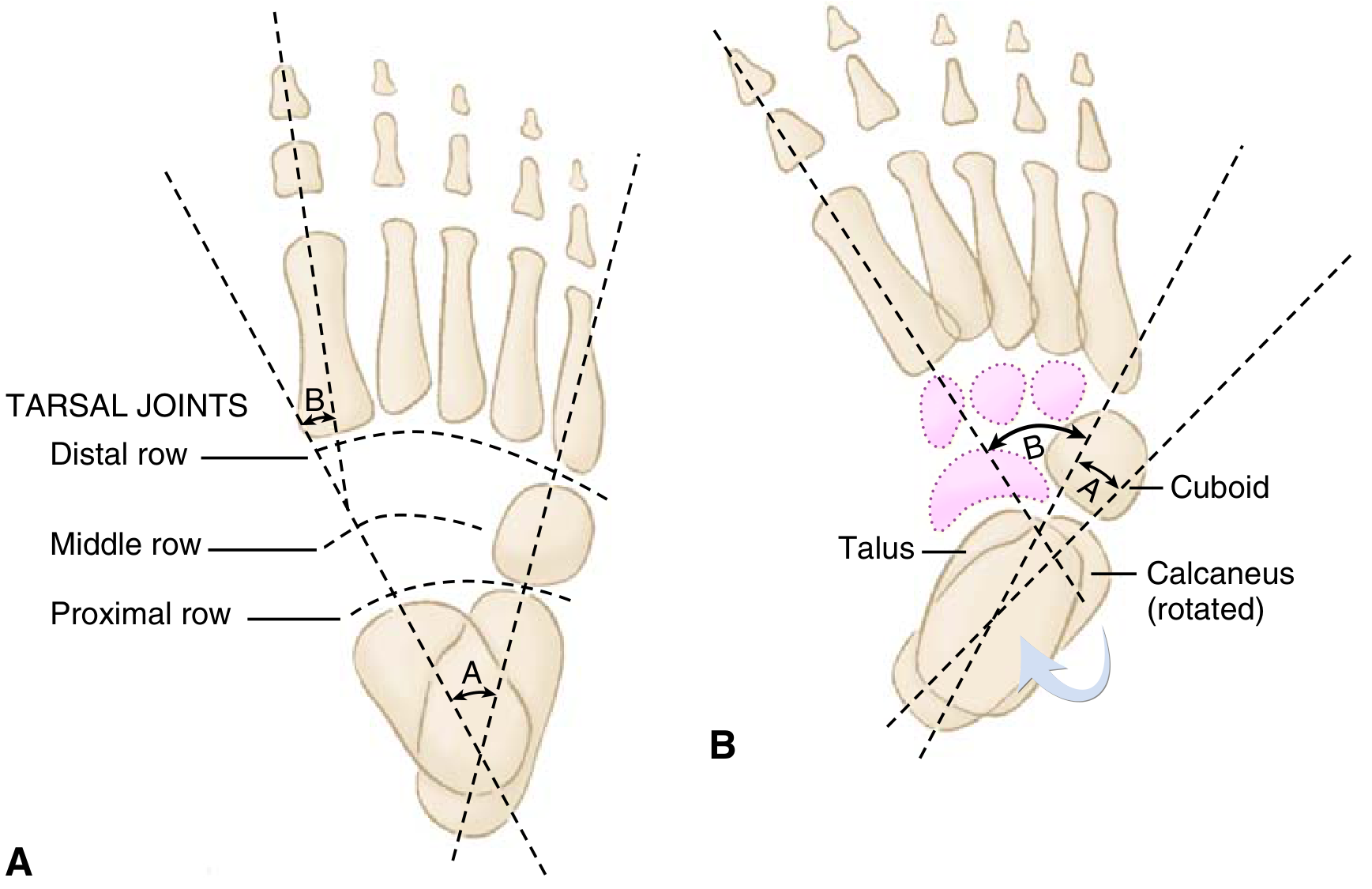
Fig. 3.20 - (A) Normal foot. (B) Clubfoot showing "parallelism" of talus and calcaneus.
| Measurement | Normal | Clubfoot |
|---|---|---|
| Talocalcaneal angle (AP/Kite angle) | 20-40° | < 20° (parallelism) |
| Talocalcaneal angle (lateral/Turco - dorsiflexion view) | ~35° | < 35° |
| Talus-1st metatarsal angle (AP) | 0-20° | Negative |
The key X-ray finding is parallelism of the talus and calcaneus (normally they diverge).
Treatment
1. Ponseti Method (Gold Standard - First Line)
Serial weekly manipulation and casting using long-leg plaster casts.
Order of correction follows CAVE (same mnemonic, but correction sequence):
- Cavus - corrected first by supinating the forefoot and dorsiflexing the first ray
- Adductus - corrected using lateral pressure on distal talar head as a fulcrum
- Varus - corrected simultaneously with adductus
- Equinus - addressed last (after varus is fully corrected to avoid rocker-bottom deformity)
Key steps:
- ~90% of patients require percutaneous Achilles tendon lengthening (tenotomy) to address hindfoot equinus
- Last cast placed in 70° of abduction
- Treatment typically complete in 1-5 months
2. Foot Abduction Brace (FAB) - Mandatory Post-Casting
- Full-time wear for 3 months post-casting
- Then during naps and nighttime for 3 years
- Brace non-compliance is the #1 cause of recurrence
- No impairment in athletic ability after successful treatment
Complications of Treatment
| Complication | Cause |
|---|---|
| Recurrence / undercorrection | Most common; strongly associated with brace non-compliance |
| Rocker-bottom deformity | Attempting to dorsiflex hindfoot before varus is corrected |
| Flat-top talus | Aggressive dorsiflexion causing flattening of the talar dome |
| Dorsal bunion | After clubfoot surgery; tibialis anterior + FHB/abductor hallucis |
Operative Treatment
Reserved for only ~5% of idiopathic clubfeet (resistant/refractory cases):
-
Posteromedial soft tissue release (PMSTR): tendon lengthening + subtalar, tibiotalar, and talonavicular release
- Must protect posterior tibial artery (dorsalis pedis often insufficient)
- Used in severe disease or when treatment initiated after 9 months of age
-
Older children (3-10 years): medial opening-wedge or lateral column-shortening osteotomy, or cuboidal decancellation for persistent adductus
-
Late presentation: Triple arthrodesis
- Contraindicated in insensate feet (risk of ulceration from rigidity)
Recent Evidence (2024-2026)
Recent systematic reviews confirm the Ponseti method as the standard of care:
- A 2024 systematic review (PMID 39365806) found Ponseti method is also effective for myelodysplastic (non-idiopathic) clubfoot.
- A 2026 meta-analysis (PMID 40888800) compared Plaster of Paris vs. semirigid fiberglass casting - POP remains the reference standard.
- A 2024 systematic review (PMID 39134994) examined anterior tibial tendon transfer fixation techniques for dynamic supination relapse.
Sources: Miller's Review of Orthopaedics, 9th Ed., p. 282; Schwartz's Principles of Surgery, 11th Ed., p. 1946
This is a shared conversation. Sign in to Orris to start your own chat.