
Visual learning for probable signs of pregnancy
probable signs of pregnancy Hegar sign Chadwick sign Goodell sign
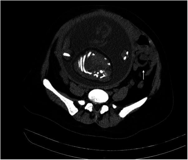
This axial computed tomography (CT) scan of the abdomen and pelvis demonstrates a classic 'whirl sign' or 'vortex sign' in the left lower quadrant, indicated by a white arrow. This sign is characterized by the swirling appearance of the small intestinal loops, mesenteric fat, and vasculature, which is highly suggestive of midgut volvulus or small bowel torsion. Adjacent to the site of torsion, the intestinal lumen shows signs of localized dilation. The scan also reveals a large, gravid uterus occupying the central and anterior abdominal cavity, containing a fetus with visible hyperdense skeletal elements. This clinical scenario represents a rare but critical surgical emergency: small bowel volvulus occurring during pregnancy. The surrounding anatomical structures include the posterior vertebral column and the pelvic girdle. This image is an essential educational resource for understanding the radiological manifestations of acute mechanical bowel obstruction in the setting of advanced gestation.
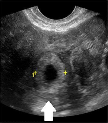
This diagnostic image is a transvaginal ultrasound (TVUS) in the sagittal plane, demonstrating a tubal ectopic pregnancy. The primary finding is the 'bagel sign' or 'tubal ring sign,' which consists of an extrauterine gestational sac—a small, dark, anechoic fluid collection—surrounded by a thick, hyperechoic ring of trophoblastic tissue. This structure is highlighted by a white arrow and yellow calibers (plus signs). The surrounding adnexal tissue and stroma appear heterogeneous in echogenicity. Clinically, this visual finding is highly suggestive of an ectopic gestation located within the Fallopian tube. It is a critical diagnostic marker for medical professionals in evaluating patients presenting with early pregnancy bleeding or abdominal pain when no intrauterine pregnancy (IUP) is identified.
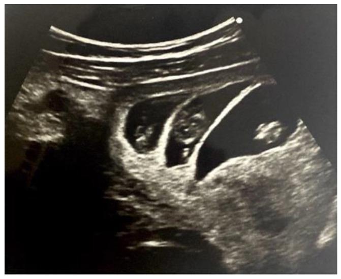
This diagnostic ultrasound image demonstrates a first-trimester trichorionic triamniotic (TCTA) triplet pregnancy at approximately 9 weeks of gestation. The transabdominal scan reveals three distinct gestational sacs within the uterus, each containing a visible embryo. Key diagnostic features include the presence of two 'lambda signs' (also known as the 'twin peak' sign), which are triangular, echogenic projections of chorionic tissue extending into the base of the intertwin membranes. The intertwin membranes themselves are notably thick and multi-layered, consisting of a central layer of chorion sandwiched between two amniotic layers, indicating high chorionicity. These findings are critical for establishing chorionicity and amnionicity in multiple gestations, which is essential for risk stratification and clinical management. The presence of three separate sacs and thick separating membranes confirms that each fetus has its own placenta and amniotic sac, distinguishing this from monochorionic triplet pregnancies.

This clinical photograph shows a close-up view of a newborn infant's abdomen, specifically highlighting the periumbilical region. The image demonstrates Cullen's sign, characterized by periumbilical ecchymosis appearing as a diffuse, bluish-purple discoloration on the pinkish-red abdominal skin. The surrounding skin exhibits a slightly rough texture with areas of fine desquamation or peeling. The umbilical cord is visible at the lower portion of the frame, showing a healthy, moist, and gelatinous (Wharton's jelly) appearance with a twisted, braided structure and characteristic white, pale blue, and yellowish-green hues. The umbilical stump itself is erythematous and slightly edematous. In a clinical context, Cullen's sign in a neonate or adult is a significant diagnostic finding indicating retroperitoneal hemorrhage, often associated with conditions such as acute pancreatitis, ruptured ectopic pregnancy, or, in newborns, potential abdominal trauma or visceral hemorrhage. This image serves as a high-fidelity educational resource for identifying classic dermatologic signs of internal bleeding.
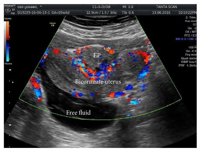
This color Doppler ultrasound image illustrates a complex gynecological presentation involving a bicornuate uterus and an ectopic pregnancy (EP). The transabdominal scan reveals the distinct morphology of the bicornuate uterus, characterized by two separate uterine horns. Positioned centrally between these horns is a gestational sac labeled as 'EP'. A classic 'Ring of Fire' sign is visible—a dense circumferential pattern of red and blue color Doppler signals surrounding the ectopic sac, which indicates hypervascularity and active trophoblastic peripheral blood flow. In the dependent portion of the pelvic cavity, a significant anechoic (dark) area is labeled as 'Free fluid', suggesting hemoperitoneum or inflammatory exudate in the context of a ruptured or leaking ectopic pregnancy. This diagnostic image is highly clinically significant for demonstrating uterine anomalies, the ultrasound signs of extrauterine gestation, and associated complications like pelvic fluid accumulation, which are critical concepts in emergency obstetrics and radiology.
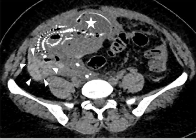
This axial abdomino-pelvic computed tomography (CT) scan illustrates a rare case of advanced extrauterine abdominal pregnancy. The imaging shows a fetus located within the abdominal cavity, separate from the uterus. A white star marks the fetal skull, which exhibits signs of fetal demise such as Spalding's sign (overlapping of cranial bones). The ossified fetal spine is visible as a series of hyperdense vertebral bodies curving through the mid-abdomen. White arrowheads delineate the placenta, which is abnormally implanted onto abdominal structures rather than the uterine wall. There is evidence of significant hemoperitoneum, visualized as high-attenuation free fluid surrounding the abdominal viscera and collecting in the paracolic gutters. This diagnostic image serves as an educational example of the radiological presentation of a secondary abdominal pregnancy, highlighting the displacement of bowel loops and the absence of surrounding myometrium around the gestational sac.
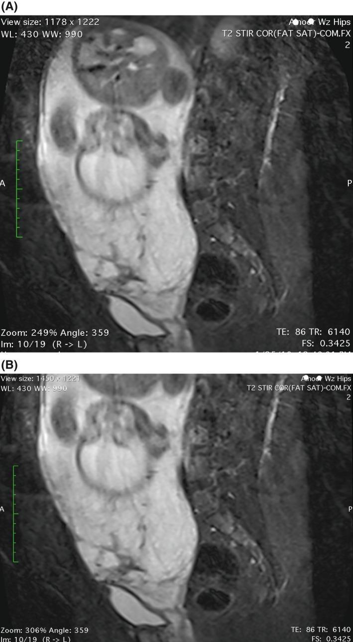
This clinical imaging composite presents two sagittal T2-weighted magnetic resonance imaging (MRI) scans of a pregnant pelvis without contrast, demonstrating placenta previa and signs of invasive placentation. Image (A) highlights significant uterine bulging and the 'tenting sign' of the bladder, where the posterior bladder wall is pulled superiorly and posteriorly toward the lower uterine segment, suggesting abnormal placental adherence or invasion. Image (B) focuses on the lower uterine segment, showing the placenta positioned anteriorly and completely covering the internal cervical os (placenta previa totalis). Key features include the loss of the normal retroplacental T2-hypointense zone and thinning of the myometrium, which are indicative of the placenta accreta spectrum (PAS). These images are pedagogically significant for teaching radiological signs of abnormal placentation, identifying the relationship between placental tissue and the bladder-uterine serosa interface, and evaluating secondary signs of invasive pregnancy complications in obstetric imaging.
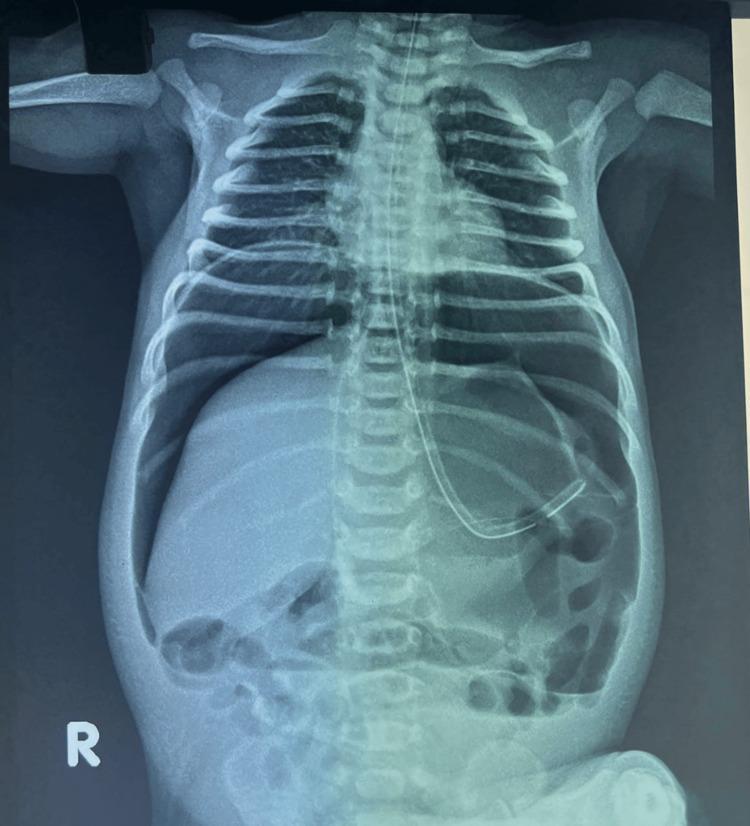
This diagnostic image is an erect abdominal X-ray of a neonate demonstrating massive pneumoperitoneum. Key radiological signs include the 'continuous diaphragm sign,' characterized by free gas outlining the entire lower surface of the diaphragm, and the 'football sign,' seen as a large, oval-shaped radiolucency within the abdominal cavity resembling an American football. Additionally, the 'falciform ligament sign' is visible as a vertical linear density in the upper right quadrant, highlighted by gas on both sides. A radiopaque orogastric tube is positioned with its distal tip in the stomach. The thoracic cavity shows clear lung fields and normal bony structures. These findings are highly suggestive of a hollow viscus perforation, such as neonatal gastric perforation. The image serves as a classic educational example of extra-luminal free air in a pediatric patient, emphasizing the importance of recognizing specific signs of intestinal or gastric emergency on plain radiography.
Hegar sign softening of uterine isthmus pregnancy examination
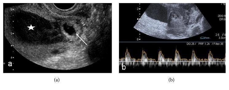
This composite diagnostic image showcases a transvaginal ultrasound (TVUS) examination of a Cesarean Scar Ectopic Pregnancy (CSEP). Panel (a) presents a sagittal grayscale ultrasound view of the uterus. A gestational sac (marked by an arrow) is seen implanted low in the uterine isthmus, specifically at the site of a previous cesarean section scar, distinct from the main endometrial cavity. The endometrial cavity itself is distended with heterogeneous, low-level echoes (marked with a star), consistent with hemorrhagic fluid. Panel (b) displays a spectral Doppler ultrasound. The top portion is a B-mode image showing the fetal pole within the eccentrically located gestational sac. The bottom portion displays a pulsatile spectral Doppler waveform, confirming viable fetal heart activity. This visual sequence is pedagogically significant for illustrating the diagnostic criteria for CSEP, emphasizing the importance of anatomical localization of the gestational sac relative to the cervical canal, endometrial cavity, and previous surgical scars.
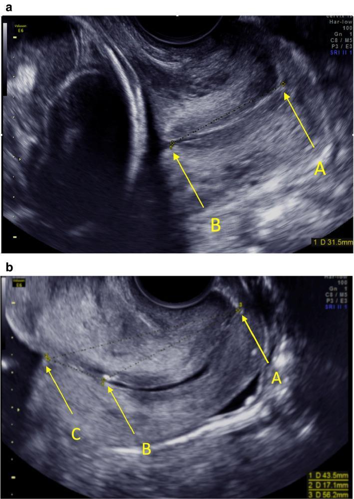
This diagnostic visual consists of two transvaginal ultrasound images (a and b) in the sagittal plane, illustrating methods for measuring cervical length during pregnancy. Image (a) demonstrates a standard measurement when the uterine isthmus is absent; yellow calipers and arrows mark 'A' (external os) and 'B' (internal os), with a linear dotted line representing the endocervical length (31.5 mm). Image (b) displays a scenario where the isthmus is present, introducing a third landmark 'C' designated as the 'virtual inner os.' In this view, distance A-B (43.5 mm) represents the endocervical canal length, B-C (17.1 mm) denotes the isthmus length, and A-C (56.2 mm) represents the total length from the external os to the innermost end of the isthmus. The images highlight key anatomical landmarks used in obstetrics to assess the lower uterine segment and predict preterm birth risk. The hypoechoic endocervical canal and the varying morphology of the internal os region are clearly visualized against the more echogenic cervical stroma.
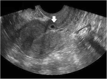
**Imaging Modality:** Transvaginal Ultrasound (TVUS), B-mode. **Anatomical Region:** Sagittal view of the uterus, specifically the lower uterine segment and the anterior uterine wall at the level of the isthmus. **Observed Pathology:** Cesarean scar ectopic pregnancy (CSEP). **Characteristic Visual Features:** * **Gestational Sac (GS):** An anechoic, rounded structure (indicated by a white arrow) is visible embedded within the anterior myometrium of the lower uterine segment. * **Localization:** The sac is positioned at the site of a previous cesarean section scar, anterior to the cervical-isthmic junction. * **Uterine Cavity:** The endometrial canal and the superior uterine cavity are empty, showing no evidence of intra-cavitary pregnancy. * **Myometrial Thinning:** There is a visible reduction in the thickness of the myometrium between the gestational sac and the bladder interface (serosa). **Diagnostic Differentiating Features:** The primary diagnostic cue is the "empty uterus sign" combined with the low implantation of the gestational sac within the anterior wall defect, distinctly separate from the endometrial canal. This distinguishes the finding from a cervical ectopic pregnancy or a low intrauterine pregnancy.
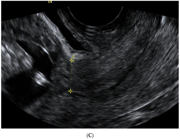
A transvaginal sagittal ultrasound image of the lower uterine segment in a first-trimester pregnancy. The myometrium exhibits a relatively homogenous, mid-gray echogenicity. Centered in the field is a caliper measurement (indicated by two yellow plus signs and a dashed line) used to assess the thickness of the isthmus uteri at the level of the internal cervical os. This image demonstrates a 'non-visible' Cesarean section (CS) scar, characterized by the absence of a distinct hypoechoic niche, indentation, or tissue defect at the site of the previous surgical incision. The anatomical relationship between the anterior uterine wall and the neighboring bladder (visible as a darker, hypoechoic space superiorly) is depicted. This diagnostic imaging serves as a clinical reference for evaluating uterine wall integrity and scar healing in patients with a history of prior Cesarean sections.
Chadwick sign bluish discoloration cervix vagina pregnancy

This clinical photograph displays a medium-to-dark-toned adult abdomen, highlighting a distinct pathological sign known as Cullen's sign. The primary finding is a periumbilical ecchymosis, characterized by an irregular, bluish-purple area of subcutaneous hemorrhage surrounding the umbilicus. The discoloration is most prominent on the right side of the umbilical ring, presenting as a mottled, bruised patch. Anatomically, the image covers the umbilical, epigastric, and portions of the lateral abdominal regions. In addition to the ecchymosis, a faint, linear surgical scar is visible in the upper left quadrant, along with scattered benign-appearing nevi. Clinically, Cullen's sign is an important diagnostic indicator of retroperitoneal hemorrhage, often associated with severe acute pancreatitis, ruptured ectopic pregnancy, or aortic aneurysm. This image serves as a teaching tool for medical students and clinicians to recognize superficial manifestations of internal abdominal pathology during a physical examination.
This clinical photograph shows a close-up view of a newborn infant's abdomen, specifically highlighting the periumbilical region. The image demonstrates Cullen's sign, characterized by periumbilical ecchymosis appearing as a diffuse, bluish-purple discoloration on the pinkish-red abdominal skin. The surrounding skin exhibits a slightly rough texture with areas of fine desquamation or peeling. The umbilical cord is visible at the lower portion of the frame, showing a healthy, moist, and gelatinous (Wharton's jelly) appearance with a twisted, braided structure and characteristic white, pale blue, and yellowish-green hues. The umbilical stump itself is erythematous and slightly edematous. In a clinical context, Cullen's sign in a neonate or adult is a significant diagnostic finding indicating retroperitoneal hemorrhage, often associated with conditions such as acute pancreatitis, ruptured ectopic pregnancy, or, in newborns, potential abdominal trauma or visceral hemorrhage. This image serves as a high-fidelity educational resource for identifying classic dermatologic signs of internal bleeding.
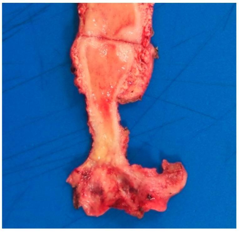
This clinical photograph displays a gross surgical specimen of the uterus, cervix, and upper vagina following a total hysterectomy. The cervix exhibits a prominent, pathological yellowish lesion that is indurated and demonstrates poorly defined, infiltrative margins. This discoloration significantly contrasts with the surrounding healthy pinkish-red cervical tissue. The lesion involves the entirety of the uterine cervix and extends focally into the upper vaginal mucosa. The surface texture of the affected area appears irregular and distorted, suggesting a chronic inflammatory or neoplastic process. Clinically, such presentations can mimic cervical carcinoma, although this specific specimen relates to malakoplakia, a rare chronic granulomatous condition. The image is an essential educational tool for understanding gross pathological manifestations in the female reproductive tract, emphasizing the importance of histological confirmation when distinguishing between infectious/inflammatory masses and malignancy.
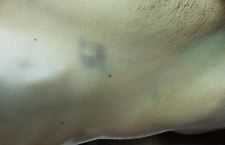
This clinical photograph shows a close-up view of the right flank of a patient, exhibiting distinctive skin discoloration. The primary finding is a livid, bluish-purple ecchymosis with a central yellowish hue, localized near the costal arch. This morphological pattern is characteristic of Grey Turner sign (and its variations like Fox sign), which represents subcutaneous retroperitoneal hemorrhage. The discoloration is irregular in shape, with a notable ring-shaped intensity in the central-left portion of the image. The surrounding skin appears pale, suggesting systemic anemia. Several small, benign-appearing melanocytic nevi are scattered across the torso. In an educational context, this visual finding is a clinical marker of severe underlying pathology, most commonly associated with acute necrotizing pancreatitis, where pancreatic enzymes cause retroperitoneal bleeding that track through fascial planes to the skin. The image serves as an important diagnostic sign for medical students and clinicians to recognize potential abdominal emergencies and internal hemorrhaging.
uterine enlargement abdominal distension early pregnancy signs
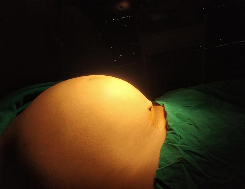
Clinical photograph of a patient in a supine position showing massive abdominal distension. The abdomen is symmetrically enlarged, presenting a rounded, protruding contour clinically comparable to a 32-week gravid uterus. The overlying skin is smooth, taut, and under tension, with a visible umbilicus. A green surgical drape is positioned over the lower abdomen, indicating a preoperative or intraoperative setting. This image illustrates severe abdominal enlargement caused by a large uterine leiomyoma (fibroid), demonstrating the clinical presentation of significant pelvic-abdominal masses that require surgical intervention such as laparotomy and hysterectomy. The photograph serves as an educational example of the physical examination findings associated with giant uterine fibroids and the resulting mass effect on the abdominal cavity.
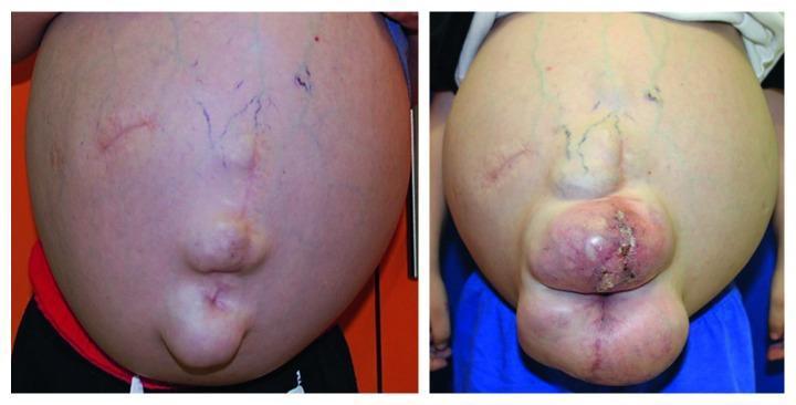
This comparison clinical photograph documents the progressive abdominal enlargement in a patient over a 17-month period. The initial visit (left) shows severe abdominal distension, consistent with massive ascites, featuring visible superficial reticular veins (caput medusae-like appearance), surgical scars, and a midline umbilical protrusion. The follow-up image (right) demonstrates a significant worsening of the condition. The abdomen is more globular and tense, with more prominent, tortuous green-blue superficial vasculature. Notably, the umbilical region now exhibits a large, multi-lobulated protrusion, likely representing a massive umbilical or ventral hernia. This mass shows signs of skin tension, erythema, and localized ulceration with crusting, indicating potential skin breakdown or incarceration. These findings are clinically relevant for chronic liver disease with portal hypertension or complex congenital anomalies, illustrating the physical manifestations of progressive intra-abdominal pressure and the associated risk of herniation and integumentary compromise.
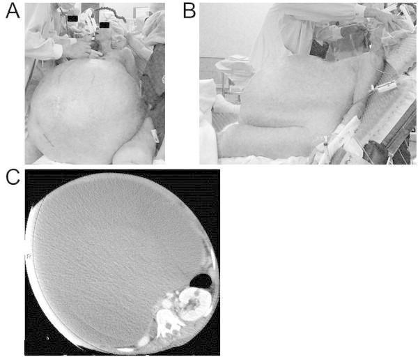
This composite image consists of clinical photographs and a diagnostic scan illustrating a massive abdominal distension. Panels A and B are preoperative clinical photographs in frontal and lateral views, respectively, showing a female patient with extreme abdominal enlargement. Visible clinical signs include taut, shiny skin and prominent, dilated subcutaneous veins (caput medusae) across the abdominal wall. The lateral view demonstrates the significant anterior protrusion of the abdomen, necessitating a semirecumbent position. Panel C is an axial abdominal computed tomography (CT) scan that confirms the etiology as a giant cystic mass occupying nearly the entire abdominal cavity. The mass is homogeneous and well-defined, severely compressing and displacing intra-abdominal viscera, including the bowel and kidneys, against the spine and posterior abdominal wall. This case demonstrates the clinical and radiological presentation of a giant ovarian tumor, highlighting its impact on abdominal anatomy and the potential for respiratory compromise due to diaphragmatic elevation.
linea nigra striae gravidarum skin changes pregnancy
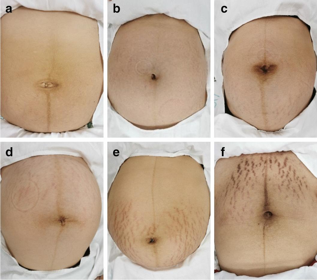
This set of six clinical photographs (a-f) displays the dermatological presentation and classification of striae gravidarum (pregnancy stretch marks) on the human abdomen. The images demonstrate a progression of severity and are organized into three clinical categories. Panels (a) and (b) represent 'mild' cases, characterized by a near-complete absence or sparse distribution of faint, thin striae that closely match the surrounding skin tone. Panels (c) and (d) represent 'moderate' cases, showing an increased density and wider distribution of visible marks across the periumbilical and lower abdominal regions. Panels (e) and (f) depict 'severe' manifestations, where the striae appear markedly more pronounced, wider, and darker (erythematous or violaceous) in color. In these severe cases, the lesions exhibit a high-density distribution pattern, extending across a larger surface area of the abdomen. A distinct linea nigra is also visible vertically across the midline in several images. This visual series is used in obstetrics and dermatology to illustrate the morphological diversity and clinical grading of striae distensae during or after pregnancy.
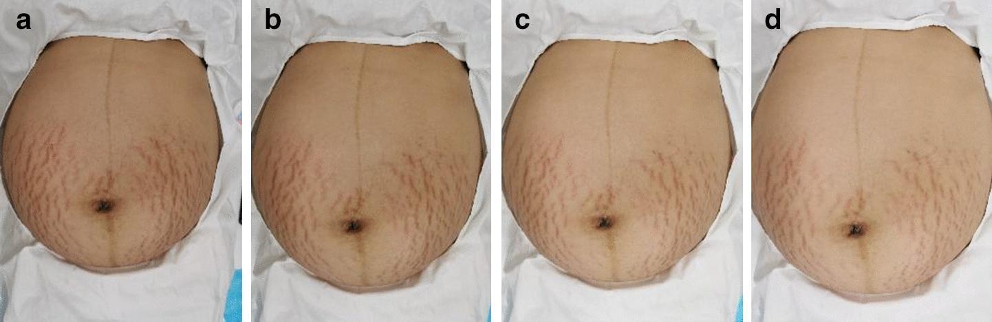
This clinical photographic series consists of four sequential images (a-d) showing the progression of striae gravidarum (stretch marks) on a female abdomen during a therapeutic intervention. Image 'a' demonstrates the baseline state with numerous erythematous (reddish-purple) striae rubra, primarily concentrated in the lower quadrants and periumbilical region. The lesions are characterized by high density, moderate width, and distinct longitudinal morphology against a slightly inflamed skin tone. A visible linea nigra is also present along the midline. Panel 'b' (week 2) and 'c' (week 4) show a gradual reduction in erythema, with the lesions becoming lighter pink and less visually prominent. By panel 'd' (week 8), the striae exhibit significant clinical improvement, characterized by a transition to a paler, more subtle appearance with reduced lesion width and overall density. The surrounding skin texture appears smoother, reflecting a decrease in local inflammation and a shift toward the appearance of striae alba.
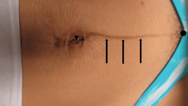
Clinical photograph of a female patient's abdomen demonstrating linea nigra, a common form of physiological hyperpigmentation. The image shows a vertically oriented, tan-to-brown linear streak extending along the midline. Black markers highlight key anatomical landmarks: a triangle at the umbilicus, arrows pointing to the hyperpigmented line, and a circle indicating the termination point near the pubic symphysis. The pigmentation is also visible within and slightly superior to the umbilical region. Secondary skin findings include subtle, light-colored striae distensae (stretch marks) radiating from the umbilicus and periumbilical area. The clinical presentation is characteristic of pregnancy-induced cutaneous changes, specifically the darkening of the linea alba due to increased melanocyte-stimulating hormone. This photograph serves as an educational resource for identifying benign pregnancy-related dermatological conditions and their characteristic anatomical distribution.
ballottement sign fetus pregnancy uterus examination fourth month
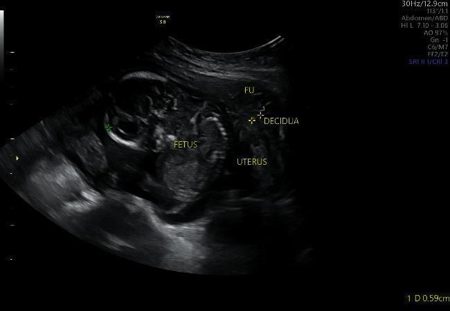
A diagnostic ultrasound image of an abdominal pregnancy, demonstrating a fetus located outside of the uterine cavity. The image captures the spatial relationship between the extrauterine fetus, the fundus (labeled 'FU'), and the uterine wall. The 'FETUS' label is placed over a region showing mixed echogenicity, where fetal structures and possibly limb buds are visible. Adjacent to the gestational sac, the 'UTERUS' is identified separately, showing a thickened endometrial lining or 'DECIDUA' (labeled with calipers). This sonographic finding is characteristic of an ectopic abdominal pregnancy, where the gestational sac is typically situated in the pouch of Douglas or attached to peritoneal surfaces rather than within the endometrial cavity. The surrounding tissues exhibit varying hypoechoic and hyperechoic patterns. The educational value of this image lies in its clear demonstration of the 'empty uterus sign' in the presence of an advanced extrauterine pregnancy, which is critical for obstetric triage and diagnosis of rare ectopic implantations.
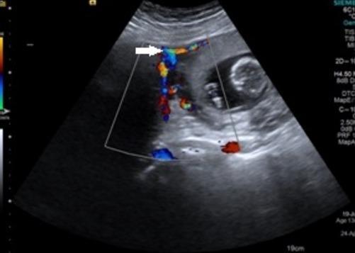
This diagnostic image is a transabdominal color Doppler ultrasound in a sagittal plane, depicting a gravid uterus at approximately 13-14 weeks of gestation. Key anatomical features include a visible fetus within the intrauterine cavity and a large, well-defined, relatively hypoechoic mass adjacent to the uterine fundus. A critical diagnostic sign is the presence of a 'bridging vessel,' indicated by a white arrow. The color Doppler overlay demonstrates prominent vascular flow (red and blue signals) traveling through this bridging vessel from the uterine fundal wall to the mass. In obstetric imaging, the bridging vessel sign is a classic sonographic marker used to differentiate a pedunculated subserosal uterine leiomyoma (fibroid) from an adnexal or ovarian mass. The presence of this vessel confirms the uterine origin of the lesion by visualizing the vascular supply derived from the uterine arteries. This clinical visualization is essential for surgical planning and distinguishing between benign uterine myomas and potential ovarian malignancies during pregnancy.
This axial abdomino-pelvic computed tomography (CT) scan illustrates a rare case of advanced extrauterine abdominal pregnancy. The imaging shows a fetus located within the abdominal cavity, separate from the uterus. A white star marks the fetal skull, which exhibits signs of fetal demise such as Spalding's sign (overlapping of cranial bones). The ossified fetal spine is visible as a series of hyperdense vertebral bodies curving through the mid-abdomen. White arrowheads delineate the placenta, which is abnormally implanted onto abdominal structures rather than the uterine wall. There is evidence of significant hemoperitoneum, visualized as high-attenuation free fluid surrounding the abdominal viscera and collecting in the paracolic gutters. This diagnostic image serves as an educational example of the radiological presentation of a secondary abdominal pregnancy, highlighting the displacement of bowel loops and the absence of surrounding myometrium around the gestational sac.
Braxton Hicks contractions uterine palpation pregnancy intermittent
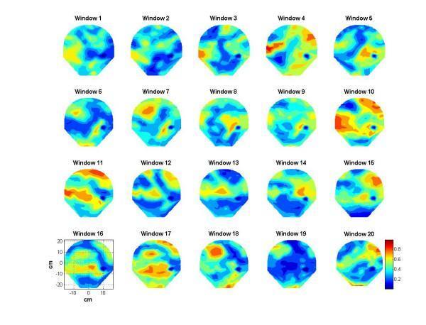
This figure presents a series of twenty sequential contour plots mapping phase synchronization indices derived from transabdominal magnetomyography (MMG) data. Each plot, labeled 'Window 1' through 'Window 20', represents a 20-second stepping interval of uterine activity in a pregnant subject. The spatial maps utilize a color scale ranging from blue (0.0, low synchronization/quiescence) to red (1.0, high synchronization/peak contraction). Centimeters (cm) on the axes in Window 16 indicate the spatial distribution over the maternal abdomen. The series illustrates the spatio-temporal evolution of uterine electrical activity, where windows 10, 11, and 17 show significant clusters of orange and red, indicating localized regions of high synchronization characteristic of uterine contractions. Other windows, such as 6 and 19, are dominated by blue and green shades, representing baseline relaxation phases. This visualization is used in reproductive assessment to study the emergence of coordinated electrical activity in the myometrium, which is critical for identifying labor progression and distinguishing between Braxton-Hicks and true labor contractions.
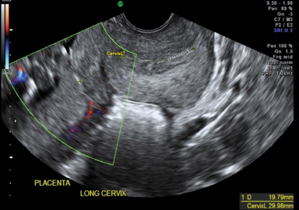
**Imaging Modality:** Transvaginal Ultrasound (TVUS) with Color Doppler. **Anatomical Region:** Lower uterine segment, cervix, and posterior placenta. **Visual Features:** - **Cervix:** The endocervical canal is clearly delineated and measured (CervixL 29.98 mm), appearing elongated and closed. - **Myometrium:** There is a focal, hypoechoic thickening of the lower uterine segment adjacent to the internal os, consistent with a localized myometrial contraction (Braxton Hicks). - **Placenta:** A posterior placenta is visible, positioned superior to the cervical os. - **Doppler Findings:** Limited color flow mapping is visible in the periphery, likely representing maternal vasculature. **Diagnostic Considerations:** The image illustrates a dynamic lower uterine segment contraction. Such contractions can transiently thicken the myometrium and shift the apparent position of the placental edge or internal os. This physiological finding is a known pitfall in obstetric imaging that can mimic the appearance of a short cervix or placenta previa. Distinguishing the homogeneous, transient nature of the contraction from the granular texture of the placenta is critical for accurate diagnosis.
pregnant uterus fundal height levels symphysis umbilicus xiphisternum diagram
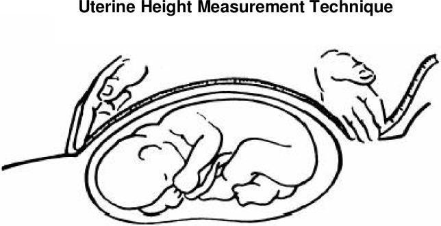
This medical illustration demonstrates the standardized technique for measuring symphysis-fundal height (SFH) during pregnancy. The diagram shows a sagittal-style cross-section of a gravid uterus containing a fetus in a cephalic presentation. A flexible, non-stretchable measuring tape is used to quantify the uterine height. One hand of the clinician is shown anchoring the 'zero' mark of the tape at the upper border of the symphysis pubis. The second hand is positioned at the uterine fundus, the highest point of the uterus, ensuring the tape follows the longitudinal curve of the abdominal wall. This clinical measurement is a primary screening tool in obstetrics used to monitor fetal growth, estimate gestational age, and screen for conditions such as intrauterine growth restriction (IUGR) or macrosomia. The visual emphasizes the proper placement of hands and the orientation of the measurement tool relative to maternal anatomical landmarks and the internal fetal position.
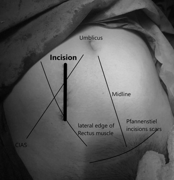
This monochrome clinical photograph and procedural diagram illustrates a novel surgical approach for adnexal pathologies during pregnancy. The image shows a pregnant abdomen with a thick vertical line marking a mediolateral (near vertical) skin incision located in the right lower quadrant. Anatomical landmarks are labeled for orientation: the umbilicus is superior and medial to the incision; the abdominal midline is shown to the left of the incision; and the lateral edge of the rectus muscle is indicated. The incision is positioned approximately 3 cm lateral to the midline, near the lateral third of the rectus muscle. Additional landmarks include the Crista Iliaca Anterior Superior (CIAS) located inferiorly and laterally, and pre-existing Pfannenstiel incision scars in the suprapubic region. This surgical illustration is designed for obstetricians and surgeons to demonstrate a targeted paramedian laparotomy entry point that provides access to the adnexa while accommodating the displaced pregnant uterus, minimizing surgical trauma and avoiding previous scar tissue.
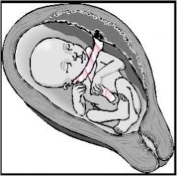
This medical illustration depicts an anatomical diagram of a fetus in utero demonstrating a nuchal cord with a 'free sliding pattern'. The fetus is shown in a flexed, cephalic presentation within the uterine cavity. The umbilical cord is visibly coiled around the fetal neck in at least two loose loops. These loops are characterized by a lack of constriction or indentation of the fetal soft tissues, representing a 'Type A' nuchal cord configuration where the cord can move freely and potentially undo itself spontaneously. The placenta is positioned at the fundal/posterior aspect of the uterus, with the cord extending toward the neck before reaching the umbilicus. This diagram serves as an educational tool in obstetrics to differentiate between non-constricting (Type A) and locked/constricting (Type B) nuchal cord patterns, highlighting the clinical relevance of cord entanglement and its impact on fetal well-being during gestation and labor.
Medical educational diagram showing the probable signs of pregnancy: 1) Hegar's sign - bimanual examination showing two fingers in posterior fornix and hand on abdomen meeting through soft isthmus at 6-8 weeks; 2) Goodell's sign - cervix softening like lips vs firm cartilage in non-pregnant state; 3) Chadwick's sign - bluish-violet discoloration of cervix and vaginal walls; 4) Ballottement - fetus floating in liquor amnii being tossed like a ball at 4-5 months; 5) Uterine fundal height levels at different months (symphysis to xiphisternum); 6) Braxton-Hicks contractions - intermittent painless uterine contractions; 7) Striae gravidarum and linea nigra on abdomen. Clean clinical illustration style with labels.
Probable Signs of Pregnancy - Visual Learning Guide
Classification of Signs of Pregnancy:
- Presumptive (subjective symptoms - e.g. amenorrhea, nausea, breast changes)
- Probable (objective signs on examination - suggestive but not confirmatory)
- Positive/Definitive (confirm pregnancy - fetal heart sounds, USG, fetal movements)
Overview Diagram
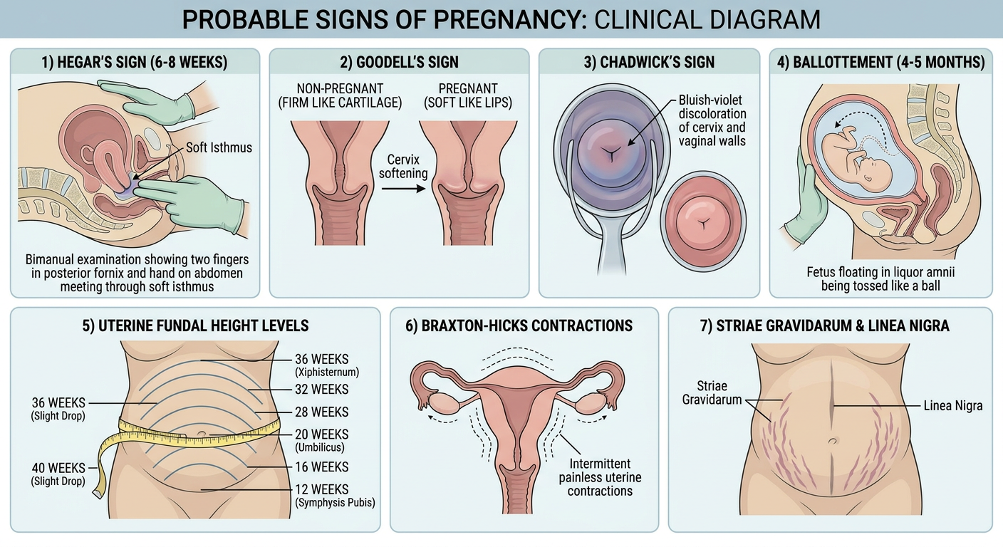
1. Changes in the Abdomen - Uterine Enlargement
| Month | Uterine Fundal Level |
|---|---|
| Up to 3rd month | Within the pelvic cavity |
| 4th month | Just above the symphysis pubis |
| 5th month | Midway between symphysis pubis and umbilicus |
| 6th month | At the level of the umbilicus |
| 7th month | Midway between umbilicus and xiphisternum |
| 8th month | At the level of xiphisternum |
| 9th-10th month | Sinks back below xiphisternum (lightening) |
2. Striae Gravidarum and Linea Nigra
- Umbilicus becomes flush with the abdominal surface by ~7th month
- Umbilicus becomes everted by ~9th month
3. Hegar's Sign
"The cervix is felt as firm and hard whereas the uterus is felt as an elastic body. In between the two, the isthmus is felt as a soft compressible area." - P C Dikshit Textbook of Forensic Medicine
4. Goodell's Sign
- In non-pregnant state: Cervix feels as hard as the convex surface of a nail (or tip of nose)
- In pregnancy: Cervix feels as soft as the lips
"The cervix is felt as soft as the lips. In the non-pregnant state, it is felt as hard as the convex surface of the nails." - Parikh's Textbook of Medical Jurisprudence
5. Chadwick's Sign (Jacquemier's Sign)
6. Cervical Mucus Plug
7. Braxton-Hicks Sign (Intermittent Uterine Contractions)
- Painless (unlike true labor)
- Irregular frequency
- Do not increase in intensity or frequency over time
8. Ballottement
Two Types:
| Type | Technique |
|---|---|
| Internal ballottement | Two fingers inserted into the anterior fornix give a sudden upward impulse; the fetus moves up and can be felt rebounding by the hand on the abdomen, or by the same fingers after a moment |
| External ballottement | Palms placed on either side of the midline on the abdomen; sudden impulse by fingers of one hand - fetus moves and is felt by the other hand |
9. Uterine Souffle
10. Biological Tests (Pregnancy Tests)
| Test | Animal Used | Observation |
|---|---|---|
| Aschheim-Zondek test | Immature female mice | Haemorrhagic follicles/corpus luteum in ovary |
| Friedman's test | Adult female rabbit | Haemorrhagic Graafian follicle |
| Hogben test (female frog) | Mature African toad | Ovulation within 12-18 hrs |
| Galli-Mainini test (male frog) | Rana tigrina | Spermatozoa in cloacal urine within 2-3 hrs |
| Rapid rat test | Immature female albino rats | Congested ovaries |
Modern equivalent: Urine/serum beta-hCG (immunoassay) has replaced all biological tests in clinical practice - detectable as early as 10-12 days post-fertilization.
Quick Reference Summary Table
| Sign | Timing | Mechanism | Clinical Method |
|---|---|---|---|
| Hegar's sign | 6-10 weeks | Softening of isthmus | Bimanual examination |
| Goodell's sign | 2nd month+ | Cervical softening | Per vaginum |
| Chadwick's sign | 6-8 weeks | Venous congestion (bluish cervix/vagina) | Speculum exam |
| Cervical mucus plug | Early pregnancy | Glandular secretion | Speculum exam |
| Braxton-Hicks | 4th month+ | Intermittent myometrial contractions | Abdominal palpation |
| Ballottement | 4th-5th month | Fetus floating in liquor amnii | Bimanual/abdominal |
| Uterine souffle | 4th month+ | Blood in dilated uterine vessels | Auscultation |
| Abdominal enlargement | Progressive | Uterine growth | Inspection/measurement |
| Striae gravidarum | 2nd trimester | Subcutaneous rupture | Inspection |
| Linea nigra | 2nd trimester | MSH-induced pigmentation | Inspection |
| Biological tests | 10-12 days | hCG in blood/urine | Lab test |