
What are red cell ghosts
Red cell ghosts are the remnant membranes left behind after red blood cells (erythrocytes) have been lysed and their hemoglobin content has been washed out. They appear as faint, pale, "empty" cell outlines on a blood smear - hence the name "ghosts."
Searching sources...
Reading File
Searching the Web
red cell ghosts erythrocyte pathology significance
Finding Sources
Finding Sources
Finding Sources
Finding Sources
Finding Sources
Finding Sources
Reading Web Page
https://eclinpath.com/hematology/morphologic-features/red-bl…
Loading Image
Finding Sources
Here is a complete answer on red cell ghosts:
Red Cell Ghosts (Erythrocyte Ghosts)
Definition
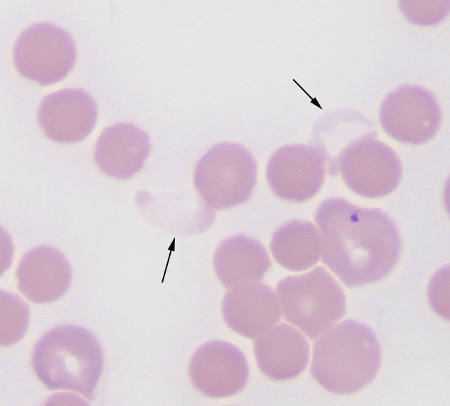
Red cell ghosts are the empty, translucent remnants of red blood cells (RBCs) after they have been lysed and their hemoglobin has leaked out. On a peripheral blood smear they appear as faint, pale outlines - barely staining "shells" of a cell, with no pink hemoglobin content. The name comes from their ghost-like appearance under the microscope.
Here is what they look like on a blood smear (arrows point to ghost RBCs alongside normal, pink-stained erythrocytes):
Mechanism of Formation
Ghost cells form when the RBC membrane is physically breached in the circulation (true intravascular hemolysis). The key sequence is:
- Something attacks or disrupts the RBC lipid bilayer membrane
- Hemoglobin spills out into the plasma (causing hemoglobinemia and hemoglobinuria)
- The empty membrane sac persists briefly in circulation before being cleared by the reticuloendothelial system
- This deflated membrane remnant is what appears as a ghost cell on the smear
The most common in-vivo mechanism involves the membrane attack complex (MAC) of complement - specifically C5b-6789 - which punches pores into the RBC membrane, allowing hemoglobin to escape while the lipoprotein shell remains intact.
Causes (Clinical Associations)
| Cause | Mechanism |
|---|---|
| Immune-mediated hemolytic anemia (IMHA) | Complement-fixing antibodies (IgG, IgM) activate MAC |
| Transfusion reactions (ABO incompatibility) | Complement activation against foreign RBCs |
| Microangiopathic hemolytic anemia (TTP, HUS, DIC) | Mechanical shearing of RBCs |
| Clostridial sepsis | Bacterial phospholipases destroy the membrane |
| Burns | Thermal damage to RBC membrane |
| Paroxysmal nocturnal hemoglobinuria (PNH) | Complement-mediated lysis due to absent GPI-anchored proteins |
| Snake/spider envenomation | Direct lytic toxins |
| In vitro artifact | Delayed smear preparation, incorrect EDTA concentration |
What They Signify Clinically
The presence of ghost cells on a peripheral smear is a marker of intravascular hemolysis - a more severe form of hemolysis compared to extravascular (spleen-mediated) hemolysis. The accompanying laboratory findings typically include:
- Hemoglobinemia - pink/red plasma
- Hemoglobinuria - pink or brown/cola-colored urine
- Decreased haptoglobin (haptoglobin binds free Hb and is consumed)
- Elevated LDH (released from lysed cells)
- Elevated indirect bilirubin
- Reticulocytosis (bone marrow compensatory response)
Ghost cells are often accompanied on the smear by spherocytes (IMHA), schistocytes (microangiopathic), or polychromasia (reticulocytes from regenerative response).
Laboratory/Research Use
In the research setting, red cell ghosts are also deliberately created in the lab by hypotonic lysis - placing RBCs in distilled water or very dilute saline causes them to swell and burst, releasing hemoglobin. The resulting ghost membranes are used to study:
- RBC membrane protein composition (spectrin, ankyrin, band 3, etc.)
- Membrane permeability and osmotic behavior (erythrocyte ghosts behave as near-perfect osmometers)
- Drug/molecule encapsulation (ghosts can be "resealed" to trap substances inside)
Key Takeaway
Red cell ghosts on a blood smear = evidence of intravascular hemolysis. They are the "empty husks" left after hemoglobin escapes a breached RBC membrane, and their presence should prompt investigation for complement-mediated, mechanical, or toxic causes of hemolysis.
Types of polypeptide chains in normal blood
Normal adult blood contains several types of hemoglobin, each made of different polypeptide (globin) chain combinations: alpha (α), beta (β), gamma (γ), delta (δ), epsilon (ε), and zeta (ζ) chains.
Searching sources...
Finding Sources
Searching the Web
hemoglobin polypeptide chains types normal human blood alpha beta gamma delta
Reading File
Reading File
Loading Image
Loading Image
Excellent - the textbooks provide everything needed. Here is a complete answer:
Polypeptide (Globin) Chains in Normal Human Blood
Each hemoglobin molecule is a tetramer - two pairs of unlike globin polypeptide chains, each wrapped around an iron-containing heme group. The chain types expressed change across development.
The Globin Chain Types
| Chain | Gene | Chromosome | Notes |
|---|---|---|---|
| Alpha (α) | HBA1, HBA2 | 16p | Present in ALL normal hemoglobins throughout life; 141 amino acids |
| Beta (β) | HBB | 11p15.5 | Major adult chain; 146 amino acids |
| Delta (δ) | HBD | 11p15.5 | Minor adult chain; structurally similar to β |
| Gamma (γ) | HBG1 (Aγ), HBG2 (Gγ) | 11p15.5 | Fetal chain; differs from β at 39 amino acid positions |
| Epsilon (ε) | HBE1 | 11p15.5 | Embryonic only (yolk sac stage) |
| Zeta (ζ) | HBZ | 16p | Embryonic α-like chain (yolk sac stage only) |
The Normal Hemoglobins They Form
| Hemoglobin | Chains | Stage | % in Normal Adult |
|---|---|---|---|
| HbA | α₂β₂ | Adult (predominant) | >95% |
| HbA₂ | α₂δ₂ | Adult (minor) | 2-3% |
| HbF | α₂γ₂ | Fetal; persists in small amount | <1% after 6 months of life |
| Hb Gower I | ζ₂ε₂ | Embryonic only | 0 in normal adults |
| Hb Gower II | α₂ε₂ | Embryonic only | 0 in normal adults |
| Hb Portland | ζ₂γ₂ | Embryonic only | 0 in normal adults |
So in normal adult blood, only three chains are actually present: α, β, and δ (with a trace of γ from HbF).
Structure of Hemoglobin
Each heme group binds one O₂ molecule; with four chains per molecule, each hemoglobin can carry four O₂ molecules.
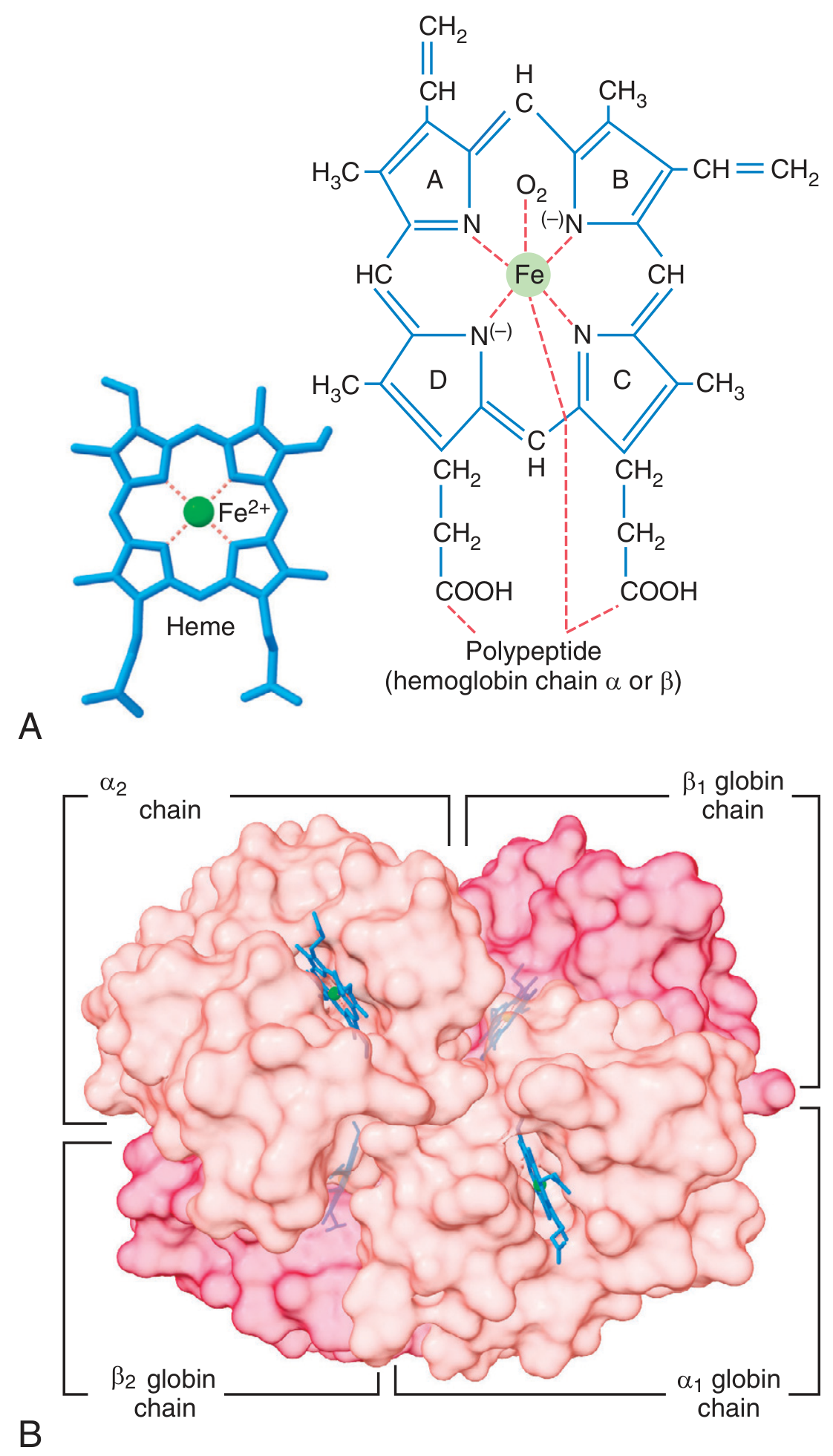
Figure: (A) The heme moiety with its central Fe²⁺ atom bound to the polypeptide chain. (B) The full HbA tetramer showing two α and two β globin chains. - Guyton & Hall Medical Physiology
Developmental Switching of Globin Chains
The gene clusters and their switching pattern over development are shown below:
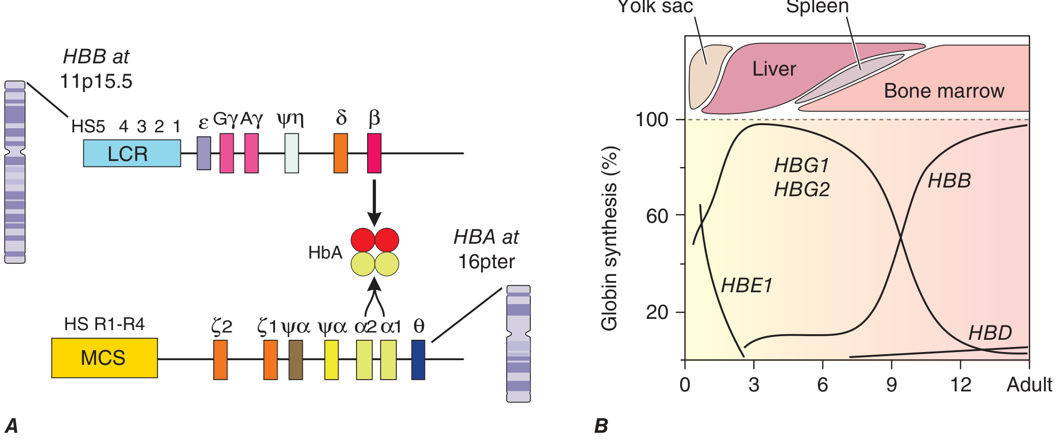
Figure: (A) β-globin gene cluster on chromosome 11 (ε, Gγ, Aγ, δ, β) and α-globin gene cluster on chromosome 16 (ζ, α2, α1). (B) Progressive switching from embryonic → fetal → adult globin chains over gestation and into adulthood. - Harrison's Principles of Internal Medicine, 22e
Key points from this diagram:
- ε (epsilon) and ζ (zeta) are active only in the yolk sac (first ~3 months)
- γ (gamma) dominates during fetal life, then is silenced after birth
- β (beta) rises sharply after birth to become the dominant non-α chain in adults
- δ (delta) appears in small amounts and stays low throughout adult life
- α (alpha) is expressed continuously from early fetal life through adulthood
Clinical Relevance
Mutations or deletions in these globin genes cause the hemoglobinopathies:
-
Abnormal β chain → Sickle cell anemia (β^S), β-thalassemia
-
Abnormal α chains → α-thalassemia
-
Failure of γ→β switching → Hereditary persistence of fetal hemoglobin (HPFH)
-
Elevated HbA₂ (α₂δ₂ >3.5%) → diagnostic marker for β-thalassemia trait
-
Guyton & Hall Textbook of Medical Physiology, p. 445
-
Harrison's Principles of Internal Medicine, 22e, Chapter 103
What happens during thalassemia and sickle cell
Both are inherited hemoglobin disorders but with different mechanisms: thalassemia involves reduced/absent globin chain production, while sickle cell involves a structural defect in the beta chain causing RBCs to deform under low oxygen conditions.
Searching sources...
Finding Sources
Finding Sources
Reading File
Finding Sources
Reading File
Finding Sources
Reading File
Reading File
Reading File
Loading Image
Finding Sources
Loading Image
Here is a thorough, textbook-based breakdown of both conditions:
Thalassemia and Sickle Cell Disease
PART 1: THALASSEMIA
The Core Problem
Thalassemia is caused by mutations that reduce or abolish the synthesis of one globin chain type (α or β). The result is a chain imbalance - too much of the other chain - which is the root cause of almost all the pathology.
"Thalassemia is a genetically heterogeneous disorder caused by germline mutations that decrease the synthesis of either α-globin or β-globin, leading to anemia, tissue hypoxia, and red cell hemolysis related to the imbalance in globin chain synthesis." - Robbins Pathology
β-Thalassemia (most common form)
Mutation types on chromosome 11:
| Mutation Type | Effect | Class |
|---|---|---|
| Splicing mutations | Disrupt normal/create ectopic splice sites | β⁺ (reduced) or β⁰ (absent) |
| Promoter mutations | Reduce transcription by 75-80% | β⁺ |
| Chain terminator mutations (nonsense/frameshift) | Block translation entirely | β⁰ (most common β⁰ cause) |
Pathophysiology of β-thalassemia:
When β-globin production falls, excess α-chains accumulate. These α-chains are insoluble and precipitate inside developing red cell precursors, forming toxic inclusions that:
- Damage the RBC membrane
- Cause massive apoptosis of erythroid precursors (ineffective erythropoiesis) - up to 70-85% of red cell precursors die before leaving the marrow in severe disease
- Red cells that do survive are fragile, hypochromic, microcytic, and get destroyed in the spleen (extravascular hemolysis)
Downstream consequences of ineffective erythropoiesis:
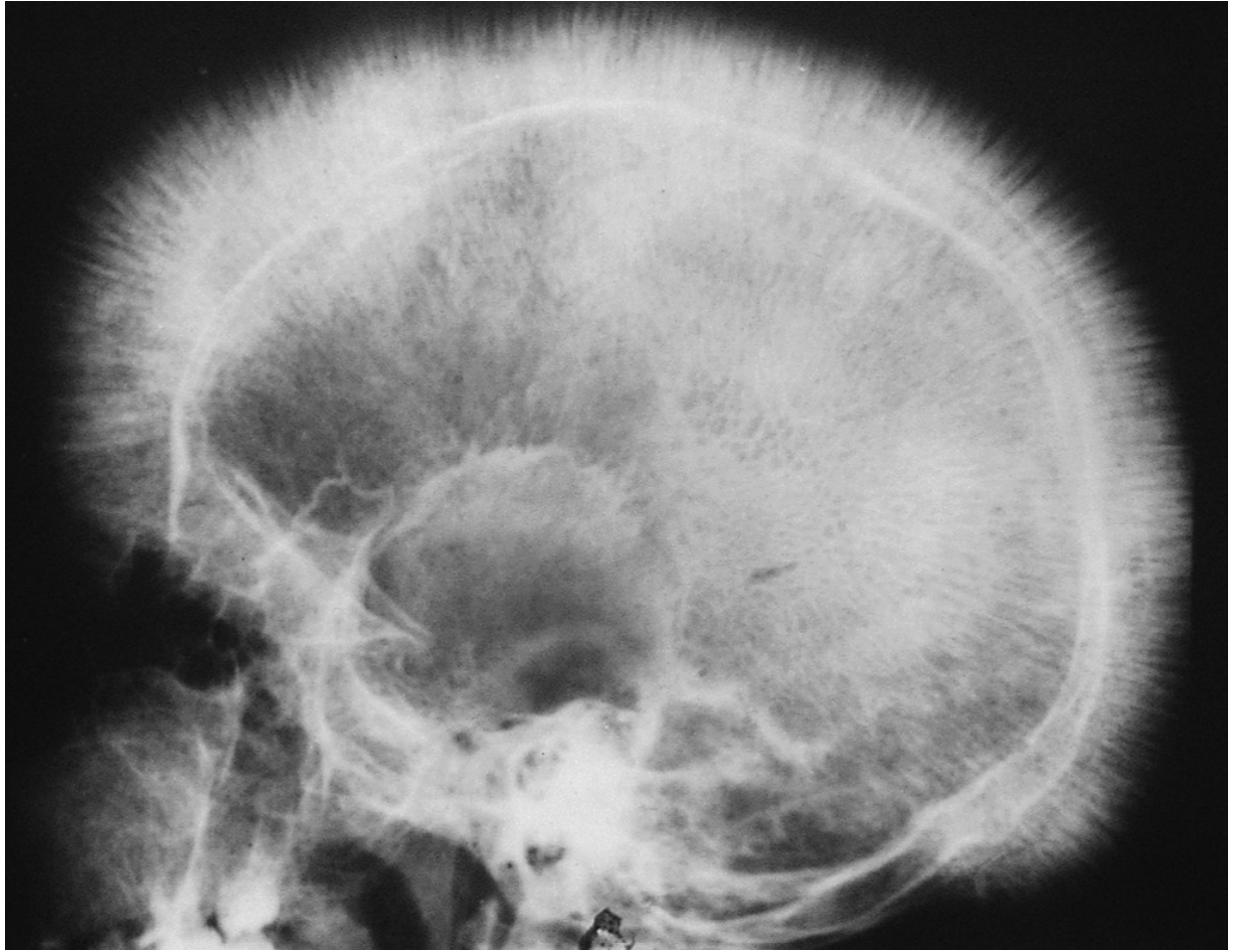
- Erythroid hyperplasia - bone marrow expands massively, eroding the bony cortex
- Skeletal deformity - "crew cut" appearance on skull X-ray (new bone laid down on outer table)
- Extramedullary hematopoiesis - liver, spleen, lymph nodes all attempt to compensate
- Iron overload - erythroid precursors release erythroferrone, which suppresses hepcidin, unleashing gut iron absorption. This, plus repeated transfusions, causes secondary hemochromatosis and organ damage (heart, liver)
"Crew cut" skull X-ray in β-thalassemia major due to expansion of marrow space from erythroid hyperplasia:
β-Thalassemia Clinical Spectrum
| Syndrome | Genotype | Severity |
|---|---|---|
| β-Thalassemia major (Cooley's anemia) | β⁰/β⁰ or β⁰/β⁺ | Severe, transfusion-dependent |
| β-Thalassemia intermedia | Various β⁺/β⁻ or mild β⁻/β⁰ | Moderate; does not require regular transfusions |
| β-Thalassemia minor/trait | β⁺/β or β⁰/β (heterozygous) | Mild microcytic anemia; usually asymptomatic |
α-Thalassemia
Caused mainly by gene deletions on chromosome 16. Each of the 4 α-globin genes contributes 25% of α-chain output:
| Syndrome | Genes deleted | Clinical effect |
|---|---|---|
| Silent carrier | 1 gene (-/α α/α) | Asymptomatic |
| α-Thal trait | 2 genes | Mild microcytic anemia |
| HbH disease | 3 genes | Severe hemolytic anemia; excess β-chains form HbH (β₄) tetramers |
| Hydrops fetalis | 4 genes (--/--) | Lethal in utero; only Hb Barts (γ₄) present, which cannot deliver O₂ |
PART 2: SICKLE CELL DISEASE
The Core Mutation
A single point mutation on chromosome 11 in the β-globin gene:
- Codon 6: GAG → GTG (mRNA)
- Result: Glutamic acid → Valine at position 6 of the β-chain
- This produces HbS (α₂β^S₂) instead of normal HbA (α₂β₂)
The substitution of a charged, hydrophilic amino acid (glutamate) with a nonpolar, hydrophobic one (valine) creates a "sticky patch" on the surface of the deoxygenated β-chain.
Pathophysiology: HbS Polymerization
When HbS gives up oxygen (deoxygenation in tissues), the valine residue at β6 fits into a hydrophobic pocket on an adjacent HbS molecule. This triggers:
- Linear polymer formation - HbS molecules stack into long, rigid fibers
- Sickling - these fibers distort the RBC into a sickle/crescent shape
- Irreversibly sickled cells (ISCs) - after repeated sickling/unsickling cycles, the membrane becomes permanently damaged and the cell stays sickled even when reoxygenated
Key point from Harrison's: "Polymerization is dependent on the 30th power of hemoglobin concentration" - meaning even tiny decreases in cell hydration or tiny increases in Hb concentration dramatically accelerate sickling.
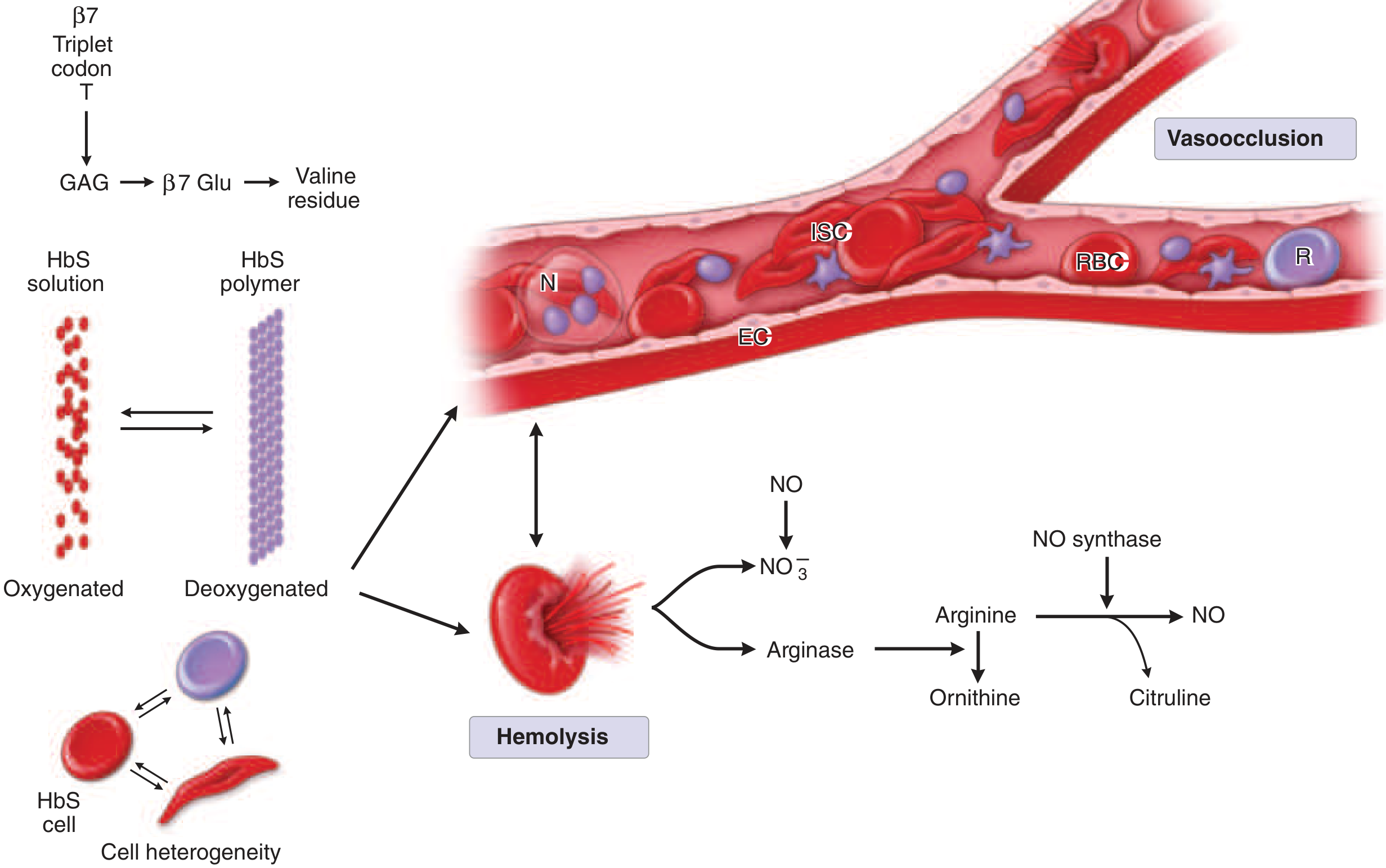
The two main disease arms are: (1) vasoocclusion and (2) hemolysis. Both stem from HbS polymerization on deoxygenation.
Two Major Pathologic Arms
1. Vasoocclusion
- Sickled RBCs are rigid and sticky; they adhere to endothelial cells and interact with leukocytes and platelets
- This blocks flow in small vessels → ischemia and infarction in multiple organs
- Sickled cells live only ~20 days (vs normal 120 days)
2. Hemolysis + NO depletion
- Intravascular hemolysis releases free hemoglobin, arginase, and heme into plasma
- Free Hb scavenges nitric oxide (NO), causing vasoconstriction
- Arginase depletes arginine (the substrate for NO synthesis), further reducing NO
- This promotes a pro-inflammatory, prothrombotic state
Clinical Manifestations
| Complication | Mechanism |
|---|---|
| Vaso-occlusive pain crises | Vessel obstruction → bone/joint ischemia; most common acute event |
| Acute chest syndrome | Sickling in pulmonary vasculature + fat emboli → hypoxia |
| Stroke | Large or small vessel occlusion |
| Splenic sequestration/autosplenectomy | Repeated infarction → functional asplenia → susceptibility to encapsulated organisms |
| Avascular necrosis (femoral head) | Vascular occlusion of bone blood supply |
| Chronic hemolytic anemia | RBC lifespan ~20 days; Hb typically 6-9 g/dL |
| Aplastic crisis | Parvovirus B19 infects erythroid precursors, stops production |
| Renal papillary necrosis | Medullary hypoxia due to sickling |
| Pulmonary hypertension | Chronic NO depletion + hemolysis |
Sickle Cell Trait vs. Disease
| Trait (HbAS) | Disease (HbSS) | |
|---|---|---|
| Genotype | One β^S, one normal β | Two β^S alleles |
| HbS level | ~40% | ~80-90% |
| Symptoms | Usually none | Full clinical disease |
| Protection | Against severe malaria | - |
Side-by-Side Comparison
| Feature | Thalassemia | Sickle Cell Disease |
|---|---|---|
| Defect type | Quantitative - reduced chain production | Qualitative - structurally abnormal chain |
| Mechanism | Chain imbalance → toxic precipitates | HbS polymerization on deoxygenation |
| RBC morphology | Hypochromic, microcytic, target cells | Sickle cells, irreversibly sickled cells |
| Primary problem | Ineffective erythropoiesis + hemolysis | Vasoocclusion + hemolysis |
| Major complication | Iron overload, skeletal deformity | Painful crises, stroke, organ infarction |
| Key lab finding | Low MCV, elevated HbA₂ (β-thal) | HbS on electrophoresis |
- Robbins, Cotran & Kumar Pathologic Basis of Disease, Ch. 14
- Harrison's Principles of Internal Medicine, 22e, Ch. 103
This is a shared conversation. Sign in to Orris to start your own chat.