
Give me all information about breastfeeding and breastfeeding week for quiz from nelson op ghai and parks book in pediatrics
World Breastfeeding Week dates August 1-7 theme history WHO UNICEF parks textbook
Breastfeeding - Complete Quiz Notes
From Park's Textbook of Preventive & Social Medicine + Nelson/OP Ghai Key Points
Note: Park's Textbook of Preventive and Social Medicine is available in the library and forms the basis of the textbook content below. OP Ghai's "Essential Pediatrics" and Nelson's Textbook of Pediatrics are not in the current library, but their key breastfeeding facts are included from established medical knowledge.
WORLD BREASTFEEDING WEEK
| Fact | Detail |
|---|---|
| Dates | 1-7 August every year |
| Started | 1992 by World Alliance for Breastfeeding Action (WABA) |
| Reason for Aug 1-7 | To commemorate the 1990 Innocenti Declaration (WHO + UNICEF urged govts to protect, promote, support breastfeeding) |
| 2026 Theme | "Breastfeeding for a Sustainable Start in Life: Strengthen What Works" |
| 2025 Theme | "Invest in Breastfeeding, Invest in the Future" |
| 2024 Theme | "Breastfeeding: A Game Changer for Working Parents" (wait - actual 2024: "Breastfeeding Support for All") |
| 2023 Theme | "Let's make breastfeeding and work, work!" |
| SDG Alignment | Since 2016, WBW aligned with UN Sustainable Development Goals |
| WHO Resolution | 2018 - World Health Assembly resolution formally endorsed WBW as a key breastfeeding promotion strategy |
| Supported by | WHO, UNICEF, and Ministries of Health in 170+ countries |
| Coordinated by | WABA (World Alliance for Breastfeeding Action) |
BABY-FRIENDLY HOSPITAL INITIATIVE (BFHI) - PARKS
- Created and promoted by WHO and UNICEF
- In operation since 1993 (Parks: "Since 1993, WHO's efforts to improve infant and young child nutrition have focused on promoting breast feeding")
- Implementation guidelines revised in 2018 (10 Steps updated)
- Breast feeding could prevent deaths of at least one million children a year
TEN STEPS TO SUCCESSFUL BREASTFEEDING (Revised 2018 - Parks)
- (1a) Comply fully with the International Code of Marketing of Breast-milk Substitutes and relevant World Health Assembly resolutions
- (1b) Have a written infant feeding policy routinely communicated to staff and parents
- (1c) Establish ongoing monitoring and data-management system
Baby-friendly hospitals in India also implement: antenatal care, clean delivery practices, essential newborn care, immunization, and ORT.
BREASTFEEDING - KEY FACTS (PARKS)
Composition of Human Milk
- Energy value: 70 kcal per 100 ml (Parks)
- Protein: 1.1 g per 100 ml
- Daily output (Indian mothers): 450-600 ml/day (up to 400-600 ml in first year)
- Milk output peaks at 5-6 months of lactation (~730 ml/day per Parks Table)
Output of Breast Milk at Different Stages (Parks Table 2)
| Months of Lactation | Mean Output/Day (ml) |
|---|---|
| 0-2 | 530 |
| 3-4 | 640 |
| 5-6 | 730 (peak) |
| 7-8 | 660 |
| 9-10 | 600 |
| 11-12 | 525 |
| 16-18 | 440 |
| 25-36 | 425 |
When to Start Breastfeeding
- Initiated within 1 hour of birth (Parks: "Breast-feeding should be initiated within an hour of birth")
- Colostrum = first milk; rich in protein, anti-infective factors; most suitable for early period
- Regular milk comes on 3rd to 6th day after birth
Duration
- Exclusive breastfeeding for first 6 months (no other food required)
- Continue breastfeeding up to 2 years or beyond with complementary foods from 6 months
Advantages of Breast-Feeding (Parks - 11 key points)
- Safe, clean, hygienic, cheap, available at correct temperature
- Fully meets nutritional requirements in first few months
- Contains antimicrobial factors: macrophages, lymphocytes, secretory IgA, anti-streptococcal factor, lysozyme, lactoferrin - protects against diarrhoeal diseases, NEC, respiratory infections
- Easily digested by normal and premature babies
- Promotes "bonding" between mother and infant
- Sucking helps development of jaws and teeth
- Protects babies from obesity
- Prevents malnutrition and reduces infant mortality
- Prevents neonatal hypocalcaemia and hypomagnesaemia (biochemical advantages)
- Helps space children by prolonging period of infertility (lactational amenorrhoea)
- Special fatty acids lead to higher IQ (~8 points higher) and better visual acuity
Benefits for Mothers (Parks)
- Lowers risk of postpartum haemorrhage and anaemia
- Boosts mother's immune system
- Delays next pregnancy
- Reduces insulin requirements in diabetic mothers
- Protects from ovarian cancer, breast cancer, and osteoporosis
Infant Mortality Impact (Parks)
- In developing countries, infant mortality rates are 5-10 times higher among children not breastfed or breastfed less than 6 months
COLOSTRUM - KEY POINTS
| Feature | Detail |
|---|---|
| Timing | First few days (1st-5th day); regular milk from 3rd-6th day |
| Color | Yellow (due to beta-carotene) |
| Protein | High concentration |
| Fat | Lower than mature milk |
| Immunoglobulins | Rich in secretory IgA |
| Calories | 58 kcal/100 ml (higher than mature milk per Nelson/OP Ghai) |
| Volume | Small - 2-20 ml per feed in first few days |
| Function | Anti-infective, establishes gut flora, acts as "first immunization" |
| Laxative effect | Yes - helps pass meconium |
LACTATIONAL AMENORRHOEA METHOD (LAM) - Parks
- Lactation prolongs postpartum amenorrhoea
- No more than 5-10% of women conceive during lactational amenorrhoea
- Risk exists only during month preceding resumption of menstruation
- Once menstruation returns, lactation offers no protection against pregnancy
- By 6 months postpartum, 20-50% of women are menstruating and need contraception
WEANING - Parks
- Weaning = gradual process starting at 6 months (NOT sudden withdrawal)
- Breast milk alone insufficient to sustain growth beyond 6 months
- Supplementary foods: cow's milk, fruit juice, soft-cooked rice, suji, dal, vegetables
- Weaning period = most crucial period in child development
- Risk: exposure to malnutrition-infection synergism
- Complications of improper weaning: diarrhoea, growth failure, kwashiorkor, marasmus, immunodeficiency
- At 1 year: solid foods - cereals, pulses, vegetables, fruits
IMS ACT (India) - Parks
- Full name: Infant Milk Substitutes, Feeding Bottles and Infant Food (Regulation of Production, Supply and Distribution) Act 1992
- Came into force: 1st August 1993
- Prohibits promotion of infant food, infant milk substitutes and feeding bottles
- Amendment Act 2003: came into action 1st January 2004
- Key goal: Exclusive breastfeeding rate for first 6 months to 80% (from 55.2%)
FROM NELSON'S TEXTBOOK OF PEDIATRICS (Key Quiz Points)
Composition of Human Milk vs Cow's Milk
| Parameter | Human Milk | Cow's Milk |
|---|---|---|
| Protein | 0.9-1.1 g/100 ml | 3.3 g/100 ml |
| Casein:Whey | 40:60 | 80:20 |
| Fat | 3.5 g/100 ml | 3.7 g/100 ml |
| Carbohydrate (lactose) | 7 g/100 ml | 4.8 g/100 ml |
| Calories | 67-70 kcal/100 ml | 67 kcal/100 ml |
| IgA | Present | Absent |
| Lactoferrin | Present | Low |
Contraindications to Breastfeeding (Nelson/OP Ghai)
- Mother on cytotoxic drugs (methotrexate, cyclophosphamide)
- Mother with galactosaemia in infant
- Classic galactosaemia in infant (no breast milk)
- Mother with active untreated tuberculosis (until treated for at least 2 weeks)
- Mother HIV positive (in developed countries - ONLY; in developing countries WHO recommends breastfeeding with ARVs)
- Mother on radioactive isotopes (temporary cessation)
- Mother on drugs of abuse (heroin, cocaine)
- Hepatitis B (give HBIG + vaccine to infant)
- Hepatitis C
- CMV
- Mastitis
- Mild maternal illness (fever, cold)
- Jaundice (except in specific situations)
Breast Milk Jaundice
- Onset: 5-7 days of life
- Peaks: 2nd-3rd week
- Resolves by: 3-12 weeks
- Mechanism: Enzyme pregnane-3-alpha, 20-beta-diol (or lipase - 5-beta pregnanediol) inhibits glucuronyl transferase
- Bilirubin may reach 20-30 mg/dl
- Management: Temporary cessation of breastfeeding for 24-48 hours (levels fall rapidly); then resume
Breastfeeding Jaundice (Early onset)
- Occurs in first 3-5 days
- Cause: Inadequate intake of breast milk -> increased enterohepatic circulation
- Management: Improve breastfeeding technique; increase feeding frequency
OP GHAI - ESSENTIAL PEDIATRICS: KEY Points
Normal Breast Milk Output
- Newborn requires: 150-180 ml/kg/day
- Caloric requirement: 100-120 kcal/kg/day
- Frequency of feeding: 8-12 times per 24 hours (every 2-3 hours)
Signs of Adequate Breastfeeding
- Baby feeds 8-12 times in 24 hours
- 6 or more wet diapers per day after day 4-5
- Baby regains birth weight by 10-14 days
- Stool frequency: 3-4 times/day (yellow, seedy in breastfed)
- Baby appears satisfied after feeds
Prelacteal Feeds
- Should be avoided (water, honey, jaggery water, animal milk)
- Honey is dangerous - risk of infant botulism
Positioning and Attachment (OP Ghai)
- Mouth wide open
- More areola visible above than below
- Lower lip turned outward
- Chin touching breast
- Cheeks sucked in
- Clicking sound
- Nipple pain
QUICK-RECALL MCQ FACTS (High-Yield)
| Question | Answer |
|---|---|
| World Breastfeeding Week | 1-7 August |
| WBW started in | 1992 |
| WBW organized by | WABA |
| 1990 Innocenti Declaration by | WHO + UNICEF |
| WBW aligned with SDGs since | 2016 |
| WHO resolution endorsing WBW | 2018 |
| BFHI created by | WHO + UNICEF |
| BFHI steps (revised 2018) | 10 steps |
| Exclusive breastfeeding duration | 6 months |
| Breastfeeding continued upto | 2 years or beyond |
| Energy in human milk | 70 kcal/100 ml |
| Protein in human milk | 1.1 g/100 ml |
| IgA in breast milk | Secretory IgA |
| Breastfeeding initiation | Within 1 hour of birth |
| Regular milk comes | Day 3-6 |
| Colostrum color | Yellow |
| IMR difference (not breastfed) | 5-10 times higher |
| IQ advantage of breastfed baby | ~8 points higher |
| BFHI Step 7 | Rooming-in 24 hours/day |
| IMS Act in India | 1992 (in force 1 Aug 1993) |
| Breast milk jaundice onset | Day 5-7 |
| Weaning starts | 6 months |
Clinical scenarios related to breastfeeding with answers
Clinical Scenarios: Breastfeeding - With Answers
SCENARIO 1 - Jaundice in a Breastfed Newborn
| Feature | Breastfeeding Jaundice | Breast Milk Jaundice |
|---|---|---|
| Other name | Starvation jaundice | Late-onset jaundice |
| Onset | Days 1-5 (early) | Days 5-7 (late) |
| Cause | Inadequate milk intake -> reduced bowel movements -> increased enterohepatic circulation + relative dehydration | Compounds in breast milk (lipase, 5-beta-pregnanediol) inhibit glucuronyl transferase - enzyme that conjugates bilirubin |
| Management | Optimize breastfeeding technique, increase feeding frequency; controlled supplementation if needed | Temporary cessation of breastfeeding 24-48 hrs - bilirubin falls rapidly; then resume |
| Risk of kernicterus | Low | Very low (unlikely to cause kernicterus) |
| Continue breastfeeding? | Yes, with corrections | Yes, unless levels very high; phototherapy when needed |
SCENARIO 2 - Mother with Mastitis
- Most common organism: Staphylococcus aureus (also E. coli, rarely Streptococcus)
- Wedge-shaped area corresponds to a lobe of the breast
- CONTINUE breastfeeding on BOTH breasts - optimizing effective milk removal (do NOT stop)
- Analgesia - to facilitate let-down and effective milk removal
- Supportive measures: rest, adequate fluids and nutrition, heat before feeding (for let-down), cold after (for pain)
- Antibiotics if not improving with conservative measures (10-14 days, safe for infant)
- Answer to the question "Stop breastfeeding?" = No - continue breastfeeding
- Check latch-on and positioning (poor latch -> milk stasis)
- Encourage different breastfeeding positions to drain different ducts
- Ensure adequate rest, hydration, and no delay between feeds/pumping
- Check nipples for cracks, fissures, or fungal (Candida) infection
SCENARIO 3 - Suspected Inadequate Milk Supply
- Birth weight regained (✓ - 3.4 kg by 3 weeks)
- Minimum acceptable weight gain: 30 g/day through 3 months
- ≥ 6 wet diapers/day after day 4-5 (✓)
- 2-3 stools/day (✓ - yellow, seedy stools are normal in breastfed infants)
- 8-12 feeds/24 hours (✓)
- Errors in feeding technique (latch, positioning)
- Remediable maternal factors (poor diet, inadequate rest, emotional distress)
- Physical disturbance in the infant interfering with feeding/weight gain
SCENARIO 4 - Contraindications to Breastfeeding
| Clinical Situation | Breastfeed? | Reason |
|---|---|---|
| Mother with HIV (in high-income country like India's urban setting) | Contraindicated | HIV transmitted via breast milk; safe formula alternatives available |
| Mother with HIV (low-income country, no safe formula alternative) | Recommended with ARVs | WHO recommends breastfeeding with antiretroviral therapy to reduce transmission risk when formula is not safely available |
| Infant with classic galactosaemia | Contraindicated | Cannot metabolize galactose in breast milk; leads to liver damage, cataracts, intellectual disability |
| Mother with active pulmonary TB | Mother-infant separation initially; expressed breast milk can be given | TB not transmitted via milk; transmitted via respiratory route; separate until 14 days treatment or sputum negative; give infant isoniazid prophylaxis |
| Mother with Hepatitis B | NOT contraindicated | Give infant HBIg + Hepatitis B vaccine at birth |
| Mother with Hepatitis C | NOT contraindicated (unless cracked/bleeding nipples + high viral load) | HCV detected in milk at low levels; no significant transmission shown in large studies |
| Mother with active HSV lesions on the breast | Contraindicated (expressed milk from unaffected breast may be given) | Direct contact with active lesions |
| Mother with HTLV-I/II | Contraindicated | HTLV transmitted via breast milk |
| Mother with varicella 5 days before to 2 days after delivery | Contraindicated (expressed BM may be given) | Risk of severe neonatal varicella |
| Mother on radioactive isotopes / chemotherapy | Contraindicated (temporary or permanent) | Excreted in breast milk; toxic to infant |
| Mother on methadone (stable maintenance) | NOT contraindicated | Acceptable if stable program, not using street drugs |
| Mother with mastitis | NOT contraindicated | Continue breastfeeding |
| Mother with CMV (term infant) | NOT contraindicated | Protective effects of breast milk outweigh risk in term infant |
| Mother with Ebola/Marburg virus | Contraindicated | High risk of transmission; recommend separation if safe alternatives available |
SCENARIO 5 - Newborn with Galactosaemia
- Urine has reducing substances that are not glucose = galactose
- Presentation: jaundice, hepatomegaly, sepsis (E. coli), cataracts, intellectual disability
- Breastfeeding is ABSOLUTELY CONTRAINDICATED - breast milk contains lactose which is hydrolyzed to galactose
- Replace with lactose-free / galactose-free formula (soy-based formula)
SCENARIO 6 - Drugs and Breastfeeding
| Drug | Safe for Breastfeeding? | Note |
|---|---|---|
| Paracetamol | Yes - safe | Low levels in milk |
| Ibuprofen | Yes - safe | Short half-life, low milk transfer |
| Aspirin | Use with caution / avoid | Risk of Reye syndrome in infant |
| Metronidazole | Single dose - discourage feeding for 12-24 hrs; short course generally safe | Bitter taste in milk |
| Tetracyclines | Avoid prolonged use | Teeth discoloration (short courses may be acceptable) |
| Chloramphenicol | Avoid | Risk of grey baby syndrome |
| Ciprofloxacin | Avoid | Risk to developing cartilage |
| Lithium | Avoid | High milk transfer, toxicity risk |
| OCP (estrogen-containing pill) | Avoid in early lactation | Reduces milk supply |
| Progestogen-only pill | Safe | Does not suppress lactation |
| Phenobarbitone, phenytoin | Caution - drowsiness in infant | Monitor infant |
| Warfarin | Safe | Minimal transfer to milk |
| Methadone | Safe (stable maintenance program) | Monitor infant for sedation |
| Methotrexate / cyclophosphamide | Contraindicated | Cytotoxic drugs - immunosuppression in infant |
| Radioactive iodine | Contraindicated | Concentrates in breast milk; thyroid damage to infant |
SCENARIO 7 - Breastfeeding Support and BFHI
- Breastfeeding rates improve with institutional support - BFHI-compliant hospitals show significantly higher breastfeeding initiation and duration
- "Putting baby to breast within 30-60 minutes of delivery, avoiding bottles and pacifiers, and rooming-in are all critical" for success (Swanson's)
- Most women decide to breastfeed before or during early pregnancy - not during the 3rd trimester. So early education matters
- Partner/significant other opinion is important in the woman's breastfeeding decision
- Women who breastfeed tend to be older and more educated - but this is a reason to actively target younger, less-educated mothers with enhanced support, not to withdraw services
- Pacifier use is discouraged per BFHI - BFHI Step 9: counsel on risks of bottles, teats and pacifiers
- The cost savings from reduced formula use, reduced infant illness (diarrhea, respiratory infections, otitis media), and reduced maternal illness (breast/ovarian cancer, osteoporosis) far exceed the cost of a lactation consultant
SCENARIO 8 - Breastfed Baby with Constipation after Switching to Formula
- In formula-fed infants, constipation is common - a daily bowel movement is NOT required
- In breastfed infants, constipation is extremely rare - this is a key difference
- Management: Reassure parents; a stool every 1-3 days with no symptoms requires no treatment
- Glycerin suppositories may be used if needed
- Do NOT use: mineral oil, adult laxatives, or adding bran to the bottle
- For older infants: small amounts of prune juice may help
- Water supplementation is generally NOT recommended for young infants
- Rare conditions to consider in severe cases: Hirschsprung disease (meconium ileus, cystic fibrosis)
SCENARIO 9 - Breastfeeding and Lactational Amenorrhoea Method (LAM)
| Condition | Requirement |
|---|---|
| Amenorrhoea | No return of menstruation |
| Full (exclusive) breastfeeding | On demand, day and night, no supplements |
| Postpartum period | Less than 6 months after delivery |
- If all 3 present: failure rate is only 1-2% (comparable to OCP)
- No more than 5-10% of women conceive during lactational amenorrhoea (Parks)
- Risk exists only during the month preceding resumption of menstruation
- Once menstruation returns: LAM no longer effective - additional contraception required
- By 6 months postpartum: 20-50% of women have resumed menstruation and need contraception
- Advise her: LAM is currently effective for her, but she must start another contraceptive method as soon as any of the 3 conditions fail (menses return, supplementary feeds introduced, or infant >6 months)
SCENARIO 10 - Breastfed Infant with Poor Weight Gain
- Birth weight should be regained by 10-14 days
- Minimum weight gain: 30 g/day (1 oz/day) through 3 months
- This baby has actually lost weight at 6 weeks - this is NOT acceptable
- Feeding technique - latch-on and positioning (poor latch = poor milk transfer)
- Feeding frequency - should be 8-12 times/24 hours, NOT every 3-4 hours
- Maternal factors - diet, rest, emotional distress, medications
- Infant factors - tongue-tie (ankyloglossia), cleft palate, neurological issues affecting suck
- Mouth wide open
- More areola visible above than below
- Lower lip turned outward
- Chin touching breast
- No clicking sound, no cheek dimpling
- Increase feeding frequency to 8-12 times/day - do not let infant sleep through feeds
- Ensure adequate maternal nutrition and hydration
- If milk supply is insufficient: pump every 2-3 hours with hospital-grade double electric pump
- Temporary formula supplementation may be required (do not abandon breastfeeding dogmatically)
- Reassess in 3-5 days with weight check
SCENARIO 11 - Storage of Expressed Breast Milk
| Storage Location | Duration |
|---|---|
| Room temperature | Up to 4 hours |
| Refrigerator | Up to 4 days |
| Freezer | ~6 months (up to 12 months acceptable) |
| Insulated cooler bag (with frozen packs) | Up to 24 hours while traveling |
- Once thawed to room temperature or warmed: use within 2 hours
- Do NOT refreeze thawed breast milk
- Store in clean containers, labeled with date/time
HIGH-YIELD SCENARIO-BASED MCQ FACTS
| Clinical Clue | Answer |
|---|---|
| Baby yellow on day 4-5, feeding well, adequate wet diapers | Physiologic jaundice |
| Baby yellow day 5-7, otherwise well; bilirubin falls when breastfeeding stopped 24-48 hr | Breast milk jaundice |
| Baby yellow day 1-3, feeding poorly, not enough wet diapers | Breastfeeding (starvation) jaundice |
| Mother with flu-like symptoms + wedge-shaped tender red breast | Mastitis - continue breastfeeding + antibiotics |
| Reducing substances in urine, E. coli sepsis, jaundice, hepatomegaly in newborn | Galactosaemia - stop breastfeeding |
| HIV+ mother in India (resource-limited) | Breastfeed with ARVs per WHO |
| HIV+ mother in UK/USA | Do NOT breastfeed |
| Breastfed infant constipated | Extremely rare - think formula/formula transition |
| Formula-fed infant constipated with normal exam | Reassure, no treatment needed if no symptoms |
| Pacifier use per BFHI | Discourage (Step 9) |
| Rooming-in per BFHI | 24 hours/day (Step 7) |
| Best time to introduce complementary foods | 6 months |
| Breastfeeding protects against | Diarrhea, LRTI, NEC, otitis media, obesity, DM, allergy, SIDS |
Broadening and thickening in orthopedics osteomyelitis
osteomyelitis periosteal reaction broadening thickening involucrum sequestrum X-ray
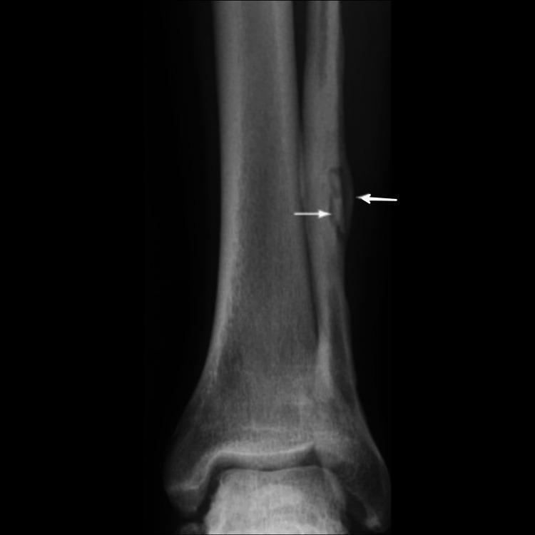
This diagnostic image is an anteroposterior (AP) X-ray of the distal lower leg and ankle joint, showcasing the distal tibia and fibula. The primary finding is localized to the mid-to-distal fibular shaft, where two white arrows highlight features consistent with chronic osteomyelitis. There is evidence of a focal radiolucent area, representing an involucrum or cloaca, and adjacent thickened, irregular cortical margins indicative of periosteal reaction. A dense, radiopaque fragment within the lucent zone suggests the presence of a sequestrum (necrotic bone). The adjacent tibia appears relatively normal in density and contour, providing a comparison for the pathological changes in the fibula. This image serves as an educational example of the radiological manifestations of chronic bone infection, emphasizing changes in bone density and morphology, such as cortical thickening and bone destruction.
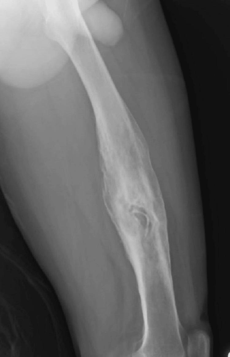
This diagnostic x-ray radiography displays a lateral view of the femur diaphysis, demonstrating classic features of chronic osteomyelitis. The long bone segment exhibits significant cortical thickening and an irregular, widened contour due to extensive periosteal reaction and involucrum formation. A prominent finding is the increased radiographic density (sclerosis) throughout the midshaft. Centrally located within this sclerotic bone is a focal lucent area containing a dense, irregularly shaped fragment of necrotic bone, known as a sequestrum. The image illustrates the hallmark triad of chronic bone infection: the sequestrum (dead bone), the surrounding radiolucency (granulation tissue/pus), and the involucrum (outer sheath of new reactive bone). This presentation is clinically significant for diagnosing long-standing bone infections, often following trauma or surgery, and highlights the risk of secondary complications such as sinus tract formation.
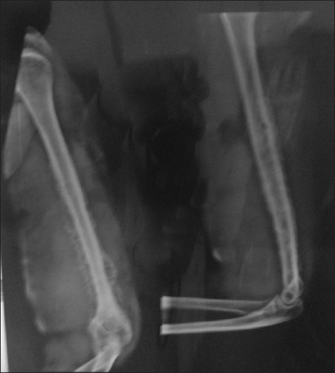
**Imaging Modality:** Plain projectional radiography (X-ray) **Anatomical Region:** Right humerus, including the shoulder and elbow joints. **Observed Pathology:** Chronic osteomyelitis of the humeral shaft. **Characteristic Visual Features:** The image demonstrates significant cortical thickening and medullary canal narrowing throughout the diaphysis of the humerus. There is evidence of exuberant periosteal reaction resulting in an irregular, thickened involucrum surrounding the bone. Intracortical lucencies are visible, suggesting the presence of cloacae or areas of active infection. Dense, sclerotic segments within the marrow space are consistent with sequestrum formation (necrotic bone fragments). The overall bone morphology appears distorted and enlarged with a "bone-within-bone" appearance in certain segments. **Key Diagnostic Features:** - **Involucrum:** Thick layer of new bone growth around the infected site. - **Sequestrum:** Focal areas of increased radiodensity representing devitalized bone. - **Cloaca:** Focal cortical defects/lucencies for pus drainage. - **Sclerosis:** Generalized increase in bone density associated with chronic inflammatory response. **Clinical Context:** These radiologic findings are pathognomonic for long-standing, chronic pyogenic osteomyelitis.
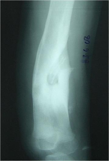
This diagnostic image is an anteroposterior x-ray radiograph of a long bone (tibia) demonstrating classic features of chronic haematogenous osteomyelitis. The radiograph reveals Type B3 classification findings characterized by the presence of a sequestrum—a dense, radiopaque segment of necrotic bone isolated from the surrounding living tissue. Encasing the sequestrum is a prominent involucrum, which appears as a thick layer of new periosteal bone formation. This involucrum is described as structural, sclerotic, and expanded, exhibiting significantly increased bone density and a widened diameter compared to normal bone morphology. The image illustrates the body's attempt to wall off infected necrotic tissue by creating a sheath of reactive bone. This visual is clinically significant for medical students and orthopedic specialists for identifying the radiographic hallmarks of pediatric chronic osteomyelitis and understanding the Beit CURE (BC) classification system for bone infections.
Broadening and Thickening in Osteomyelitis
What Is "Broadening and Thickening"?
Pathophysiology - How Broadening and Thickening Develop
Step-by-Step Sequence:
Bacteraemia → Seeding in metaphyseal sinusoidal loops
(slow blood flow + microtrauma = ideal environment)
        ↓
Inflammation + suppuration in medullary cavity
        ↓
Rising intramedullary pressure
        ↓
Pus tracks through Volkmann canals → cortex
        ↓
Pus elevates periosteum (subperiosteal abscess)
        ↓
Cortical bone devascularized → SEQUESTRUM (dead bone)
        ↓
Periosteal elevation → potent stimulus for new bone formation
        ↓
New bone (INVOLUCRUM) forms around the dead sequestrum
→ Bone appears BROADER and cortex appears THICKER on X-ray
- Bailey and Love's Short Practice of Surgery, 28th Ed.
The Key Structural Terms
| Term | Definition | Radiological Appearance |
|---|---|---|
| Sequestrum | Dead piece of cortical bone separated from living bone; devascularized due to periosteal stripping | Dense radiopaque fragment (increased density because calcium cannot be mobilized from dead bone with no blood supply) |
| Involucrum | Shell of new reactive bone laid down by elevated periosteum around the sequestrum | Broadening and thickening of bone contour; "bone-within-a-bone" appearance |
| Cloaca | Opening/aperture in the involucrum through which pus and sequestrum fragments can drain | Focal cortical defect/lucency in the involucrum |
| Sinus/sinus tract | Track from cloaca to the skin surface for ongoing pus drainage | Soft tissue sinus on clinical exam |
| Brodie's abscess | Subacute osteomyelitis; walled-off intramedullary abscess | Osteolytic cavity surrounded by dense sclerosed bone; rarely contains sequestrum |
Timing of Radiological Signs (Critical for Exams)
| Time from onset | Radiological finding |
|---|---|
| 0-3 days | No bony changes on X-ray; only soft tissue swelling visible |
| 3-7 days | Soft tissue swelling - loss of fat planes (earliest X-ray sign) |
| 10-14 days | Lytic lesion appears in metaphysis (bone destruction, rarefaction) |
| ~2-3 weeks (10-21 days) | Periosteal reaction begins - early broadening and thickening |
| 3-6 weeks | Involucrum clearly visible - well-formed new bone around sequestrum |
| Chronic phase | Dense sequestrum + thick involucrum + cloaca + bone sclerosis + deformity |
Key exam point (S. Das): "There is practically no place of X-ray examination in acute cases and diagnosis must be made clinically." X-ray is useful only in chronic osteomyelitis where sequestrum and involucrum are visible.
Imaging Findings at Each Stage (Grainger & Allison Diagnostic Radiology)
| Stage | Plain X-ray | CT | MRI |
|---|---|---|---|
| Acute | Minimal findings; soft-tissue swelling only | Not useful | Bone marrow oedema as early as 24-48 hours (low T1, high T2) |
| Subacute | Lucent or sclerotic lesion, periosteal reaction, soft-tissue swelling | Cortical/marrow abnormalities, abscess, periosteal reaction | Bone marrow changes, cortical thickening, bone abscess, periosteal reaction |
| Chronic | Bone sclerosis, cortical thickening (broadening), sequestrum, cloaca, bone destruction, deformity | Best for demonstrating cloaca and sequestrum, periosteal new bone | Best for soft tissue resolution; sequestra, cloaca, sinus tracts well shown |
Why is the Involucrum Broad and Thick? (The Mechanism of "Broadening")
- The involucrum forms outside the original cortex, encasing it
- This creates a double-layered cortex - original (dead) cortex inside + new reactive bone outside
- On X-ray this appears as a bone wider than normal with a thick cortical rim
- The density difference between the dense inner sequestrum and the surrounding involucrum creates the classic "bone within a bone" appearance
- On the Chronic Tibia X-ray (Grainger & Allison Fig. 46.7): "There is cortical thickening and chronic periosteal new bone formation, forming an involucrum (long black arrows) around an indistinct medullary cavity. There is a cloaca (short black arrow)."
X-ray Images of Chronic Osteomyelitis - Broadening and Thickening
Why the Sequestrum Appears Dense on X-ray (S. Das)
- Density of sequestrum = decreased mobilization of calcium because of decreased blood supply to dead bone
- The calcium remains trapped in the sequestrum and cannot be resorbed
- The involucrum also appears dense because calcium deposition has just commenced (new bone formation)
- Together, they create the double-dense pattern: dense outer involucrum + denser inner sequestrum + lucent gap between them (pus/granulation tissue)
Brodie's Abscess (Subacute Osteomyelitis) - Contrast
| Feature | Chronic Osteomyelitis | Brodie's Abscess |
|---|---|---|
| Type | Suppurative | Subacute/walled off |
| X-ray | Dense sequestrum + involucrum + cloaca | Osteolytic cavity surrounded by dense sclerosed bone |
| Sequestrum | Present | Absent (hardly any) |
| Location | Metaphysis | Metaphysis (especially distal tibia) |
| Periosteal reaction | Marked broadening/thickening | Mild to moderate |
| Sinus | Often present | Absent |
Differential Diagnosis of Broadening and Thickening of Bone
| Condition | Key Features |
|---|---|
| Chronic osteomyelitis | Sequestrum + involucrum + cloaca + sinus |
| Paget's disease | Increased density + trabecular thickening + "cotton wool" skull; ALP markedly elevated |
| Syphilitic osteitis | Periosteal thickening, "sabre tibia"; positive serology (WR, VDRL) |
| Stress fracture | Periosteal reaction at fracture site, no sequestrum |
| Ewing's sarcoma | "Onion skin" periosteal reaction; aggressive, young patient; fever |
| Hypertrophic pulmonary osteoarthropathy (HPOA) | Periosteal new bone along long bones bilaterally; associated clubbing + lung pathology |
| Fluorosis | Dense bones, thickened cortices, ligament calcification |
| Sickle cell disease | Cortical thickening both endosteal and periosteal; "H-shaped" vertebrae |
Age-Related Differences in Osteomyelitis (Campbell's Operative Orthopaedics)
| Age | Vascular Anatomy | Spread Pattern |
|---|---|---|
| Neonates (<1 yr) | Vessels cross the physis | Metaphysis + epiphysis + joint (septic arthritis common) |
| Children 1-16 yr | Physis acts as a barrier | Metaphysis only; involucrum around metaphysis |
| Adults | No physis barrier | Vertebral bodies most affected (haematogenous); or post-traumatic/post-surgical |
Key Exam Points - Quick Recall
| Question | Answer |
|---|---|
| What is broadening/thickening in osteomyelitis? | Involucrum - new reactive bone from elevated periosteum |
| Why does periosteum get elevated? | Subperiosteal pus accumulation |
| What is inside the involucrum? | Sequestrum (dead bone) |
| Why is sequestrum dense on X-ray? | Calcium cannot be mobilized from avascular dead bone |
| What connects involucrum to skin? | Cloaca (opening) → sinus tract |
| Earliest X-ray sign of osteomyelitis? | Soft tissue swelling (loss of fat planes) |
| When does lytic lesion appear on X-ray? | 10-14 days |
| When does periosteal reaction appear? | 10-21 days (subacute phase) |
| X-ray role in acute osteomyelitis? | Minimal/no role - diagnose clinically |
| Best investigation in acute osteomyelitis? | MRI (changes at 24-48 hrs) |
| Brodie's abscess X-ray appearance? | Osteolytic cavity surrounded by dense sclerosis; no sequestrum |
| "Bone within bone" appearance | Chronic osteomyelitis - sequestrum inside involucrum |
| Most common organism | Staphylococcus aureus |
| Most common site in children | Metaphysis of long bones (distal femur, proximal tibia) |
| Complication of chronic sinus in chronic osteomyelitis | Squamous cell carcinoma (Marjolin's ulcer) |
Broadening vs thickening
osteomyelitis broadening periosteal new bone outside cortex versus thickening cortex endosteal diagram
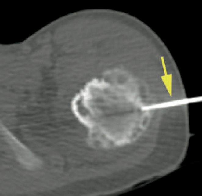
**Modality:** Axial CT-fluoroscopic image. **Anatomic Region:** Subtrochanteric region of the proximal femur. **Pathology/Observation:** The image demonstrates significant osteoproliferative changes characterized by marked periosteal and endosteal new bone formation. The cortical thickening appears robust with smooth outer contours. Internally, multiple intraosseous lytic areas are visible within the thickened bone, suggesting a chronic inflammatory or infectious process, such as chronic osteomyelitis. **Interventional Features:** A radiopaque biopsy needle (indicated by a yellow arrow) is visualized traversing the lateral soft tissues and the thickened cortex to access the medullary compartment for tissue sampling. **Key Diagnostic Features:** * **Bone Reaction:** Dense, smooth periosteal reaction and endosteal scalloping. * **Lucencies:** Multi-focal lytic lesions within the hyperostotic bone. * **Clinical Procedure:** CT-guided percutaneous bone biopsy. **Clinical Context:** These imaging features—specifically the combination of smooth cortical thickening and lytic foci—are characteristic of chronic osteomyelitis or certain benign bone-forming lesions. The image documents the procedural targeting of the lesion for histopathological correlation.
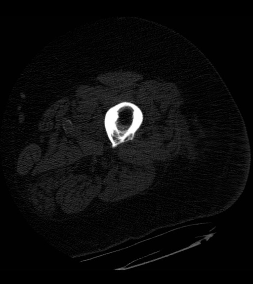
**Imaging Modality:** Non-contrast Computed Tomography (CT), axial cross-section. **Anatomical Region:** Mid-shaft of a long bone (likely femur or humerus) and surrounding soft tissue. **Observed Pathology:** Evidence of chronic osteomyelitis. The cortical bone exhibits significant circumferential thickening (endosteal and periosteal) and increased radiodensity, consistent with reactive sclerosis and involucrum formation. **Characteristic Visual Features:** * **Cortical Bone:** Marked thickening and irregularity of the cortex. The marrow cavity appears narrowed due to endosteal bone formation. * **Osteolysis:** Small, ill-defined lucent areas are visible within the thickened cortex, suggesting active inflammatory or necrotic components within a chronic process. * **Soft Tissue:** The surrounding musculature and subcutaneous fat appear slightly distorted, though no overt abscess or gas collection is visualized in this specific slice. * **Sequestrum/Involucrum:** The image highlights the classic appearance of a dense involucrum (new bone sheath) surrounding the original infected bone. **Clinical Significance:** These CT findings are diagnostic of chronic osteomyelitis. The presence of focal lucencies within the sclerotic bone helps differentiate chronic active infection from a healed, inactive state or simple cortical thickening.
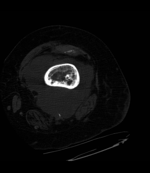
**Imaging Modality:** Axial Computed Tomography (CT) scan. **Anatomical Region:** Mid-shaft of a long bone (likely the femur or tibia) within the lower extremity, surrounded by soft tissue musculature. **Observed Pathology:** The image demonstrates features consistent with osteomyelitis. There is significant cortical thickening (hyperostosis) and endosteal scalloping. The medullary cavity shows heterogeneous attenuation with areas of osteolysis and focal sclerosis. **Characteristic Visual Features:** - **Cortical Changes:** Prominent circumferential thickening of the bony cortex, suggesting a chronic inflammatory process. - **Medullary Findings:** Presence of ill-defined lucent (osteolytic) areas within the marrow space. - **Periosteal Reaction:** Evidence of organized periosteal new bone formation. - **Soft Tissue:** Surrounding soft tissue planes appear relatively preserved in this single section, though mild effacement may be present adjacent to the bone. **Key Diagnostic Features:** The combination of cortical thickening, irregular medullary destruction, and sclerosis are hallmark radiographic signs of chronic osteomyelitis. The lack of a distinct sequestrum or involucrum in this specific slice does not exclude their presence elsewhere in the bone.
Broadening vs Thickening in Osteomyelitis
The Core Distinction
| Feature | Broadening | Thickening |
|---|---|---|
| What changes | Outer diameter / width of the whole bone | Thickness of the cortical wall |
| Direction of new bone | Outward (periosteal - added outside the cortex) | Inward (endosteal) OR outward (periosteal cortical thickening) |
| Mechanism | Periosteum is elevated by subperiosteal pus → lays new bone outside original cortex → bone becomes wider | Periosteal new bone increases the wall thickness of the cortex; endosteal reaction also narrows the medullary canal |
| What you measure on X-ray | Total transverse diameter of the bone at the affected segment | Cortical wall width on cross-section / AP view |
| Anatomical cause | Involucrum forming outside the original cortex | Involucrum + sclerotic cortex + endosteal reaction thickening the wall |
| Result | Bone looks fatter/wider than normal | Bone wall looks denser/heavier; medullary cavity looks narrower |
Visual Concept (Cross-Section of Bone)
NORMAL BONE (cross-section):
        ←— Normal diameter —→
    ___________________________
   |   Medullary cavity        |
   |   [marrow]                |
   |___________________________|
        ←— cortex →←— cortex →
         (thin, uniform)

OSTEOMYELITIS - BROADENING + THICKENING:
        ←——— BROADENED diameter ————→
   _____________________________________
  |NEW BONE     |  ORIGINAL  |NEW BONE  |  ← Involucrum outside
  |(Involucrum) |  CORTEX    |(Involucrum)|   = BROADENING
  |             |____________|          |
  |             | SEQUESTRUM |          |  ← Dead original cortex
  |             |  (dense)   |          |
  |_____________|____________|__________|
  ←— thicker wall —→ ← thicker wall—→   = THICKENING
        ← narrowed medullary cavity →
In Simple Terms
-
Broadening = the bone is wider when you look at it on X-ray (increased outer diameter) - because new bone has been deposited outside the original cortex by the elevated periosteum
-
Thickening = the cortical wall is thicker/heavier - the shell of the bone has increased in thickness, either from periosteal new bone adding to the outside, or endosteal new bone adding to the inside (or both)
They usually occur together in chronic osteomyelitis and both result from the involucrum, but:
- Broadening is a diameter change (how wide the bone is)
- Thickening is a wall change (how thick the cortex is)
The Two Sources of Thickening
1. Periosteal Thickening (outward)
- Periosteum is elevated by subperiosteal pus
- Lay down new bone on the outer surface of the cortex
- Cortex becomes thicker from outside in
- Also contributes to broadening
2. Endosteal Thickening (inward)
- Endosteal reaction lays bone inside the medullary cavity
- Cortex becomes thicker from inside out
- Medullary canal becomes narrower (not broadened - the outer diameter may stay same)
- More prominent in adults and chronic disease (Goldman-Cecil: "Endosteal thickening predominates during infancy and childhood, and periosteal new bone formation is prominent during adulthood")
How They Relate to Each Stage
| Stage | Sign | Explanation |
|---|---|---|
| Acute | Neither visible on X-ray | Too early; only soft tissue swelling on X-ray; MRI shows marrow oedema at 24-48 hrs |
| Subacute (10-21 days) | Early periosteal reaction | Haziness around bone + early periosteal line visible; earliest hint of broadening |
| Chronic | Full broadening + thickening | Dense involucrum outside original cortex → both broader AND thicker on X-ray; sequestrum inside |
"Periosteal new bone appears at 5 to 7 days" (in children) "Osteolysis - 30-50% loss of bone mineral - may not appear until 10 to 14 days"
Key Exam One-Liners
| Statement | Answer |
|---|---|
| Broadening = increase in _____ | Outer diameter of the bone (from periosteal new bone outside) |
| Thickening = increase in _____ | Cortical wall thickness (from periosteal + endosteal new bone) |
| Both are due to | Involucrum formation |
| Involucrum = | Periosteal new bone laid down around sequestrum |
| In children, thickening is mainly | Endosteal |
| In adults, thickening is mainly | Periosteal |
| Broadening narrows the medullary cavity? | No - it widens the outer diameter; medullary cavity is narrowed by endosteal thickening |
| The "bone within a bone" appearance = | Dense sequestrum inside the broader involucrum seen on X-ray |
CT View - Both Signs Together
Summary Table
| Broadening | Thickening | |
|---|---|---|
| Measurement | Total bone diameter (transverse) | Cortical wall width |
| Direction of new bone | Outside original cortex (periosteal) | Both outside (periosteal) AND inside (endosteal) |
| Effect on medullary canal | Not directly | Narrowed by endosteal component |
| Visibility on X-ray | Bone wider than contralateral | Cortex denser and heavier than normal |
| Common cause | Involucrum (chronic osteomyelitis) | Involucrum + endosteal reaction |
| Other causes | Paget's disease, syphilitic periostitis | Sclerosing osteomyelitis (Garré), fluorosis, stress |