

What is this on lower lip
aphthous ulcer minor canker sore lower lip mucosa
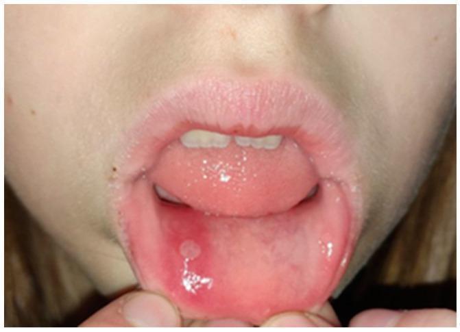
This clinical photograph displays a case of aphthae minor, a common oral ulcerative condition. The image shows the intraoral cavity of a patient with the lower lip everted to reveal a single, well-demarcated minor aphthous ulcer (canker sore). The lesion is approximately 5-8 mm in diameter, characterized by a shallow, circular-to-oval shape with a yellowish-white pseudomembranous base consisting of fibrin and cellular debris. A distinct, bright red (erythematous) halo surrounds the ulcerated area, indicating localized inflammation. The surrounding labial mucosa appears moist and otherwise healthy. The tongue is visible in the background with normal morphology, and there are no signs of generalized stomatitis. This image serves as a classic diagnostic example of Recurrent Aphthous Stomatitis (RAS) for medical and dental education, highlighting the morphological hallmarks used to differentiate minor aphthae from major or herpetiform ulcers.
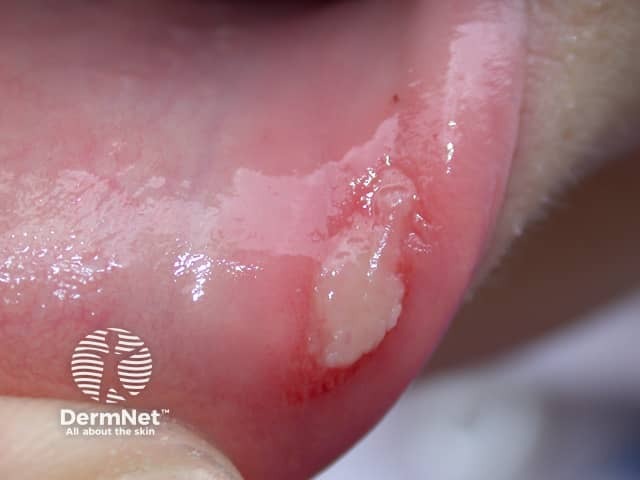
Clinical photography of the lower labial mucosa showing a solitary aphthous ulcer. Imaging modality: intraoral clinical photograph with standard white-light illumination; close-up view; high-resolution. Location: oral cavity, mucosa of the lower lip (labial mucosa) near the vestibule. Appearance: shallow, round-to-oval ulcer with a pale gray-to-yellow floor and a surrounding erythematous halo; mild mucosal edema is evident; bordered margins are distinct. The lesion is non-vesicular; there is minimal exudate and no surrounding necrosis. The specimen type is human oral mucosa; perspective is an anterior-facing close-up. It is compatible with minor aphthous stomatitis (aphthous ulcer), commonly termed canker sore. Differential diagnoses include traumatic ulcer, herpetic gingivostomatitis, erythema multiforme, and candidal ulcer in immunocompromised individuals; clinical correlation with history of oral trauma, stress, nutritional deficiency (iron, B12, folate), or underlying systemic conditions may be required. Typical prognosis is self-limited with spontaneous resolution within 7–14 days; symptomatic relief with topical steroids or protective barriers may be used. Clinically relevant in dentistry, dermatology, oral medicine, and primary care as a common mucosal inflammatory lesion. This image aids education on differential diagnosis, clinical recognition, patient counseling, and management planning. No suspicious features such as ulcerative plaques, vesicles, or necrotic shrouds are evident in this image today.
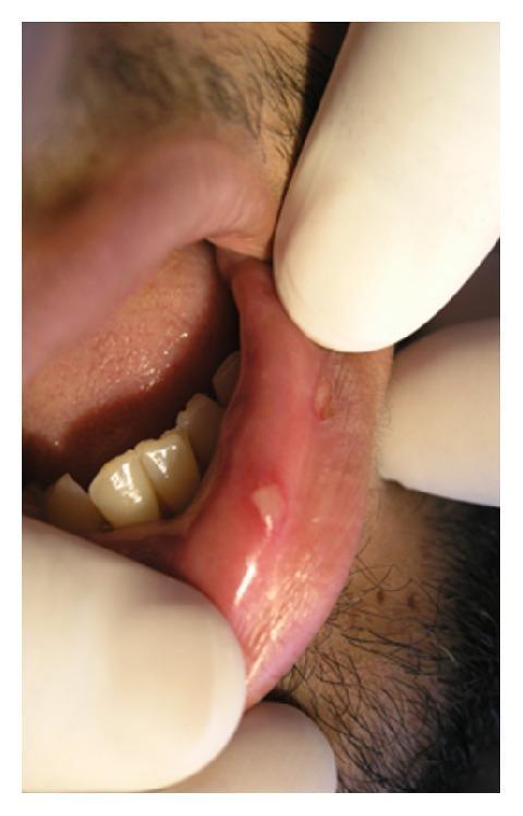
This clinical photograph displays intraoral imaging of the lower labial mucosa, demonstrating the classic presentation of recurrent aphthous stomatitis (RAS). Two distinct, small, round-to-oval ulcers are visible on the non-keratinized inner lining of the lip. The primary lesion is centrally located and characterized by a well-demarcated, yellowish-white pseudomembranous base consisting of fibrin. It is surrounded by a prominent, symmetrical erythematous halo, indicating localized inflammation. A secondary, smaller ulcer with similar morphology is noted more distally on the mucosa. The surrounding tissue appears moist and glistening. Such lesions are typical of minor aphthous ulcers, commonly known as canker sores, which are self-limiting but painful. This image serves as an educational example for diagnosing common inflammatory oral mucosal diseases based on morphological features like the fibrin-covered base and peripheral inflammatory redness.
This Appears to Be a Minor Aphthous Ulcer (Canker Sore)
- Single, well-defined ulcer ~5-8 mm in diameter
- Yellowish-white center (fibrinous slough covering the base)
- Bright red (erythematous) halo around it
- Location on the movable, non-keratinized labial mucosa - exactly where aphthous ulcers prefer
What is an Aphthous Ulcer?
- NOT contagious - not caused by herpes virus (unlike cold sores on the outer lip)
- Painful - especially when eating, drinking, or talking
- Self-limiting - heals on its own in 7-14 days
Common Triggers
- Accidental lip bite or minor trauma
- Stress
- Nutritional deficiencies (iron, vitamin B12, folate)
- Hormonal changes
- Certain foods (acidic, spicy, or salty)
Differential Diagnoses to Keep in Mind
| Condition | Distinguishing Features |
|---|---|
| Minor aphthous ulcer (most likely) | Single ulcer, yellow-white center, red halo, non-keratinized mucosa, very painful |
| Traumatic ulcer | History of biting/sharp food; may be irregular in shape |
| Herpetic ulcer (HSV) | Usually outer lip or hard palate, preceded by blisters, clusters |
| Major aphthous ulcer | >1 cm, deeper, heals over weeks, can scar |
What to Do
- Rinse with warm salt water (1/2 tsp salt in 1 cup warm water) 3-4 times daily
- Avoid spicy, salty, or acidic foods
- Use over-the-counter topical anesthetic gels (e.g., benzocaine gel) to numb the area before meals
- Avoid toothpaste with sodium lauryl sulfate (SLS), which can irritate sores
- Topical triamcinolone acetonide in orabase (a mild corticosteroid paste) speeds healing if available
- The ulcer does not heal within 2 weeks
- It is growing larger or is >1 cm
- You have multiple ulcers at once
- You have fever, swollen lymph nodes, or difficulty swallowing
- This is your first-ever episode and you want confirmation
Disclaimer: This is an educational assessment based on the image. A definitive diagnosis requires in-person clinical examination by a healthcare provider.
Is this something serious
Reassuring signs (your lesion has these ✓)
- Small size (under 1 cm)
- Located on movable, inner lip mucosa
- Single ulcer
- Classic yellow-white center with red border
- No hard/firm base underneath
Warning signs that would warrant a dental/medical visit
| Sign | Why it matters |
|---|---|
| Ulcer persists beyond 3 weeks | Could suggest something other than a canker sore |
| Ulcer grows larger over time | Major aphthous ulcer or rarely, malignancy |
| Hard or raised edges | Squamous cell carcinoma can mimic an ulcer |
| Painless ulcer | Painless oral ulcers are more suspicious than painful ones |
| Occurs with fever, weight loss, fatigue | May signal a systemic condition (Crohn's, Behcet's, HIV) |
| Many ulcers at once | Viral infection or autoimmune condition |
| Recurs very frequently (every few weeks) | Recurrent Aphthous Stomatitis - worth investigating nutritional deficiencies |
Bottom line

What is on my lower lip
What the image shows:
- Small ulcer (~3-5 mm), slightly right of center on the inner lower lip
- Yellowish-white/grayish-white center with a bright red halo
- Round, well-defined borders
- Shallow crater - not a raised lump
- Surrounding tissue looks healthy and normal
A few things to consider since you've had two episodes:
- If it's the same ulcer from before, and it looks smaller/less inflamed - it's healing normally. Good sign.
- If it's a new ulcer appearing shortly after the last one healed, that puts you in the category of Recurrent Aphthous Stomatitis (RAS) - worth investigating.
If you get canker sores repeatedly, check for:
| Possible cause | What to do |
|---|---|
| Iron / B12 / Folate deficiency | Simple blood test with your doctor |
| Stress or poor sleep | Very common trigger |
| SLS in toothpaste | Switch to SLS-free toothpaste |
| Certain foods (nuts, chocolate, citrus) | Try eliminating them |
| Celiac disease | Blood test (anti-tTG IgA) if gut symptoms present |
| Hormonal changes | Common in young adults |
Bottom line
Disclaimer: This is an educational assessment based on visual analysis. For a definitive diagnosis, please consult a dentist or physician in person.
It is from last 3 months
⚠️ 3 Months is a Red Flag - Please See a Doctor
"Of major concern are chronic, relatively painless ulcers... of >2 weeks' duration. Squamous cell carcinoma and premalignant dysplasia should be considered early and a diagnostic biopsy performed. This awareness is critically important because early-stage malignancy is vastly more treatable than late-stage disease. High-risk sites include the lower lip..."
What could cause a 3-month ulcer on the lower lip?
| Condition | Key features |
|---|---|
| Major aphthous ulcer | Benign but large, very painful, heals over weeks - still shouldn't last 3 months |
| Oral squamous cell carcinoma | Most important to rule out - can mimic a simple ulcer; lower lip is a high-risk site |
| Traumatic ulcer | From a sharp tooth or denture rubbing - persists as long as the irritant remains |
| Tuberculosis | Rare but can cause chronic oral ulcers identical to carcinoma |
| Syphilis (chancre or gumma) | Painless ulcer - syphilis is rising in prevalence |
| Crohn's disease | Oral ulcers can occur, especially with gut symptoms |
| HIV/AIDS | Persistent deep aphthous ulcers are a known presentation |
| Fungal infection | Rare, but possible in immunocompromised individuals |
Quick self-check - tell your doctor about these:
- Is the ulcer painful or painless? (Painless ulcers are more suspicious)
- Has it grown in size over 3 months?
- Does it have hard or raised edges when you press around it?
- Do you smoke, use tobacco (any form), or drink alcohol regularly?
- Any unexplained weight loss or fatigue?
- Any swollen lymph nodes under your jaw or in your neck?
What you should do - urgently
- See a dentist or oral surgeon this week - do not wait further
- They will examine it clinically and almost certainly recommend a biopsy (a tiny tissue sample sent to a lab) - this is the only definitive way to rule out cancer
- A biopsy is a simple, quick procedure done under local anesthetic
Disclaimer: This is an educational assessment only. A 3-month non-healing oral ulcer requires in-person clinical evaluation and biopsy to reach a definitive diagnosis. Please consult a dentist or physician promptly.
I am having this but painless only pain when I drink something hot
🔴 Painless + 3 Months = Please See a Doctor This Week
Why painless matters so much
| Condition | Pain level | Duration |
|---|---|---|
| Minor aphthous ulcer (canker sore) | Very painful | 7-14 days |
| Major aphthous ulcer | Painful | Up to 6 weeks |
| Oral squamous cell carcinoma | Often painless early on | Weeks to months, grows |
| Syphilitic chancre | Classically painless | 3-6 weeks if untreated |
| Traumatic ulcer (from tooth/sharp edge) | Mild discomfort | Persists while irritant present |
| Tuberculous ulcer | Less painful than it looks | Chronic |
"Of major concern are chronic, relatively painless ulcers... Squamous cell carcinoma and premalignant dysplasia should be considered early and a diagnostic biopsy performed."
Your full picture puts you in the "must be evaluated" category:
- ✅ Lower lip location (highest risk site for oral cancer)
- ✅ Lasting 3 months (well beyond any benign self-healing lesion)
- ✅ Painless (the most concerning feature)
- ⚠️ Only hurts with hot liquids (heat sensitivity of exposed tissue, not true pain)
What you need to do - do not delay
- Done under local anesthetic (numbing injection)
- Takes about 5-10 minutes
- A tiny piece of tissue is sent to a lab
- Results come back in 5-7 days
- The only way to definitively rule out cancer
One important question to ask yourself:
Disclaimer: This is educational information only and not a diagnosis. Please see a dentist or oral and maxillofacial surgeon in person as soon as possible.
No I don’t eat tobacco and I am also virgin so how can I have syphills
Updated Assessment - No Tobacco, No Sexual Activity
Most likely causes in your situation
- A sharp or broken tooth, rough dental restoration, or hard food repeatedly rubbing the same spot on your inner lip
- Stays painless because the area becomes desensitized over time
- Will NOT heal as long as the irritant keeps touching it
- Fix: A dentist identifies and smooths the sharp tooth/edge - ulcer heals within 2 weeks
- Iron, Vitamin B12, or Folate deficiency are well-documented causes of persistent, recurring oral ulcers
- Very common in young people, especially with restricted diets
- Simple blood test confirms this
- Fix: Supplements - ulcer resolves within weeks
- A more severe form of canker sore - can last weeks to months
- Usually painful, but can become relatively painless over time as the tissue adapts
- Associated with stress, hormonal changes, or immune fluctuations
- Both can cause persistent oral ulcers even without obvious gut symptoms
- Worth testing if blood work is otherwise normal
- Ask yourself: Any unexplained bloating, loose stools, or stomach discomfort?
- Rare under age 40 without tobacco/alcohol, but not impossible
- Cannot be fully excluded without a biopsy
- Does not mean you have it - just means it must be formally excluded
The most practical next steps for you
- They will check if a sharp tooth or rough surface is rubbing that exact spot
- This is the single most common and easily fixable cause in a young non-smoking person
- Complete blood count (CBC)
- Serum iron + ferritin
- Vitamin B12
- Folate
- These are cheap, routine tests
- A biopsy should be done to get a definitive answer
- This is the responsible next step after 3 months of no healing
Reassurance for you
Disclaimer: Educational information only. Please see a dentist or doctor in person for proper evaluation.
What is traumatic ulcer
traumatic oral ulcer lower lip sharp tooth irritation
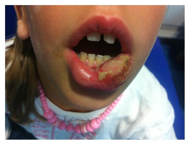
A clinical photograph of a pediatric patient's face, focusing on a self-inflicted traumatic injury to the lower lip. The left side of the lower lip exhibits a large, irregular traumatic ulcer characterized by a central yellowish-white fibrinous slough and a granular texture, indicative of soft tissue necrosis or deep ulceration. The surrounding labial mucosa and vermilion border are erythematous and edematous. Visible within the oral cavity are primary teeth with mild plaque accumulation and gingival inflammation adjacent to the injury site. The photograph demonstrates a classic presentation of post-anesthetic lip biting, a common complication in pediatric dentistry following mandibular nerve blocks or long-acting local anesthesia where numbness leads to inadvertent masticatory trauma. This image serves as an educational tool for identifying traumatic oral ulcers and emphasizes the importance of postoperative caregiver education regarding numbing effects in children.
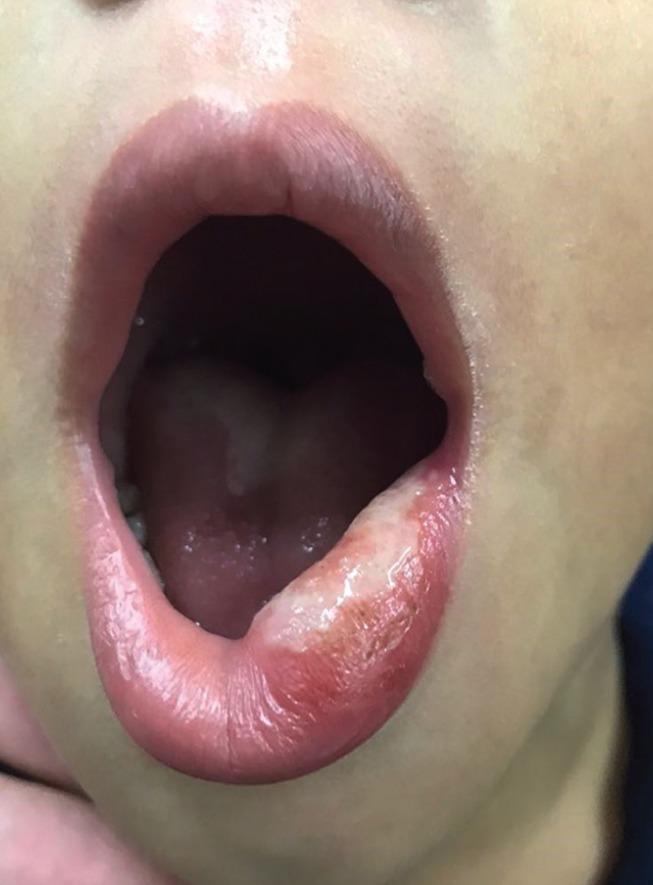
This clinical photograph displays a large traumatic ulcer located on the left side of the lower lip in a pediatric patient. The lesion is characterized by a prominent, irregularly shaped whitish-yellow fibrinopurulent plaque covering the mucosal and vermilion surface. The surrounding tissue exhibits significant erythema and edema, indicating an active inflammatory response. The ulceration appears localized to the area of mechanical trauma (lip-biting), with the adjacent upper lip and intraoral mucosa appearing healthy and pink. This image serves as a clinical example of post-anesthetic lip-biting, a common complication in pediatric dentistry. It is an educational tool for identifying traumatic oral lesions and understanding the clinical presentation of secondary pseudomembranous formation during the healing phase.
Traumatic Ulcer - Explained Simply
What it is
What causes it
| Cause | How it happens |
|---|---|
| Sharp or broken tooth | A jagged tooth edge rubs the same spot on your inner lip repeatedly every time you talk, chew, or swallow |
| Rough dental filling | An uneven restoration scratches the mucosa constantly |
| Accidental lip bite | Biting the inside of your lip once - if the tooth keeps catching the same spot, it won't heal |
| Hard or sharp foods | Crusty bread, chips, bones - a one-time injury that gets re-traumatized |
| Ill-fitting denture/brace | Rubbing from orthodontic wires or braces |
| Toothbrush injury | Aggressive brushing in the same area |
Why it stays painless
- When the same tissue is irritated over and over for months, the nerve endings in that area become desensitized
- The ulcer becomes "numb" to everyday irritation
- You only feel it with temperature extremes (like hot drinks) because heat bypasses that desensitization
- This is exactly what you described
How it looks
- Yellow-white base (just like an aphthous ulcer)
- Red border around it
- Usually in a location that corresponds to a nearby sharp tooth or dental edge
- Does NOT change much in size over time - it stays roughly the same
The critical difference from a canker sore
| Feature | Canker sore | Traumatic ulcer |
|---|---|---|
| Cause | Immune/nutritional | Physical damage |
| Pain | Very painful | Mild or painless (chronic) |
| Heals on its own? | Yes, 7-14 days | No - keeps returning while irritant present |
| Fix | Supportive care | Remove the cause |
A simple self-test you can do right now
- A sharp, chipped, or jagged tooth edge
- A rough or uneven filling
- Anything that feels like it could be catching or scraping the same spot