
Cervical spondylosis features and treatment
cervical spondylosis X-ray MRI features
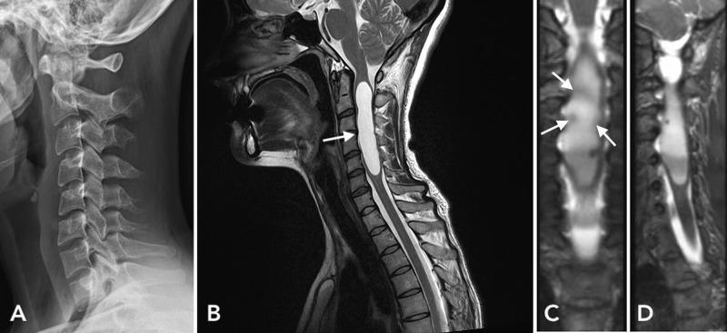
This composite figure presents multimodality clinical imaging of the cervical spine. (A) Lateral X-ray radiography demonstrates widening of the cervical spinal canal between the C3 and C5 vertebral levels, accompanied by mild degenerative spondylosis at C6-C7 and a slight kyphotic curvature. (B) A sagittal T2-weighted MRI reveals a significant hyperintense intramedullary lesion, consistent with a large syrinx, extending from C2 to C6. (C) Coronal T2-weighted MRI view identifies multiple small mural nodules along the margin of the cystic lesion, with arrows highlighting areas of potential contrast enhancement. (D) Corresponding sagittal MRI view provides longitudinal visualization of the cystic intramedullary mass. Collectively, these images illustrate the diagnostic features of a spinal intramedullary mass, specifically a cystic tumor with mural nodules (later pathologically confirmed as pilocytic astrocytoma) and its associated secondary effects on the spinal canal and vertebral alignment.
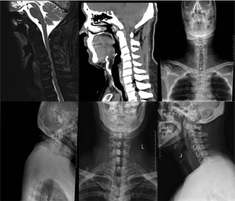
A multi-modal radiographic composite illustrating the cervical spine and surgical outcomes for cervical spondylosis. The upper panels display a sagittal T2-weighted MRI, a sagittal CT scan, and an anteroposterior (AP) X-ray of the head, neck, and upper thorax. The MRI highlights soft tissue structures, specifically showing cerebrospinal fluid (bright white) and the spinal cord. The CT emphasizes bony morphology and vertebral alignment. The lower panels provide follow-up radiographs, including AP and lateral X-rays, demonstrating post-surgical status. These images reveal multi-level internal fixation, specifically ROI-C interbody spacers placed within the intervertebral disc spaces of the middle to lower cervical spine. On the lateral view, the radiopaque fixators are visible across four segments, restoring disc space height and maintaining cervical lordosis (Cobb angle). This educational material demonstrates the combined use of diagnostic imaging (MRI/CT) and postoperative monitoring (radiography) in managing multi-segment spinal cord compression and evaluating the stability of anterior cervical discectomy and fusion (ACDF) instrumentation.
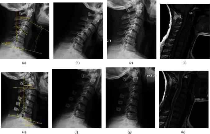
A comparative radiographic and MRI study of the cervical spine in a 42-year-old female patient with cervical spondylosis, highlighting pre-operative (a-d) and two-year post-operative (e-h) results. The pre-operative lateral X-ray (a) demonstrates a kyphotic alignment (Cobb angle ̑ = -13.3°) and measurement of T1 slope (β). Dynamic flexion (b) and extension (c) views assess the range of motion. The pre-operative sagittal MRI (d) shows baseline spinal cord compression. Post-operative lateral X-ray (e) following double-door laminoplasty shows improved cervical lordosis (̑1 = 10.6°) and the presence of radiopaque hydroxyapatite spacers within the expanded posterior elements. Post-operative dynamic X-rays (f, g) and sagittal MRI (h) illustrate preserved spinal stability and increased space for the spinal cord (decompression). Annotations on lateral views depict Cobb angles (C2-C7), T1 slope, and C2-C7 Sagittal Vertical Axis (SVA), providing quantitative metrics for surgical correction and sagittal balance.
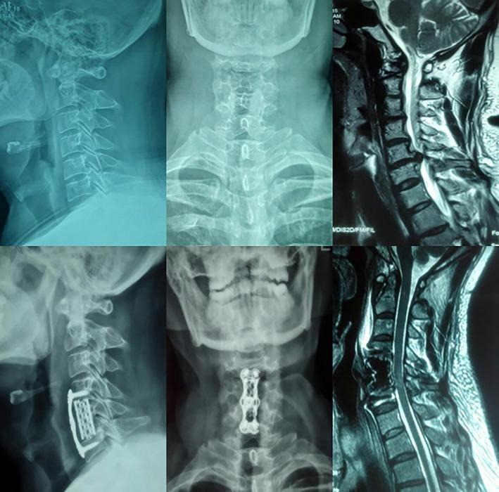
A six-panel medical image composite showing preoperative and postoperative radiographic and MRI views of the cervical spine. The top row displays preoperative status: lateral X-ray, anteroposterior (AP) X-ray, and sagittal T2-weighted MRI, showing cervical spondylosis and intervertebral disc narrowing. The bottom row presents postoperative imaging after anterior cervical decompression and fusion (ACDF): a lateral X-ray (left) shows a radiopaque metallic anterior cervical plate and screw system spanning two motion segments, with an interbody cage visible; an AP X-ray (center) displays the rectangular plate with a central perspective window and four-screw fixation; and a sagittal MRI (right) shows the decompressed spinal canal. The titanium alloy hardware appears as highly radiopaque structures on X-ray and signal voids on MRI. This visual demonstrates surgical stabilization using a dynamic anterior cervical nail plate system for multilevel cervical spine pathologies.
"cervical spondylosis" treatment management
Cervical Spondylosis: Features and Treatment
Epidemiology
- Peak incidence between age 40-60 years; nearly universal radiographic changes by age 70
- Men are affected more than women
- Most common levels: C5-C6 (most frequent), followed by C6-C7
- By age >50, approximately 75% of asymptomatic individuals have radiologic evidence of canal narrowing; only half develop signs of root or cord involvement
- Adams and Victor's Principles of Neurology, p. 1268
- Miller's Review of Orthopaedics, p. 758
Pathoanatomy
- Two uncovertebral joints (of Luschka)
- Two facet joints - their capsules contain sensory receptors contributing to pain and proprioception
Sequence of changes
- Disc degeneration - annular fraying, nucleus pulposus extrusion or bulging
- Osteophyte/spondylotic bar formation - "hard disc" with disc-osteophytic spurs; posterior ridging compresses cord and roots
- Progressive disc collapse - loss of normal cervical lordosis, chronic anterior cord compression
- Facet and uncovertebral joint loading - spondylotic foraminal narrowing restricts motion and compresses nerve roots
- Ligamentum flavum hypertrophy and posterior compression (especially in extension)
- Dural thickening and adhesion to posterior longitudinal ligament
- Spinal cord changes - demyelination, focal necrosis at dentate ligament attachment zones, posterior and lateral column rarefaction
Canal dimensions (lateral plain radiograph)
| Classification | Canal diameter |
|---|---|
| Normal | ≥14 mm |
| Relative stenosis | 10-13 mm |
| Absolute stenosis | <10 mm |
-
Pavlov (Torg) ratio (canal width / vertebral body width): normal = 1.0; ratio <0.8 is considered a risk factor for neurologic involvement
-
Dynamic compression: neck extension worsens cord compression (cord squeezed between anterior spondylotic bar and posterior infolded ligamentum flavum); neck flexion transiently relieves it
-
Miller's Review of Orthopaedics, p. 759
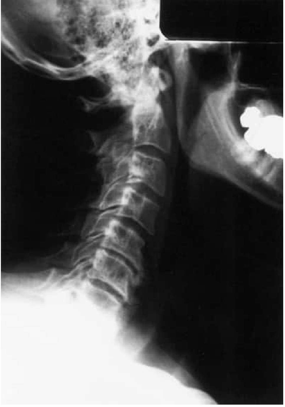
Lateral X-ray: Typical Spondylosis Changes
Clinical Entities
1. Discogenic (Axial) Neck Pain
- Insidious onset of neck pain without neurologic deficits
- Exacerbated by excess vertebral motion
- Occipital headache is common
- Normal neurologic examination
2. Cervical Radiculopathy
- Nerve root compression - often C5, C6, or C7
- Sharp, radiating pain from neck down the arm; may be acute in onset
- Paresthesias in specific dermatomal distribution (fingers, palm, forearm band)
- Depressed biceps/brachioradialis reflexes (C5/C6) or triceps reflex (C7)
- Wasting of hand or forearm muscles in severe cases
- Spurling test (neck extension + lateral rotation toward affected side) reproduces radicular pain
- Younger patients (<45 yrs): more often "soft disc" herniation; older patients: more often foraminal osteophytic stenosis
3. Cervical Myelopathy (Spondylotic Myelopathy) - the Most Serious
- Neck pain / brachialgia - painful stiff neck, pain radiating to shoulders and upper arms, aching or sharp radicular pain on movement
- Numbness and paresthesias of the hands - often earliest symptom; "feeling as if wearing gloves," "hands coated with glue," "swollen" feeling
- Spastic leg weakness with Babinski signs, unsteady gait, Romberg sign
- Lhermitte's sign (neck flexion causes electrical feelings down the spine)
- Impaired vibration and position sense in toes and feet (posterior column)
- Asymmetric sensory deficits; rarely Brown-Sequard pattern
- Mirror movements of hands in advanced cases
- Bladder hesitancy/precipitancy; frank incontinence is late
- Brisk triceps and finger reflexes with depressed biceps/brachioradialis (inverted reflex pattern)
- Fasciculations in proximal arm muscles
4. Myeloradiculopathy
-
Combined spinal cord and nerve root compression at multiple levels
-
Adams and Victor's Principles of Neurology, p. 1268-1269
-
Miller's Review of Orthopaedics, p. 758
Investigations
| Modality | Findings |
|---|---|
| Plain X-ray (lateral) | Disc space narrowing, osteophytes, loss of lordosis, foraminal narrowing; measure canal diameter and Torg ratio |
| MRI | Investigation of choice; shows disc herniation, cord signal change (T2 hyperintensity = myelomalacia), foraminal stenosis, ligamentum flavum hypertrophy |
| CT myelography | Alternative when MRI contraindicated; delineates bony canal encroachment |
| EMG/NCS | Differentiates radiculopathy from peripheral nerve pathology |
Treatment
Conservative (Non-surgical) Management
- Rest and activity modification - brief period of collar use (soft cervical collar) to limit motion; prolonged immobilization not recommended
- Analgesics and NSAIDs - for pain relief
- Physiotherapy - gentle cervical traction, strengthening, and postural correction exercises
- Epidural corticosteroid injections - for radicular pain not responding to oral medications
- Cervical manipulation - used cautiously (risk of worsening neurologic compromise)
- The 2025 systematic review on health qigong found benefit from exercise-based programs for symptom relief
Surgical Management
- Cervical myelopathy (progressive or moderate-to-severe)
- Radiculopathy with severe or progressive neurologic deficit failing conservative treatment
- Intractable pain
| Approach | Procedure | Indication |
|---|---|---|
| Anterior | ACDF (Anterior Cervical Discectomy and Fusion) | Single or multi-level disc/osteophyte disease; direct decompression |
| Anterior | Cervical Disc Replacement (arthroplasty) | Single-level disease in younger patients; preserves motion |
| Posterior | Laminectomy ± fusion | Multilevel cord compression with preserved lordosis |
| Posterior | Laminoplasty (open/double-door) | Multilevel cord compression; preserves motion; common in Asia |
Key Distinguishing Points
-
Radiologic spondylosis is ubiquitous and does not always explain symptoms - the clinical and imaging findings must correlate
-
Symmetric sensory symptoms mimicking spondylotic myelopathy are also seen in subacute combined degeneration (B12 deficiency) - always check B12
-
Acute worsening or paraplegia/quadriplegia can follow forceful flexion-extension neck injuries in patients with pre-existing canal stenosis
-
Cervical spine surgery in purely degenerative disease is not justified for headache or neck pain in the absence of radiculopathy or myelopathy
-
Adams and Victor's Principles of Neurology 12th Ed., p. 1268-1270
-
Miller's Review of Orthopaedics 9th Ed., p. 758-760
-
Bradley and Daroff's Neurology in Clinical Practice, p. 2567-2568