
teach me everything i needf to know about ischemic stroke use all kinds of sourses and mainly harrisons latest edition and also add images
ischemic stroke brain CT MRI infarction
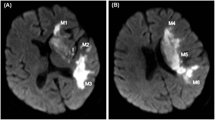
This diagnostic imaging set consists of two axial Diffusion-Weighted Imaging (DWI) MRI scans of the brain (A and B) demonstrating acute ischemic stroke. The images utilize the Alberta Stroke Program Early CT Score (ASPECTS) topographic system to map the extent of cerebral infarction within the Middle Cerebral Artery (MCA) territory. Image A, at the level of the basal ganglia, shows significant hyperintense signals (diffusion restriction) involving the insula (labeled 'I'), lentiform nucleus, internal capsule, and the M1, M2, and M3 cortical zones. Image B, at the suprabasal ganglion level, reveals contiguous infarct expansion into the M4, M5, and M6 cortical regions. The extensive hyperintensity across these zones indicates a large-territory MCA infarction. This clinical example serves as a teaching tool for neuroradiological assessment of stroke severity, infarct growth monitoring, and the application of standardized anatomical labeling (M1-M6) in acute stroke management.
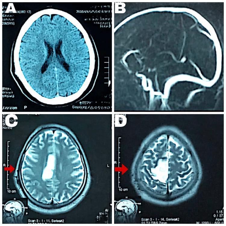
This composite of diagnostic neuroimaging displays a comparison between CT and MRI findings in a case of cerebral infarction. Panel A shows an axial non-contrast CT scan of the brain with no acute intracranial abnormalities visible. Panel B is a sagittal magnetic resonance venography (MRV) showing major venous sinuses. Panels C and D are axial MRI Fluid-Attenuated Inversion Recovery (FLAIR) sequences at different superior levels. These FLAIR images reveal a hyperintense, well-demarcated lesion in the right parietal region, specifically within the vascular territory of the anterior cerebral artery (ACA). Red arrows highlight this area of high signal intensity, which is characteristic of an acute to subacute ischemic infarct. The imaging demonstrates the superior sensitivity of MRI FLAIR sequences over standard CT for detecting early ischemic changes and edema. This resource is suitable for medical education regarding stroke localization, neuroimaging modalities, and the radiographic appearance of vascular insults in the brain.
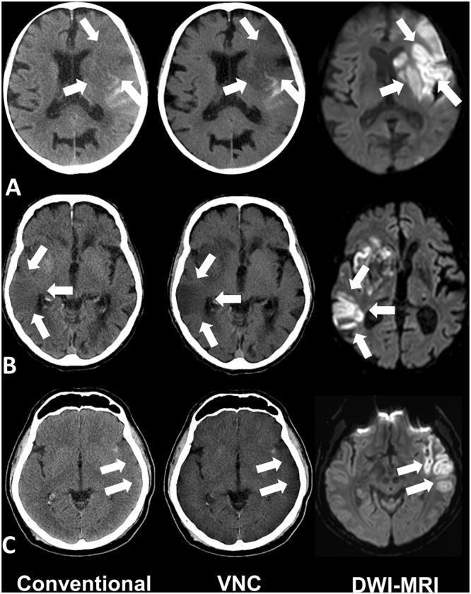
This diagnostic imaging panel displays axial brain scans from three different patients (A, B, and C) comparing three modalities: Conventional Non-Contrast CT, Virtual Non-Contrast (VNC) spectral CT, and Diffusion-Weighted Imaging (DWI-MRI). The images illustrate acute ischemic stroke following mechanical thrombectomy. White arrows indicate regions of acute infarction. In the 'Conventional' CT column, the ischemic lesions appear as subtle, poorly defined areas of hypodensity (low attenuation) within the brain parenchyma. In the 'VNC' CT column, these same lesions demonstrate more pronounced hypodensity and increased contrast-to-noise ratio, making the infarct boundaries clearer compared to conventional CT by removing potential iodine contrast masking. The 'DWI-MRI' column serves as the gold standard, showing corresponding regions of high signal intensity (hyperintensity), which represents restricted water diffusion characteristic of acute cytotoxic edema. The cases highlight infarctions in various territories, including the left and right middle cerebral artery (MCA) regions. This comparison demonstrates the clinical utility of spectral CT VNC reconstructions in improving the detection of early ischemic changes that may be subtle on standard CT.
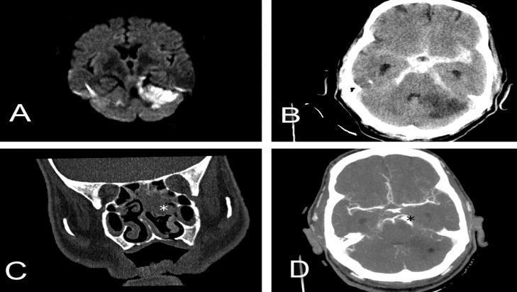
This composite of diagnostic imaging illustrates various neurovascular and otorhinolaryngological pathologies across multiple modalities. Panel A: Axial diffusion-weighted magnetic resonance imaging (DWI MRI) demonstrating hyperintensity in the left cerebellar hemisphere, consistent with restricted diffusion in acute ischemic infarction. Panel B: Non-contrast axial head CT showing diffuse hyperdensity within the basal cisterns and sulci, characteristic of extensive subarachnoid hemorrhage (SAH), alongside a hypodense region in the cerebellum representing secondary ischemic stroke. Panel C: Coronal CT scan of the paranasal sinuses revealing opacification of the nasal cavity and ethmoid cells (marked with a white star), indicating an inflammatory or infectious process such as sinusitis. Panel D: Axial brain CT angiography (CTA) showing vascular morphology, specifically highlighting two consecutive fusiform aneurysms localized in the superior cerebellar artery (marked with a black star). The set provides a comprehensive visual comparison of acute stroke, intracranial hemorrhage, infectious sequelae, and vascular malformations for medical education in radiology and neurology.
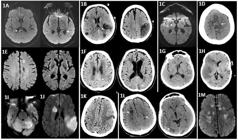
A multi-panel figure consisting of axial non-contrast Computed Tomography (CT) scans and Diffusion-Weighted Magnetic Resonance Imaging (DWI-MRI) showing various patterns of acute and subacute ischemic stroke in the brain. The CT images (B, D, F, G, H, K, L) demonstrate areas of hypodensity indicating cytotoxic edema or established infarction, notably involving the middle cerebral artery (MCA) and anterior cerebral artery (ACA) territories, with some cases showing large territorial infarcts (H) and others showing bilateral involvement (B, L). The MRI panels (A, C, E, I, J, M) utilize diffusion-weighted sequences to highlight acute ischemic lesions as bright hyperintensities, reflecting restricted water diffusion. Anatomical regions affected include the frontal, temporal, parietal, and occipital lobes, as well as the cerebellum (I), corpus callosum (A), and deep white matter structures such as the corona radiata and centrum semiovale (M). These images illustrate both focal and multifocal (embolic-type) patterns of cerebral infarction, serving as a comparative educational resource for neuroimaging modalities in vascular neurology.
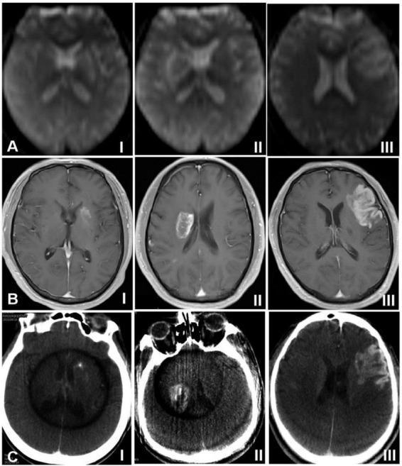
This diagnostic imaging composite illustrates the radiological classification of hemorrhagic transformation (HT) in the brain across three modalities: (A) Arterial Spin Labeling (ASL) MRI, (B) Dynamic Contrast-Enhanced (DCE) MRI, and (C) Xper CT. The images are categorized into three types based on the morphology and volume of the lesion relative to the cerebral infarction focus. Type I demonstrates focal HT, characterized by high signal (MRI) or high density (CT) distributed in small dots or stripes along the periphery of the infarct. Type II shows confluent patches of high signal/density within the infarct area, involving less than 30% of the total infarction volume. Type III represents the most severe form, where high-intensity signals or high-density contrast extravasation appear as dense sheets occupying 30% or more of the infarction focus. These findings are critical for post-endovascular treatment monitoring, where high signals on ASL and DCE indicate increased regional cerebral blood flow and blood-brain barrier permeability, respectively. The images provide a standardized visual reference for neuroradiology and neurology education regarding post-ischemic stroke complications.
"ischemic stroke"[MeSH Terms] AND "management"[MeSH Terms]
stroke pathophysiology cerebral blood flow penumbra thrombus
stroke syndromes middle cerebral artery territory clinical deficits
ischemic stroke 2024 2025 updated guidelines tPA thrombectomy AHA ASA
mechanical thrombectomy endovascular stroke treatment stent retriever
Ischemic Stroke - A Comprehensive Guide
Primary Source: Harrison's Principles of Internal Medicine, 22nd Edition (2025)
1. DEFINITION & EPIDEMIOLOGY
2. PATHOPHYSIOLOGY
The Ischemic Cascade
"A decrease in cerebral blood flow to zero causes death of brain tissue within 4-10 min; values <16-18 mL/100 g tissue per min cause infarction within an hour."
- Harrison's 22E, p. 3484
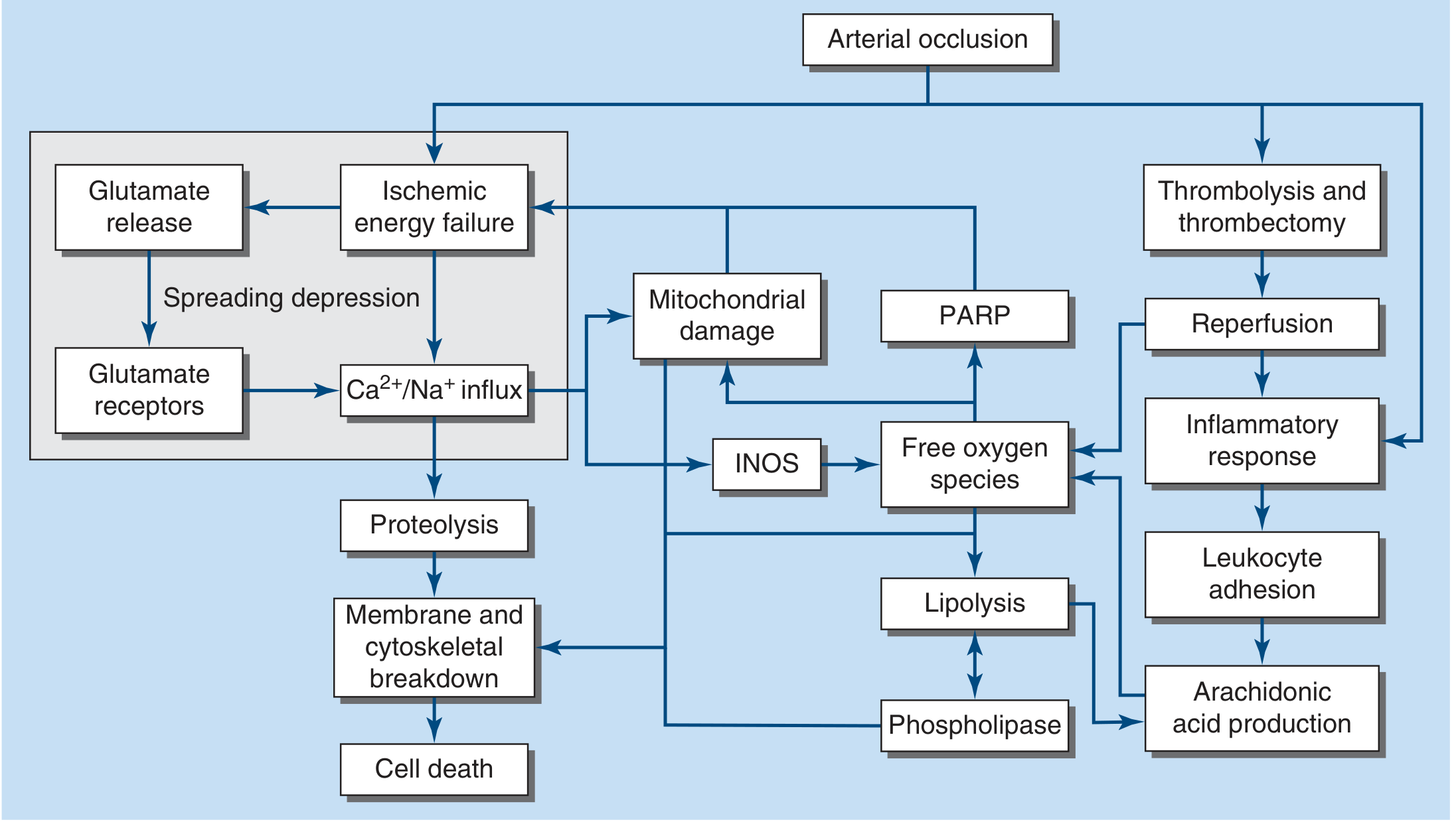
The Ischemic Core vs. Penumbra
- Ischemic core: Irreversibly infarcted tissue (CBF near zero)
- Ischemic penumbra: Surrounding tissue with severely reduced but not zero flow - electrically silent but still viable. This is the therapeutic target.
- The penumbra can survive for hours if reperfusion is achieved - this is the biological rationale for tPA and thrombectomy.
3. ETIOLOGY
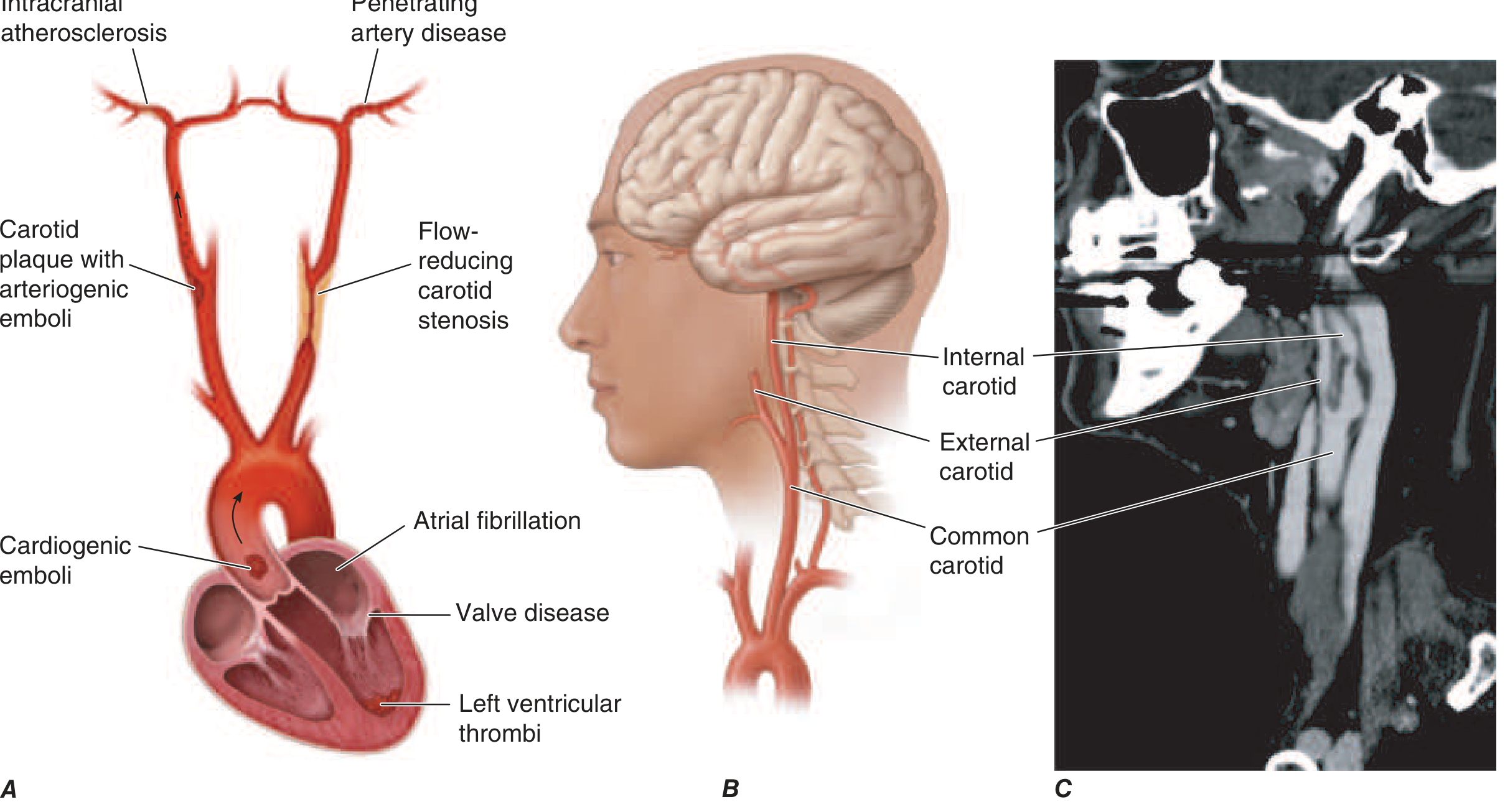
Causes Summary (Harrison's Table 438-2)
| Common Causes | Uncommon Causes |
|---|---|
| Lacunar (small vessel) stroke | Hypercoagulable states (protein C/S deficiency, APS, Factor V Leiden) |
| Large-vessel thrombosis | Sickle cell anemia |
| Carotid bifurcation embolism | Systemic lupus erythematosus |
| Aortic arch embolism | Homocysteinemia |
| Atrial fibrillation (most common cardiac source) | CADASIL |
| Mural thrombus post-MI | Moyamoya disease |
| Dilated cardiomyopathy | Fibromuscular dysplasia |
| Mitral stenosis / mechanical valve | Drug-induced (cocaine, methamphetamine) |
| Bacterial endocarditis | Oral contraceptive-related |
A. Cardioembolic Stroke
B. Large-Vessel Atherothrombosis
C. Small-Vessel (Lacunar) Stroke
"The term lacunar infarction refers to infarction following atherothrom-botic or lipohyalinotic occlusion of a small artery in the brain... Small-vessel strokes account for ~20% of all strokes."
- Harrison's 22E, p. 3489
- Pure motor hemiparesis - infarct in posterior limb of internal capsule or pons
- Pure sensory stroke - infarct in ventral thalamus
- Ataxic hemiparesis - infarct in ventral pons or internal capsule
- Dysarthria-clumsy hand - infarct in ventral pons or genu of internal capsule
4. STROKE SYNDROMES BY TERRITORY
Anterior Circulation (Internal Carotid Artery system)
Middle Cerebral Artery (MCA) - Most common stroke territory
| MCA Division | Territory | Dominant Hemisphere | Non-Dominant Hemisphere |
|---|---|---|---|
| Complete MCA occlusion | Entire lateral hemisphere | Contralateral hemiplegia, hemisensory loss, hemianopia + global aphasia | Contralateral hemiplegia + anosognosia, neglect, constructional apraxia |
| Superior division | Frontal + superior parietal cortex | Broca's (non-fluent) aphasia + arm > leg weakness | Neglect |
| Inferior division | Temporal + inferior parietal cortex | Wernicke's (fluent) aphasia | Hemineglect |
| Lenticulostriate branches | Deep structures | Dysarthria, contralateral hemiplegia (capsule) | - |
Anterior Cerebral Artery (ACA)
- Supplies medial frontal and parietal lobes
- Causes contralateral leg > arm weakness (homunculus leg area is medial)
- Bilateral ACA infarction causes abulia, paraparesis, incontinence
Internal Carotid Artery (ICA)
- May be asymptomatic if good collaterals exist
- Complete occlusion: massive MCA + ACA territory infarction
- May cause monocular blindness (amaurosis fugax) from ophthalmic artery involvement
Posterior Circulation (Vertebrobasilar system)
Posterior Cerebral Artery (PCA)
- Supplies occipital lobe and inferior temporal cortex
- Contralateral homonymous hemianopia (macular sparing often present)
- Dominant PCA: alexia without agraphia, anomia
- Thalamic branches: thalamic syndrome (contralateral pain and temperature loss)
Basilar Artery
- Most devastating posterior circulation stroke
- "Top of the basilar" syndrome: bilateral visual loss, pupillary changes, somnolence, memory disturbance (bilateral thalamic/occipital infarction)
- Basilar artery thrombosis causing locked-in syndrome: complete tetraplegia + cranial nerve palsies, but preserved consciousness and vertical eye movements
Vertebral Artery / PICA - Lateral Medullary (Wallenberg) Syndrome
- Ipsilateral: facial pain/numbness (V), Horner's syndrome, ataxia, dysphagia, hoarseness
- Contralateral: body pain/temperature loss (spinothalamic tract)
- Vertigo, nausea, vomiting (vestibular nuclei)
- No motor weakness (corticospinal tract spared)
5. NEUROIMAGING
CT - First-line Imaging
- Fast and widely available
- Reliably rules out hemorrhage (critical before giving tPA)
- Early ischemic changes in <6h: loss of gray-white differentiation, sulcal effacement, hyperdense MCA sign (clot)
MRI - Superior Sensitivity
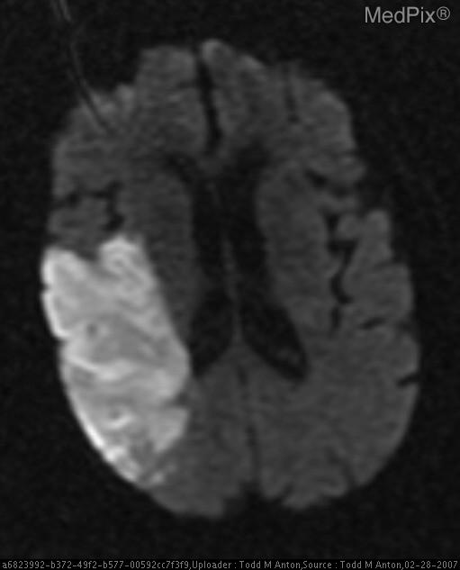
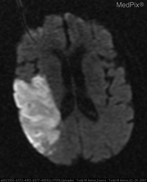
"Diffusion-weighted imaging (DWI) identifies regions of brain infarction within minutes of the stroke onset, while fluid-attenuated inversion recovery (FLAIR) imaging reliably reveals areas of prior brain infarction from a few days to years later." - Harrison's 22E, p. 3460
- Posterior fossa and cortical surface infarcts
- Small lacunar infarcts
- Distinguishing new from old infarcts (DWI bright = acute; FLAIR bright = subacute/chronic)
Vascular Imaging
- CTA: Fast, excellent for large vessel occlusion (LVO) - required for thrombectomy decision
- MRA: Good for carotid stenosis and intracranial vessels; tends to overestimate stenosis
- Conventional angiography (DSA): Gold standard, reserved for uncertain cases or intervention
6. ACUTE MANAGEMENT
"After the clinical diagnosis of stroke is made, an orderly and prompt process of evaluation and treatment should follow. The first goal is to prevent or reverse brain injury." - Harrison's 22E
1. Medical Support (Stabilization)
| Parameter | Target |
|---|---|
| Blood pressure | Do NOT lower unless >220/120 mmHg (or >185/110 if tPA candidate) |
| Blood glucose | Treat hypoglycemia AND hyperglycemia immediately |
| Temperature | Treat fever aggressively (fever is detrimental) |
| O₂ saturation | Maintain SpO₂ ≥94% |
| DVT prophylaxis | Subcutaneous heparin + pneumatic compression stockings |
2. IV Thrombolysis (tPA)
- Any prior intracranial hemorrhage
- Active internal bleeding
- Recent intracranial/spinal surgery (<3 months)
- Recent major trauma/head injury (<3 months)
- Recent MI (<3 months)
- BP >185/110 mmHg (not controllable)
- Platelet count <100,000
- Current anticoagulation with elevated INR or direct anticoagulants
"Tenecteplase (TNK) - Multiple trials including the Canadian ACT-QUICKR support non-inferiority to alteplase with potential superior reperfusion and the major practical advantage of single-bolus administration. Many systems are transitioning to TNK as first-line."
3. Endovascular Revascularization (Mechanical Thrombectomy)
- Large vessel occlusion (ICA, MCA M1/M2, basilar)
- NIHSS ≥6 typically
- Within 6 hours: Based on ESCAPE, MR CLEAN, SWIFT PRIME, EXTEND-IA trials
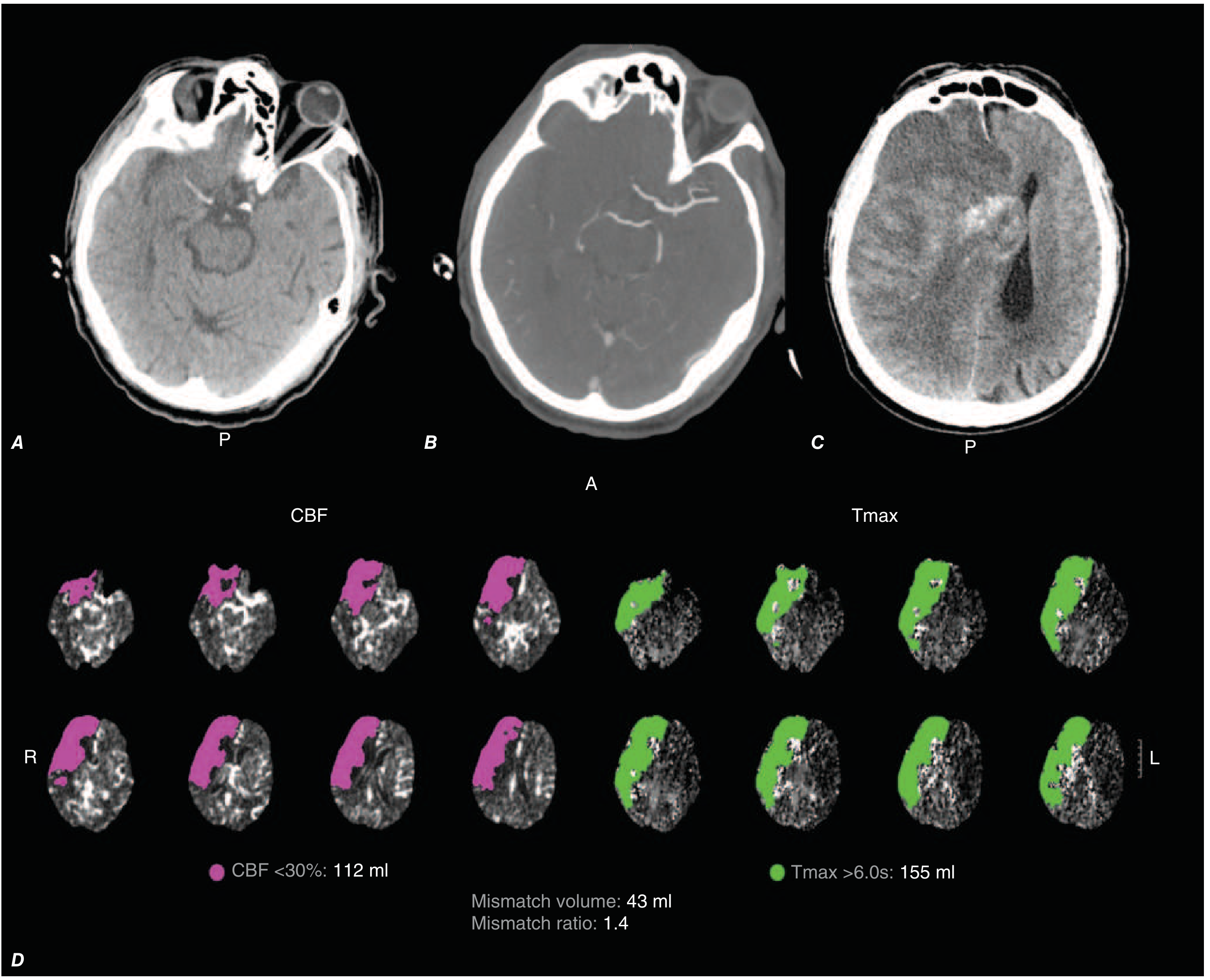
- 6-24 hours: With advanced imaging (CT perfusion or DWI-MRI) showing favorable core-penumbra mismatch (DAWN and DEFUSE-3 trials)
"EVT became standard of care with the 2015 NEJM trials (≤6h), then expanded to up to 24h based on late-window selection by advanced imaging. Across meta-analyses, NNT ≈4 for significantly improved functional outcomes."
- Ischemic core (CBF <30%, pink on CT perfusion) = already dead tissue
- Penumbra (Tmax >6s, green) = salvageable tissue
- If mismatch volume >10 mL and ratio >1.8: proceed with thrombectomy regardless of time window
4. Antithrombotic Treatment
- Non-tPA patients: Aspirin 160-325 mg within 24-48 hours
- After tPA: Withhold aspirin for 24 hours
- Minor stroke (NIHSS ≤3) / high-risk TIA: Dual antiplatelet therapy - aspirin + clopidogrel for 21 days (POINT and CHANCE trials), then single antiplatelet
- Cardioembolic stroke (AF): Start anticoagulation, but timing must balance benefit vs. hemorrhagic transformation risk (typically 2 weeks post-stroke for large infarcts)
5. Neuroprotection
6. Stroke Centers and Rehabilitation
"Patients who receive care in the setting of a stroke unit are 3-8% more likely to survive and return home than patients treated in general medical wards." - Harrison's 22E, p. 3491
- Stroke unit care is independently associated with better outcomes
- Early physiotherapy, speech therapy, and occupational therapy improve recovery
- Constraint-induced movement therapy (CIMT) is evidence-based for upper limb rehabilitation
7. HEMORRHAGIC TRANSFORMATION
- Type I: Petechial hemorrhage along infarct margins
- Type II: Confluent patches within infarct (<30% of volume)
- Type III: Dense hemorrhage involving ≥30% of infarct
8. PRIMARY & SECONDARY PREVENTION
Risk Factor Modification
| Risk Factor | Target / Intervention |
|---|---|
| Hypertension | <130/80 mmHg (thiazides + ACE inhibitors have strongest data) |
| Diabetes | Glycemic control; pioglitazone beneficial in prediabetes/insulin resistance with prior stroke |
| Dyslipidemia | LDL <70 mg/dL; statin for all patients with prior ischemic stroke |
| Smoking | Complete cessation |
| Atrial fibrillation | Anticoagulation (see below) |
Antiplatelet Therapy
- Aspirin: 50-325 mg/d - first-line for non-cardioembolic stroke
- Clopidogrel: 75 mg/d - equivalent or slightly superior to aspirin
- Aspirin + extended-release dipyridamole (Aggrenox): Superior to aspirin alone (ESPS-2, ESPRIT trials)
- Short-term DAPT (aspirin + clopidogrel): 21 days for minor stroke/high-risk TIA (CHANCE, POINT trials), reduces recurrent stroke by ~32%
Anticoagulation
| Indication | Therapy |
|---|---|
| Atrial fibrillation | Direct oral anticoagulants (DOACs) preferred over warfarin: dabigatran, rivaroxaban, apixaban, edoxaban |
| Mechanical heart valves | Warfarin (DOACs are contraindicated) |
| Patent foramen ovale (PFO) | Closure device superior to anticoagulation for cryptogenic stroke in younger patients |
| Non-cardioembolic stroke | Anticoagulation is NOT superior to antiplatelet therapy |
Carotid Revascularization
- Carotid endarterectomy (CEA): Indicated for symptomatic stenosis ≥70% (7-10 NNT to prevent one stroke at 2 years). Benefit is less clear for 50-69%.
- Carotid artery stenting (CAS): Alternative to CEA in patients with high surgical risk
9. SPECIAL SITUATIONS
CADASIL
Arterial Dissection
- Spontaneous or traumatic dissection of ICA or vertebral artery
- Often causes stroke in young patients
- Neck pain + Horner's syndrome + stroke = classic triad for carotid dissection
- Diagnosed with MRI fat saturation sequences
Cryptogenic Stroke
10. NEUROLOGICAL DEFICITS - RAPID CLINICAL LOCALIZATION
| Deficit | Location |
|---|---|
| Contralateral hemiplegia + arm > leg | MCA (lateral) |
| Contralateral hemiplegia + leg > arm | ACA (medial) |
| Global aphasia | Left MCA (dominant) |
| Wernicke's (fluent) aphasia | Left inferior MCA |
| Neglect + anosognosia | Right MCA (non-dominant) |
| Homonymous hemianopia (with macular sparing) | PCA |
| Ipsilateral face + contralateral body sensory loss + Horner's + ataxia | PICA (Wallenberg) |
| Locked-in syndrome | Basilar artery |
| Pure motor hemiplegia | Posterior internal capsule / pons (lacunar) |
| Pure sensory stroke | Ventral thalamus (lacunar) |
11. THE 2026 AHA/ASA GUIDELINE - KEY UPDATES
- Tenecteplase (TNK) now recognized as an alternative first-line thrombolytic (single-bolus, easier logistics)
- More liberal contraindications to IV thrombolysis
- Extended thrombectomy eligibility - imaging-based selection beyond 24 hours in selected patients
- Hyperglycemia management - updated targets
- Dysphagia screening - formal protocols before oral intake
- First pediatric stroke guidance included
- A recent meta-analysis (PMID 39627722) found that intensive BP management post-thrombectomy (targeting <140/90) is associated with better outcomes without increased harm