
Sebbohric dermatitis
Reading File
Now let me search the medical library for seborrhoeic dermatitis content, in parallel with a recent PubMed search.
Finding Sources
Searching PubMed
seborrheic dermatitis treatment
Excellent - multiple sources found. Let me read the most authoritative ones in parallel.
Reading File
Reading File
Reading File
Good content so far. Now let me read the main Fitzpatrick's section on seborrhoeic dermatitis and also the Andrews' dermatology section for more comprehensive coverage.
Finding Sources
Reading File
Great content. Let me read more from Andrews' (treatment section) and also the main Fitzpatrick's pathogenesis section.
Reading File
Reading File
Let me read the treatment section and more from Andrews' dermatology.
Reading File
Reading File
I now have excellent content from Fitzpatrick's and Andrews'. Let me fetch the key clinical images and also read one more treatment section from Fitzpatrick's.
Loading Image
Loading Image
Reading File
I now have a comprehensive picture from multiple authoritative textbooks. Let me compose the full answer.
Seborrhoeic Dermatitis (Seborrheic Dermatitis, SD)
Definition and Epidemiology
Seborrhoeic dermatitis (SD) is a common, chronic, relapsing inflammatory skin condition confined predominantly to areas of high sebum production and large body folds. It affects 2-5% of the general population, though prevalence rises sharply to up to 83% in HIV/AIDS patients. It occurs in two distinct age peaks: infancy (especially in the first weeks of life) and adulthood (typically 30-60 years). Men are more commonly affected than women. A 2024 systematic review and meta-analysis in JAMA Dermatology (PMID 38958996) further characterised its global prevalence.
Clinical Features
Characteristic morphology: erythematous patches or plaques covered with fine, greasy, yellow-white scales. Itching may be severe. In dark-skinned individuals, lesions often appear hypopigmented rather than erythematous.
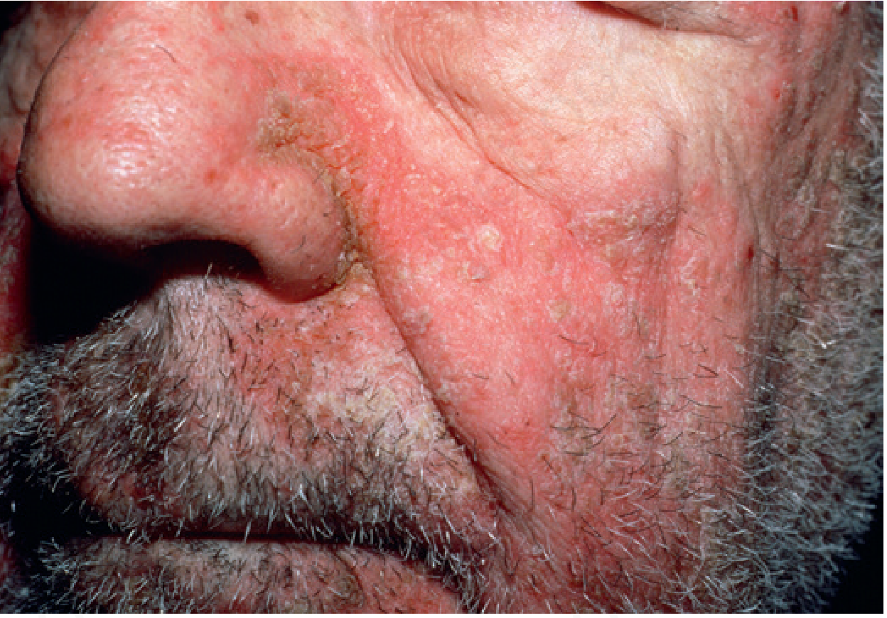
Seborrhoeic dermatitis: erythema and scaling on the face (Andrews' Diseases of the Skin)
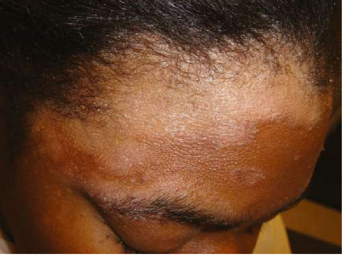
Seborrhoeic dermatitis: hypopigmentation with fine scaling at the hairline (Goldman-Cecil Medicine)
Distribution (seborrhoeic zones - high sebum output):
| Region | Features |
|---|---|
| Scalp | Dandruff (pityriasis sicca) is the mildest form; in severe cases, greasy thick crusts with offensive odour; "cradle cap" in infants |
| Face | Nasolabial folds, glabella, eyebrows, malar area, alae nasi |
| Eyelids/ears | Marginal blepharitis, otitis externa-like scaling in auditory canals |
| Trunk | Presternal/mid-chest, axillae |
| Flexures | Inframammary folds, umbilicus, groins, gluteal crease |
Folliculitis of the beard area is common in men. The presternal area is a favoured trunk site. Involvement of the groin can closely simulate tinea cruris or candidiasis.
Pathogenesis
The aetiology is multifactorial. Three main mechanisms interact:
1. Role of Malassezia
The lipophilic yeast Malassezia ovalis (Pityrosporum ovale) and related species produce bioactive metabolites - oleic acid, malassezin, indole-3-carbaldehyde, and bioactive indoles - that provoke an inflammatory response. Yeast density correlates with disease severity, and reduction occurs with antifungal therapy. However, Malassezia also colonises clinically normal scalps, so individual susceptibility is key.
2. Immune Dysregulation
- Increased T-cell activity with altered CD4+/CD8+ ratio (often decreased)
- Elevated cytokines: IL-1α, IL-1β, IL-4, IL-12, TNF-α, IFN-γ in lesional skin
- Increased NK cell activity
- Elevated IgA and IgG in serum
- Expression of cytotoxicity-activating ligands
- The dramatically increased prevalence in HIV/AIDS underscores the role of immune surveillance
3. Sebaceous Gland Activity and Epidermal Hyperproliferation
SD lesions follow the distribution of sebaceous glands; peak incidence mirrors times of maximal sebum output (infancy and adulthood). Increased epidermal turnover resembles psoriasis, and the two conditions can overlap ("seborrhiasis"). In Parkinson disease, facial immobility and elevated sebum likely create a favourable substrate for Malassezia.
Aggravating/trigger factors:
- Cold, low humidity (winter/spring flares)
- Emotional stress, fatigue
- Neurological diseases: Parkinson disease, epilepsy, mood disorders, Alzheimer disease, stroke
- Immunosuppression: HIV/AIDS, organ transplant
- Drugs: lithium, haloperidol, IFN-α, griseofulvin, gold, arsenic, methyldopa, phenothiazines
- Nutritional deficiencies: zinc (acrodermatitis enteropathica-like), biotin
- UV exposure has a variable effect
Associated Conditions
| Condition | Association |
|---|---|
| HIV/AIDS | Prevalence up to 83%; severe, extensive, refractory disease |
| Parkinson disease | Severe, waxy facial scaling; refractory; partly due to sebum overproduction and facial immobility |
| Stroke / facial nerve injury | Unilateral SD ipsilateral to the lesion |
| Diabetes mellitus (esp. obese patients) | Increased incidence |
| Neurological/psychiatric conditions | Epilepsy, Alzheimer, depression, alcoholism |
Histology
- Regular acanthosis with some thinning of suprapapillary plates
- Varying degrees of spongiosis and lymphocyte exocytosis
- Characteristic finding: focal scale-crust adjacent to follicular ostia (this is pathognomonic)
- Overlaps histologically with psoriasis in some cases
(Andrews' Diseases of the Skin, p. 229)
Diagnosis
Diagnosis is clinical, based on characteristic morphology and distribution. No laboratory tests are required.
Dermoscopy of the scalp shows:
- Twisted loop vessels
- Red dots and globules
- Glomerular vessels
Differential Diagnosis:
| Condition | Distinguishing features |
|---|---|
| Psoriasis | Heavier silvery scales peeling in layers; Auspitz sign (bleeding points on scale removal); nail pitting; less pruritus |
| Tinea capitis | KOH prep positive; lymphadenopathy |
| Rosacea | No scaling; papules and pustules; telangiectasia |
| Atopic dermatitis | Lichenification; flexural involvement; personal/family atopy history |
| Langerhans cell histiocytosis | Yellow-brown perifollicular papules; groin fissuring; systemic features |
| Pityriasis versicolor | KOH prep shows spaghetti-and-meatballs hyphae; fine scale |
| SLE | Malar rash but no scaling; ANA positive |
| Crusted scabies | Burrows; intense generalised pruritus; family contacts affected |
In infants, also consider: atopic/contact eczema, infantile psoriasis, metabolic disorders (failure to thrive, recurrent infections warrant work-up for immunodeficiency).
Treatment
Scalp
- First-line: medicated shampoos used 2-3x per week alternating with regular shampoo:
- Ketoconazole 2% (antifungal)
- Selenium sulfide
- Zinc pyrithione
- Coal tar preparations
- Ciclopirox
- Topical corticosteroids: fluocinolone acetonide 0.01% solution/gel, twice daily for 3-4 weeks; useful for rapid reduction of inflammation
- Keratolytics: salicylic acid for thick scalp crusts (avoid in neonates - risk of percutaneous absorption)
Face and Glabrous Skin
- Antifungals: ketoconazole 2% cream, ciclopirox, sertaconazole, bifonazole - safe for long-term maintenance
- Low-potency topical corticosteroids: hydrocortisone 2.5% cream twice daily for 1-2 weeks (medium-to-high potency on face risks steroid rosacea - avoid prolonged use)
- Topical calcineurin inhibitors (CNIs): pimecrolimus 1% cream or tacrolimus - equivalent efficacy to ketoconazole; good steroid-sparing option, especially on the face; may cause initial burning sensation
- Zinc pyrithione, Quassia amara extract: adjunct options
- Lithium gluconate 8% ointment: effective, especially in HIV facial disease
Ears and Blepharitis
- Ear canal: topical corticosteroids or antifungals (ketoconazole, ciclopirox); desonide otic lotion 0.05%
- Blepharitis: sodium sulfacetamide drops/ointment; daily gentle lid scrubs with diluted baby shampoo; oral tetracyclines for severe/recurrent cases
Special Populations
- HIV/AIDS: topical/systemic antifungals + topical or oral corticosteroids; antiretroviral therapy (ART) itself improves SD; WHO recommends topical ketoconazole as first-line with topical corticosteroids for severe/unresponsive cases; prolonged treatment usually needed
- Infants (cradle cap - mild): baby oil or mineral oil applied, gentle combing, then shampoo; for more severe involvement, low-potency antifungal shampoo/cream or 1% hydrocortisone; keratolytics and selenium sulfide are contraindicated in neonates
Refractory Disease / Second-line
- Oral itraconazole or terbinafine (preliminary evidence)
- Sodium sulfacetamide +/- sulfur preparations
- Narrowband UVB phototherapy (immunomodulatory/anti-inflammatory)
- Low-dose oral isotretinoin (refractory seborrhoeic blepharitis)
- Tea tree oil (some evidence for scalp disease)
- Urea + propylene glycol + lactic acid combinations
Prognosis and Course
SD is a chronic, relapsing condition - management is generally suppressive rather than curative. Infantile SD typically resolves spontaneously within the first 1-2 years of life. Adult SD tends to be lifelong, with fluctuating severity. Cold weather, stress, and immune compromise worsen flares. Maintenance therapy (e.g., twice-weekly medicated shampoo) reduces relapse frequency.
Sources:
- Andrews' Diseases of the Skin: Clinical Dermatology, p. 229
- Fitzpatrick's Dermatology, Vol. 1-2 (pathogenesis, HIV, infantile SD)
- Goldman-Cecil Medicine, p. 4276
- Global Prevalence of Seborrheic Dermatitis - JAMA Dermatology 2024 (PMID 38958996)
This is a shared conversation. Sign in to Orris to start your own chat.