
Afb stain
Reading File
AFB (Acid-Fast Bacilli) stain is used to detect mycobacteria. Let me search the textbook library for detailed information.
Searching sources...
Finding Sources
Finding Sources
Good results. Let me now read the most relevant sections from Henry's (microbial stains) and Tietz (AFB stain details), and also grab the image from Murray & Nadel.
Reading File
Reading File
Reading File
Excellent data. Let me now fetch the key images and also get Sherris microbiology section on AFB staining procedure.
Loading Image
Reading File
I now have comprehensive information. Let me compile the full answer.
Acid-Fast Bacilli (AFB) Stain
Principle
Mycobacteria have a unique cell wall that is rich in mycolic acids - long-chain, branched fatty acids that make the wall highly hydrophobic. This hydrophobicity prevents penetration of ordinary aniline dyes (like crystal violet in Gram stain), causing mycobacteria to appear as clear "ghost" shapes or faint gram-positive rods with the Gram stain.
However, when an arylmethane dye (carbol fuchsin) is driven into the cell wall using phenol and heat, it forms a stable complex with the mycolic acids. Once bound, this complex resists decolorization by mineral acids (e.g., 3% HCl in ethanol) - hence the term "acid-fast." The background is then counterstained, leaving AFB as the only colored structures. - Tietz Textbook of Laboratory Medicine, 7th Ed.
Types of AFB Stains
| Stain | Mechanism | Notes |
|---|---|---|
| Ziehl-Neelsen (ZN) | Carbol fuchsin + heat (hot stain) | Classic method; uses methylene blue counterstain |
| Kinyoun | Carbol fuchsin + increased phenol concentration (cold stain) | No heat required; slightly less sensitive |
| Auramine-rhodamine | Fluorochrome dye binds nucleic acids within AFB | ~20% more sensitive than carbol fuchsin; WHO recommended |
| Auramine-O | Fluorochrome variant | Faster screening at lower magnification |
| Fite stain | Modified acid-fast; uses weaker acid decolorization | For partially acid-fast organisms (M. leprae, Nocardia, Rhodococcus) |
- Henry's Clinical Diagnosis and Management by Laboratory Methods
Staining Appearance
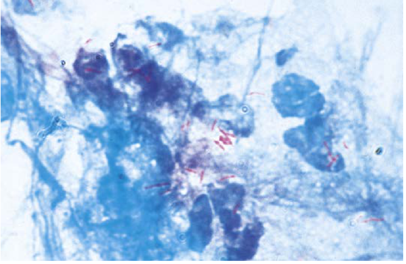
- Ziehl-Neelsen / Kinyoun: AFB appear as bright red (magenta) rods against a blue background (methylene blue counterstain)
- A characteristic beaded appearance is often seen (reflects uneven stain uptake along the cell)
- Cell size: 1 to 10 μm in length
- Fluorochrome stains: AFB fluoresce bright yellow-green or orange against a dark background; read at lower magnification (250-400x vs. 800-1000x for carbol fuchsin)
Kinyoun stain - AFB (M. tuberculosis) in sputum (400x):
Organisms Detected
Strongly acid-fast (true AFB):
- Mycobacterium tuberculosis (MTB)
- Mycobacterium leprae (requires Fite stain - more delicate)
- Non-tuberculous mycobacteria (NTM): M. avium complex, M. kansasii, etc.
- Cryptosporidium oocysts (modified AFB)
- Isospora / Cyclospora (modified AFB)
Partially/weakly acid-fast:
- Nocardia spp.
- Rhodococcus spp.
Sensitivity & Limitations
- AFB smear detects ~65% of culture-positive sputum specimens at best - Sherris & Ryan's Medical Microbiology, 8th Ed.
- Requires 1,000-10,000 CFU/mL for reliable detection
- Not species-specific: cannot distinguish MTB from NTM
- Yield from non-sputum sites (CSF, urine, bone marrow) is even lower
- Culture remains mandatory for confirmation and susceptibility testing
Reporting Scale (Carbol Fuchsin Stain, 1000x)
| AFB Seen | Report |
|---|---|
| 0 / 300 fields | No AFB seen (Negative) |
| 1-2 / 300 fields | Doubtful - repeat |
| 1-9 / 100 fields | 1+ |
| 1-9 / 10 fields | 2+ |
| 1-9 / field | 3+ |
| >9 / field | 4+ |
- Henry's Clinical Diagnosis and Management; Murray & Nadel's Textbook of Respiratory Medicine
Why Fluorochrome is Preferred
- ~20% more sensitive than carbol fuchsin
- Read at lower magnification (250-400x) → more fields scanned per unit time → faster, less labor-intensive
- WHO has endorsed global phase-out of ZN staining in favor of auramine-rhodamine, though uptake is limited in resource-poor settings (only 7% of global labs had fluorescent microscopes as of 2014) - Tietz, 7th Ed.
Special Note: Gram Stain vs. AFB Stain
With the Gram stain, mycobacteria may appear as faintly gram-positive beaded bacilli or as "Gram ghosts" (clear outlines within macrophages) because the hydrophobic wall resists crystal violet. This is why dedicated AFB staining is always required when mycobacterial infection is suspected. - Henry's Clinical Diagnosis
This is a shared conversation. Sign in to Orris to start your own chat.