
Cilinical examination of the abdomen on normal subject
I'll start by reading the medical library skill, then search for abdominal examination content.
Reading File
Now let me search the medical library for abdominal examination content in parallel:
Finding Sources
Finding Sources
Excellent results. S Das Manual on Clinical Surgery and Bailey & Love have rich sections. Let me read the most comprehensive sources in parallel:
Reading File
Reading File
Reading File
Now let me read the comprehensive S Das section on normal abdominal examination (chronic abdominal conditions chapter) and the Bailey & Love abdominal examination section:
Reading File
Reading File
The S Das chapter on chronic abdominal examination is the most relevant. Let me read the full palpation, percussion, and auscultation sections:
Reading File
Reading File
I now have excellent, comprehensive material from multiple authoritative textbooks. Let me also fetch the 9-region abdomen diagram image to include in the answer:
Loading Image
I now have comprehensive material to compose a thorough, well-structured answer. Let me deliver it:
Clinical Examination of the Abdomen (Normal Subject)
The abdominal examination follows a strict sequence: General Examination → Inspection → Palpation → Percussion → Auscultation. On a normal subject, all findings are used as the baseline against which pathology is detected.
Preparation & Positioning
-
The patient lies flat on their back, legs extended (a pillow under the head is acceptable).
-
For palpation, hips and knees are flexed to relax abdominal muscles.
-
The abdomen is fully exposed from the nipple line above to the saphenous openings (inguinal/femoral rings) below.
-
Examination is performed in good light, preferably daylight.
-
The examiner inspects first from the side, then tangentially, then from either end of the bed.
-
S Das A Manual on Clinical Surgery 13th Edition, p. 460
Surface Anatomy Reference
The abdomen is divided into nine regions by two horizontal and two vertical lines:
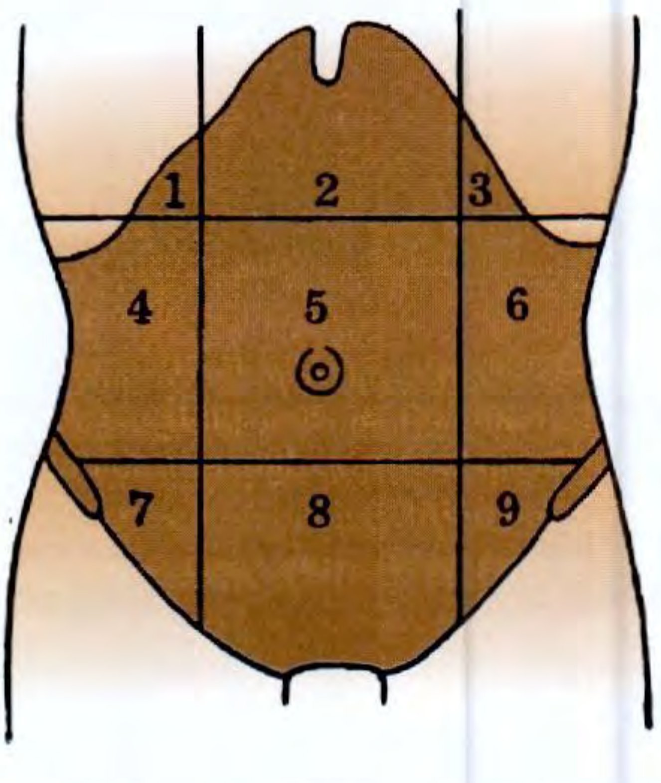
| No. | Region |
|---|---|
| 1 | Right hypochondrium |
| 2 | Epigastrium |
| 3 | Left hypochondrium |
| 4 | Right lumbar (flank) |
| 5 | Umbilical |
| 6 | Left lumbar (flank) |
| 7 | Right iliac fossa |
| 8 | Hypogastrium (suprapubic) |
| 9 | Left iliac fossa |
Alternatively, a simpler 4-quadrant scheme (RUQ, LUQ, RLQ, LLQ) is used for quick clinical description.
- S Das A Manual on Clinical Surgery 13th Edition, p. 523
Step 1: General Examination (Before the Abdomen)
Before focusing on the abdomen, a general survey is done:
-
Hands: Pallor of palmar creases (anaemia), clubbing, leukonychia, Dupuytren's contracture (liver disease), flapping tremor (hepatic encephalopathy).
-
Eyes: Jaundice (scleral icterus), anaemia (conjunctival pallor), xanthelasma.
-
Face/Mouth: Parotid enlargement, angular stomatitis, glossitis, telangiectasia (Osler-Weber-Rendu).
-
Neck: Left supraclavicular lymphadenopathy (Virchow's node / Troisier's sign) - suggests intra-abdominal malignancy.
-
Vital signs: Heart rate, BP, respiratory rate, temperature, weight/BMI.
-
Bailey and Love's Short Practice of Surgery 28th Edition, p. 1076
Step 2: Inspection
Inspect systematically:
1. Hernial Orifices (inspect first)
- Look at the inguinal, femoral, and umbilical rings. Ask the patient to cough - a cough impulse indicates a hernia.
- S Das advises inspecting hernial orifices first, as they may be the cause of acute abdominal symptoms.
2. Skin
- Normal: No scars, erythema, pigmentation, or rash.
- Note any scars - linear (healed by first intention) vs. broad/irregular (wound infection).
- Engorged superficial veins:
- Around umbilicus ("caput medusae") - portal hypertension.
- On flanks running upward - IVC obstruction.
- Hard subcutaneous periumbilical nodule (Sister Mary Joseph's nodule) - intra-abdominal carcinoma.
3. Umbilicus
- Normal position: Midway between the xiphoid tip and the pubic symphysis.
- Normal appearance: Inverted (slit-like), central.
- Displacement upward - pelvic mass; downward - ascites (Tanyol's sign).
- Eversion - ascites; deep inversion - obesity.
4. Contour / Shape of the Abdomen
- Normal: Flat or gently scaphoid (concave) in a lean person; slightly protuberant in a well-nourished adult.
- Normal umbilicus: Inverted in obesity vs. everted in intra-abdominal distension - a key distinguishing feature.
- The 5 "Fs" of distension: Fat, Fluid, Flatus, Faeces, Foetus.
5. Respiratory Movements
- Normal: The abdominal wall rises gently with inspiration (predominantly abdominal breathing in males; thoracic in females).
- Reduced or absent movement indicates peritoneal irritation (localized or diffuse).
6. Visible Peristalsis
- Normal: Not visible.
- Visible peristalsis indicates obstruction (gastric outlet - left to right; small bowel - "ladder pattern"; transverse colon - right to left).
7. Pulsations
- Normal: A faint aortic pulsation may occasionally be seen in thin individuals in the epigastrium.
- A prominent expansile pulsation suggests abdominal aortic aneurysm.
Step 3: Palpation
Technique
- The examiner's forearm must be horizontal at the level of the patient's abdomen.
- Use the volar (palmar) surfaces of the fingers - never the fingertips pointing downward.
- Begin palpation in the quadrant furthest from the site of pain (if any) and end at the painful area.
- Watch the patient's face for signs of discomfort during palpation.
Superficial (Light) Palpation
- Detect tenderness, superficial masses, rigidity, guarding.
- Normal: Soft, non-tender abdomen, no involuntary guarding.
- Voluntary guarding: Patient tenses abdomen in anticipation; can be overcome by distraction.
- Involuntary guarding/rigidity: True sign of peritoneal irritation.
Deep Palpation
Used to examine the solid organs and identify deeper masses.
Liver
- Begin in the right iliac fossa, move upward with each expiration.
- The right hand is placed flat, fingers pointing toward the right costal margin.
- Alternatively, use the bimanual method (left hand behind the right loin) or dipping method for tense ascites.
- Normal: Not palpable below the costal margin (or just palpable with a smooth, firm, regular edge on deep inspiration in thin individuals).
- Note: edge, surface, consistency, tenderness, pulsatility.
Gallbladder
- Palpable just below the liver edge at the tip of the 9th costal cartilage, right of midline.
- Normal: Not palpable.
- Murphy's sign: Deep inspiration while pressure is applied at the gallbladder point produces inspiratory arrest in acute cholecystitis.
Spleen
- Begin palpation from the right iliac fossa, move toward the left costal margin.
- Bimanual: left hand behind the left lower ribs, right hand palpating.
- The patient may be asked to roll to the right lateral decubitus (right lateral position) to bring the spleen forward.
- Normal: Not palpable (must be 2-3 times its normal size to be felt).
Kidneys
- Bimanual (ballottement): Left hand in the loin (posteriorly), right hand in the flank (anteriorly).
- The kidney can be ballotted (bounced between the two hands).
- Normal: Left kidney generally not palpable; right kidney occasionally palpable in thin individuals on deep inspiration.
Aorta
- Palpate in the midline epigastrium.
- Normal: A pulsatile, non-expansile aorta felt as a single midline structure in thin patients.
- Widened or expansile pulsation - aneurysm.
Rebound Tenderness (Blumberg's Sign)
- Press slowly and deeply, then release rapidly.
- Pain on release = peritoneal irritation.
- Normal: No rebound tenderness.
Fluid Thrill and Shifting Dullness
- Tested during percussion (see below), but sometimes elicited during palpation by the fluid thrill test.
Step 4: Percussion
- Used to delineate organ boundaries and detect free fluid or gas.
Liver Dullness
- Percuss downward from the right 4th intercostal space in the mid-clavicular line.
- Upper border: Where resonance changes to dullness (normally 5th ICS, MCL).
- Lower border: Where dullness ends (normally at the costal margin in MCL).
- Loss of liver dullness (gas over liver) is a critical sign of hollow viscus perforation (e.g., peptic ulcer perforation).
Splenic Dullness
- Traube's space (lower left chest, bounded by stomach, diaphragm, splenic flexure): normally resonant.
- Dullness in Traube's space suggests splenomegaly.
Percussion for Ascites
- Shifting dullness: Percuss from umbilicus to flank - note level where dullness begins. Patient rolls to the opposite side; the dullness shifts (fluid follows gravity). Normal: No shifting dullness.
- Fluid thrill: Place a hand flat on one flank; flick the other flank with a finger. An assistant's hand on the midline prevents conduction through fat. A transmitted impulse = free fluid. Normal: No fluid thrill.
Percussion of the Bladder
- A distended bladder produces suprapubic dullness extending upward from the symphysis pubis.
- Normal: Suprapubic area is resonant (empty bladder).
Step 5: Auscultation
- Use the diaphragm of the stethoscope, held lightly on the abdomen.
- Listen for at least 2 minutes before declaring bowel sounds absent.
Bowel Sounds
- Normal: Soft gurgling sounds every 5-10 seconds, reflecting peristalsis.
- Increased (tinkling, high-pitched): Mechanical obstruction, early peritonitis.
- Absent: Paralytic ileus, advanced peritonitis, immediately post-operative.
Vascular Bruits
- Auscultate over the aorta (epigastrium), renal arteries (just lateral to umbilicus bilaterally), and iliac arteries.
- Normal: No bruits.
- A bruit suggests arterial stenosis or aneurysm.
Succussion Splash
- The patient is asked to move rapidly from side to side while the examiner listens over the epigastrium.
- An audible splash indicates fluid + air in a hollow viscus (e.g., gastric outlet obstruction).
- Normal: No succussion splash (or a mild splash if the patient has just eaten/drunk).
Step 6: Special Tests & Completion
- Per rectal (digital rectal) examination: Should not be omitted. Assess anal sphincter tone, rectal mucosa, prostate (in males), cervix/uterus via posterior wall (in females). Note stool color/consistency.
- Hernial orifices re-examined with the patient standing and coughing.
- Examine external genitalia and scrotum (testicular malignancy can produce para-aortic masses).
Summary: Normal Findings on Abdominal Examination
| Step | Normal Finding |
|---|---|
| Inspection | Flat/scaphoid contour; inverted or centrally placed umbilicus; no scars, no dilated veins, no visible peristalsis; active respiratory movements |
| Superficial palpation | Soft, non-tender, no guarding or rigidity |
| Deep palpation | Liver, spleen, kidneys not palpable (except occasionally in thin individuals); no masses |
| Percussion | Liver dull (5th ICS to costal margin); Traube's space resonant; no shifting dullness |
| Auscultation | Normal bowel sounds every 5-10 sec; no bruits; no succussion splash |
Sources:
- S Das, A Manual on Clinical Surgery, 13th ed. - Bailey and Love's Short Practice of Surgery, 28th ed. - Textbook of Family Medicine, 9th ed.
This is a shared conversation. Sign in to Orris to start your own chat.