
A 23 year old male patient has c/o fever on touch , cold throat itching and cough generalized weakness diagnosis and management dark yellow color urine with burning sensation
infectious mononucleosis tonsils exudate pharyngitis EBV
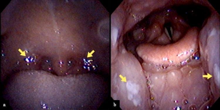
Two endoscopic images (a and b) documenting the clinical presentation of the pharynx and larynx in a patient with suspected infectious mononucleosis. Image (a) is an oropharyngeal view showing bilateral, grade II Mackenzie classification tonsillar swelling. There is notable erythema of the soft palate and localized yellow-white exudate (pus) on the tonsillar surfaces (indicated by yellow arrows), without confluent pseudomembranes. Image (b) provides a laryngeal endoscopic view looking down toward the glottis. It reveals significant edema and swelling of the lingual tonsils and posterior third of the tongue (yellow arrows). Anatomical landmarks including the epiglottis and vocal folds are visible; notably, there is an absence of laryngeal or epiglottic edema that would suggest airway compromise. These visual findings are characteristic of viral pharyngotonsillitis, often seen in Epstein-Barr virus (EBV) infections, and are used to differentiate from bacterial streptococcal pharyngitis or more severe supraglottic infections.
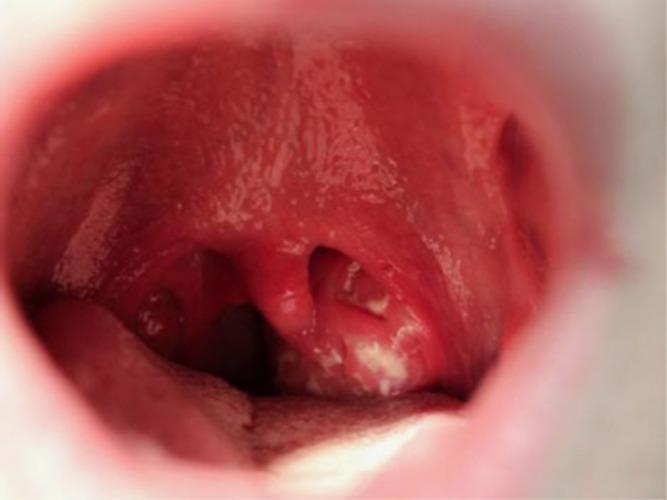
This clinical photograph provides an intraoral view of the oropharynx, demonstrating classic signs of exudative tonsillitis and pharyngitis. The palatine tonsils are bilaterally enlarged (tonsillar hypertrophy) and erythematous, featuring prominent white-to-yellowish confluent exudates on the mucosal surface. The surrounding tissues, including the posterior pharyngeal wall and soft palate, exhibit significant hyperemia. Notably, the uvula is markedly edematous and erythematous (uvulitis). These visual findings are highly characteristic of infectious mononucleosis caused by the Epstein-Barr virus (EBV), particularly in young adult populations. The image serves as an educational reference for distinguishing between simple viral pharyngitis and more severe exudative presentations that involve widespread oropharyngeal inflammation, lymphoid hyperplasia, and constitutional symptoms like cervical lymphadenopathy and fatigue.
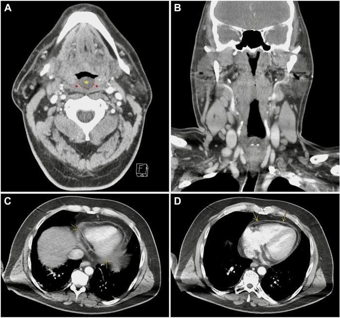
This composite diagnostic image features four panels of contrast-enhanced computed tomography (CT) scans illustrating clinical manifestations of infectious mononucleosis (Epstein-Barr virus). Panel A shows a transverse view of the oropharynx, identifying bilateral tonsillar thickening and edema (red asterisks) alongside a thickened, edematous uvula (yellow asterisk). Panel B presents a coronal view of the neck, highlighting prominent, reactive cervical lymphadenopathy. Panels C and D are transverse sections of the thorax at the level of the heart, demonstrating a trace pericardial effusion. Yellow arrows point to abnormal enhancement and thickening of the pericardial sac, indicating inflammatory pericarditis. The collection serves as an educational resource for identifying multisystem involvement of EBV, ranging from exudative pharyngitis and regional lymphadenopathy to secondary cardiac complications such as pericarditis. This material is suitable for medical education in radiology, infectious disease, and emergency medicine specialties.
Diagnosis & Management: 23-Year-Old Male with Fever, Sore Throat, Cough, Weakness, Dark Yellow Urine + Burning Sensation
Most Likely Diagnoses
- Primary: Infectious Mononucleosis (EBV) - accounting for the fever, sore throat itching/scratchiness, cough, and generalized weakness
- Secondary consideration: Hepatitis (EBV-associated or Hepatitis A/E) - accounting for dark yellow urine; and/or Urinary Tract Infection (UTI) - accounting for dark urine with burning sensation on urination
Problem 1 - Infectious Mononucleosis (EBV)
- Symptom to Diagnosis: An Evidence-Based Guide, 4th Ed.
- Enlarged, erythematous tonsils with white/gray exudate (indistinguishable from strep)
- Posterior cervical lymphadenopathy (LR+ 3.1)
- Palatal petechiae (highly specific, LR+ 5.3)
- Splenomegaly/hepatomegaly
- Periorbital edema
Problem 2 - Dark Yellow Urine + Burning Sensation
Investigations Recommended
| Investigation | Purpose |
|---|---|
| Monospot test (heterophile antibody) | Rapid screen for EBV mononucleosis |
| EBV serology (VCA IgM/IgG, EA, EBNA) | Confirm EBV if monospot negative (false negative in early disease) |
| CBC with differential | Look for lymphocytosis >50%, atypical lymphocytes >10% (highly specific for mono) |
| LFTs (ALT, AST, bilirubin) | Assess hepatitis component; explains dark urine if elevated |
| Urine routine & microscopy (R/E) | Check for bilirubin, urobilinogen, RBCs, WBCs, bacteria |
| Urine culture & sensitivity | If UTI suspected - guide antibiotic choice |
| Throat swab | Rule out concurrent Group A Strep (present in ~30% of mono cases) |
| Abdominal ultrasound | Assess for splenomegaly/hepatomegaly |
| Renal function (BUN, creatinine) | If urine output appears reduced |
Management
1. Infectious Mononucleosis
- Rest - adequate rest is important; avoid strenuous activity
- Hydration - oral fluids; IV if unable to maintain oral intake
- Antipyretics - Paracetamol (acetaminophen) for fever and pain; NSAIDs (ibuprofen) for throat pain and myalgia
- Analgesic gargles - warm saline gargles, benzydamine gargle for sore throat relief
- Do NOT prescribe amoxicillin or ampicillin - these cause a morbilliform rash in up to 30% of mono patients; use them only if Group A Strep is confirmed AND no beta-lactam allergy concerns
- Corticosteroids - NOT for routine use; reserved only for impending airway compromise or severe thrombocytopenia/hemolytic anemia
- Acyclovir - NOT recommended; reduces viral shedding but does not alter clinical course
- Cummings Otolaryngology Head and Neck Surgery
- Symptom to Diagnosis: An Evidence-Based Guide, 4th Ed.
2. Hepatitis (if LFTs confirm)
- Supportive care, avoid hepatotoxic drugs (paracetamol within dose limits is acceptable)
- Avoid alcohol completely
- Monitor LFTs every 1-2 weeks
- If Hepatitis A/E suspected (feco-oral exposure history): management is also supportive; notify public health if required
3. UTI (if confirmed on urine culture)
- First-line: Nitrofurantoin 100 mg BD x 7 days OR Trimethoprim-sulfamethoxazole (Co-trimoxazole) DS x 7-10 days for males (longer course than females)
- Alternative: Ciprofloxacin 500 mg BD x 7 days
- Hydration: Encourage 2-3 L fluids/day
- Important: Avoid fluoroquinolones if possible in young patients; guided by culture sensitivity
Differential Diagnoses to Consider
| Condition | Key Distinguishing Features |
|---|---|
| Streptococcal pharyngitis | No lymphadenopathy pattern; monospot negative; responds to penicillin |
| Acute HIV (retroviral syndrome) | Rash, mucosal ulcers; exposure history; HIV Ag/Ab test |
| CMV mononucleosis | EBV-negative mono syndrome; CMV serology |
| Hepatitis A/E | Prominent jaundice, nausea, vomiting; HAV/HEV serology |
| Influenza | Prominent myalgia, rapid onset; no prominent pharyngeal exudate |
Red Flags - Refer or Escalate If:
- Stridor or drooling (impending airway obstruction)
- Sudden severe left upper quadrant pain (splenic rupture)
- Severe thrombocytopenia or hemolytic anemia on CBC
- Neurological symptoms (encephalitis, meningitis - rare)
- LFTs >10x normal (severe hepatitis)
- Urinary symptoms not responding to antibiotics (pyelonephritis, renal involvement)