
Explain basal ganglia for me in simplest way For physiology long question
basal ganglia anatomy diagram circuit pathways
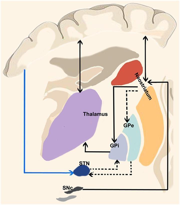
This medical schematic diagram illustrates the functional neuroanatomy of the human basal ganglia circuitry, detailing the classic motor loops. The diagram displays key anatomical structures including the cerebral cortex, thalamus (violet), neostriatum (red/orange), globus pallidus externa (GPe, light blue), globus pallidus interna (GPi, bluish-gray), subthalamic nucleus (STN, dark blue), and substantia nigra pars compacta (SNc, dark gray). Three primary functional pathways are highlighted: the Direct Pathway (solid black lines from neostriatum to GPi), which facilitates movement via disinhibition of the thalamus; the Indirect Pathway (dashed black lines connecting neostriatum to GPe, then STN, and finally GPi), which serves to inhibit motor activity; and the Hyperdirect Pathway (solid blue line from cortex directly to STN), which provides rapid inhibition of motor output by bypassing the striatum. Arrows indicate the direction of signal flow between these nuclei and the thalamocortical circuit, providing a visual model for understanding movement disorders like Parkinson's and Huntington's disease where these pathways are imbalanced.
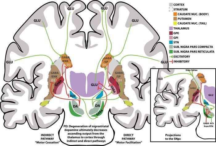
A pathophysiology diagram providing a schematic overview of the human basal ganglia motor circuits, specifically illustrating the indirect (left) and direct (right) pathways. The main illustration uses a coronal brain section to map anatomical structures: the cortex, striatum (caudate nucleus and putamen), thalamus, globus pallidus external (GPe) and internal (GPi) segments, subthalamic nucleus (STN), and substantia nigra (pars compacta, SNpc; pars reticulata, SNpr). Neurotransmission is color-coded: green arrows denote excitatory glutamatergic (GLU) or dopaminergic (DA) signals, while red T-bars signify inhibitory GABAergic (GABA/SP, GABA/ENK) signals. The direct pathway is labeled 'Motor Facilitation' and shows inhibitory projections from D1-receptor striatal neurons directly to the GPi/SNpr. The indirect pathway is labeled 'Motor Cessation' and highlights connections from D2-receptor striatal neurons to the GPe, subsequently influencing the STN and GPi/SNpr. An inset details projections to the SNpc, including inputs from the cortex, STN, and pedunculopontine nucleus (PPN). This educational material demonstrates the neuroanatomical basis for motor control and the circuit dysfunction relevant to Parkinson's disease (PD).
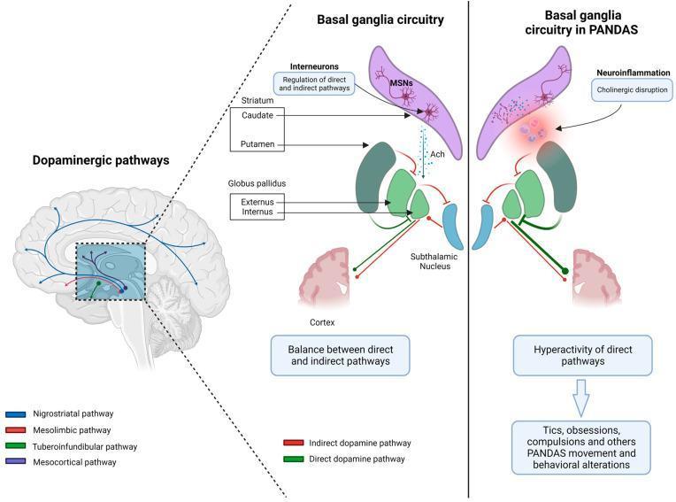
Educational medical diagram illustrating the pathophysiology of Pediatric Autoimmune Neuropsychiatric Disorders Associated with Streptococcal Infections (PANDAS) within the basal ganglia. The image is divided into three sections. On the left, a sagittal view of the human brain maps the four major dopaminergic pathways: nigrostriatal (blue), mesolimbic (red), tuberoinfundibular (green), and mesocortical (purple). The center and right panels provide a comparative schematic of basal ganglia circuitry. The 'Normal' circuit shows the functional balance between the direct and indirect pathways, highlighting the roles of the striatum (caudate and putamen), medium spiny neurons (MSNs), acetylcholine (Ach) interneurons, and the globus pallidus (externus and internus). In contrast, the 'PANDAS' circuit depicts neuroinflammation and cholinergic disruption in the dorsal striatum. This pathological state results in decreased Ach modulation and fewer distinct MSNs, leading to hyperactivity of the direct pathway. The diagram concludes by linking this physiological imbalance to clinical manifestations, including tics, obsessions, and compulsions typical of PANDAS syndrome.
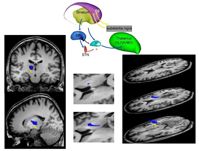
This composite educational graphic details the neuroanatomy and connectivity of the basal ganglia network, specifically focusing on the fronto-striatal circuit. It consists of a functional schematic diagram paired with MRI diffusion tractography images. The schematic illustrates key nodes including the Striatum (containing D1 and D2 pathways), Substantia Nigra, Thalamus (VL/VA/MD nuclei), Globus Pallidus (EP/IP), and the Subthalamic Nucleus (STN). Arrows indicate excitatory and inhibitory pathways, highlighting the indirect and direct pathways of motor control relevant to Parkinson's disease. The accompanying MRI panels (coronal, sagittal, and axial views) show white matter fiber reconstruction using tractography. These panels visualize axonal connections between the striatum and thalamic nuclei, color-coded in blue and yellow to represent distinct fiber bundles. The resource is designed to teach the neurobiological basis of extrapyramidal signs and the localization of basal ganglia dysfunction in clinical neurology.
Basal Ganglia - Physiology Long Answer
1. Introduction
2. Components (Nuclei)
| Structure | Part of | Neurotransmitter |
|---|---|---|
| Caudate nucleus | Striatum | GABA |
| Putamen | Striatum + Lenticular nucleus | GABA |
| Globus pallidus (external - GPe) | Lenticular nucleus | GABA (inhibitory) |
| Globus pallidus (internal - GPi) | Lenticular nucleus | GABA (inhibitory) |
| Subthalamic nucleus (STN) | Diencephalon | Glutamate (excitatory) |
| Substantia nigra | Midbrain | DA (pars compacta) / GABA (pars reticulata) |
- Caudate + Putamen = Striatum (main INPUT station)
- GPi + Substantia nigra pars reticulata (SNpr) = main OUTPUT of basal ganglia (both inhibitory)
- ~95% of striatal neurons are medium spiny neurons using GABA
3. Inputs to Basal Ganglia
- Corticostriatal pathway - from wide areas of cerebral cortex (especially motor cortex)
- Thalamostriatal pathway - from intralaminar nuclei of thalamus
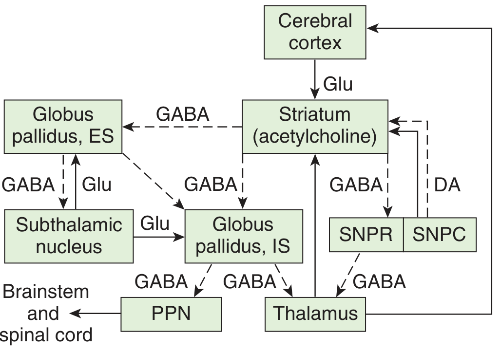
4. The Core Circuit (Cortical-BG-Thalamo-Cortical Loop)
Cortex → Striatum → (via pathways) → GPi/SNpr → Thalamus → Motor Cortex
5. The Two Pathways (THE Most Important Part for Exams)
A. Direct Pathway - "GO signal" (facilitates movement)
- Cortex excites (Glu) the striatum (D1 receptors)
- Striatum inhibits (GABA) GPi/SNpr
- GPi/SNpr is now suppressed → less inhibition on thalamus (disinhibition)
- Thalamus is now MORE active → excites motor cortex
- Result: MORE movement
B. Indirect Pathway - "STOP signal" (inhibits movement)
- Cortex excites (Glu) the striatum (D2 receptors)
- Striatum inhibits (GABA) GPe
- GPe now LESS able to inhibit STN → STN becomes more active
- STN excites (Glu) GPi/SNpr
- GPi/SNpr now MORE active → more inhibition on thalamus
- Thalamus is suppressed → motor cortex is less active
- Result: LESS movement
6. Role of Dopamine (Nigrostriatal Pathway)
| Receptor | Pathway | Effect of Dopamine |
|---|---|---|
| D1 (on direct pathway neurons) | Direct | Excites striatum → facilitates direct pathway → MORE movement |
| D2 (on indirect pathway neurons) | Indirect | Inhibits striatum → suppresses indirect pathway → also MORE movement |
Loss of dopamine (as in Parkinson's disease) → direct pathway underactive + indirect pathway overactive → net inhibition → bradykinesia and rigidity
7. Three Balanced Biochemical Systems
- Nigrostriatal dopaminergic system (SNpc → Striatum)
- Intrastriatal cholinergic system (ACh interneurons within striatum)
- GABAergic system (Striatum → GPi/SNpr)
8. Diseases of the Basal Ganglia
Hypokinetic Disorders (too little movement)
| Disease | Pathology | Features |
|---|---|---|
| Parkinson's Disease | Loss of dopamine (SNpc degeneration) | Resting tremor, rigidity, bradykinesia, shuffling gait (TRAP - Tremor, Rigidity, Akinesia, Postural instability) |
- Dopamine loss → D1 understimulated (direct pathway weak) AND D2 disinhibited (indirect pathway overactive)
- Net = excess inhibition on thalamus → reduced cortical activation → poverty of movement
Hyperkinetic Disorders (too much movement)
| Disease | Pathology | Features |
|---|---|---|
| Huntington's Disease | Loss of GABA neurons in striatum (especially indirect pathway neurons first) | Chorea (involuntary, dance-like movements), dementia |
| Hemiballismus | Lesion of subthalamic nucleus (STN) | Wild, flinging movements of one side of body |
| Athetosis | Lesion of striatum/putamen | Slow, writhing, involuntary movements |
- In Huntington's: loss of indirect pathway (GABAergic striatal neurons) → GPe is disinhibited → STN is over-inhibited → less GPi activity → thalamus over-excited → excess movement (chorea)
9. Other Functions of Basal Ganglia
- Cognitive functions - caudate nuclei connect with prefrontal cortex; lesions impair object reversal and delayed alternation tasks
- Affective behavior - via limbic connections
- Language - left caudate nucleus lesions produce a dysarthric aphasia resembling Wernicke's aphasia
10. Summary Table (Quick Revision)
| Pathway | Striatum Receptor | Net Effect on Movement | Neurotransmitters |
|---|---|---|---|
| Direct | D1 | Facilitates (MORE movement) | GABA (striatum→GPi), Glu (thalamus→cortex) |
| Indirect | D2 | Inhibits (LESS movement) | GABA (striatum→GPe), GABA (GPe→STN), Glu (STN→GPi) |
| Dopamine (SNpc) | D1 + D2 | Promotes movement | Dopamine |
11. Simple Analogy for Memory
- The direct pathway = gives the green light (go, move)
- The indirect pathway = gives the red light (stop, inhibit)
- Dopamine = biases the system toward green (movement)
- Loss of dopamine (Parkinson's) = stuck at red → poverty of movement
- Loss of indirect pathway neurons (Huntington's) = no red light → excess movement (chorea)