
Asthma based on Oxford and Davidson
asthma pathophysiology airway inflammation diagram
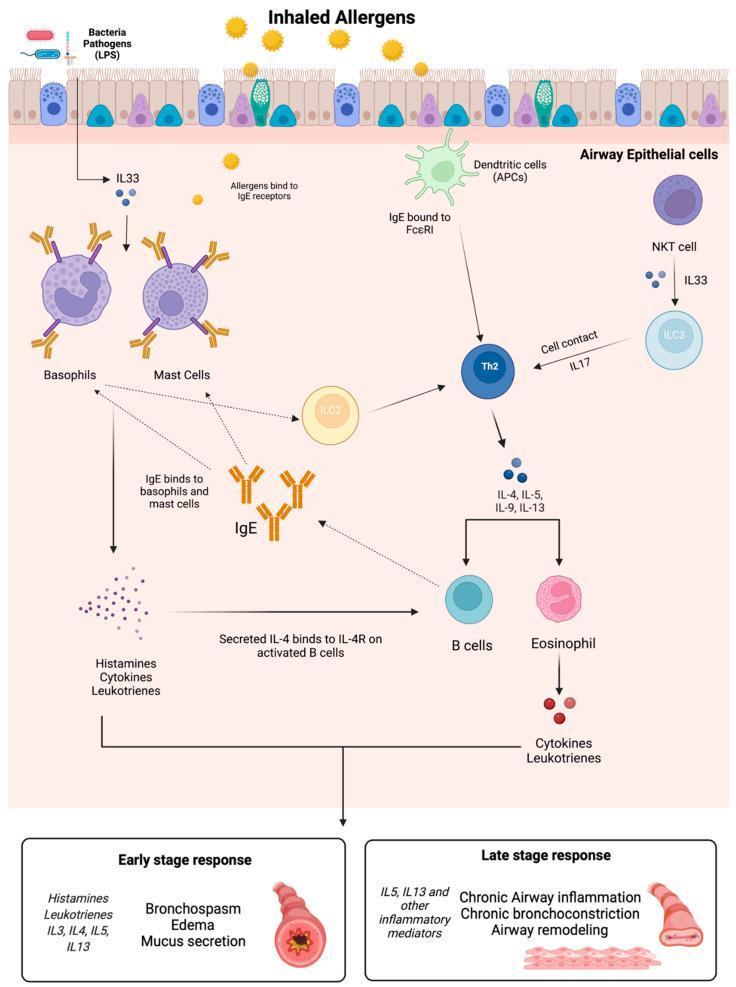
A pathophysiology diagram illustrating the cellular and molecular mechanisms of the asthma allergic response. The pathway begins with inhaled allergens and bacterial pathogens (LPS) interacting with airway epithelial cells, triggering the release of IL-33 and antigen presentation by dendritic cells (APCs). The central signaling cascade involves various immune cells, including NKT cells, ILC2/ILC3, Th2 cells, basophils, mast cells, B cells, and eosinophils. Key mediators shown include IgE (binding to FcεRI), IL-4, IL-5, IL-9, IL-13, and IL-17. The diagram divides clinical outcomes into two phases: the Early Stage Response, characterized by histamine/leukotriene release leading to bronchospasm, edema, and mucus secretion; and the Late Stage Response, driven by chronic inflammatory mediators (IL-5, IL-13) resulting in chronic airway inflammation, chronic bronchoconstriction, and airway remodeling. This schematic highlights the complex cellular cross-talk and cytokine signaling responsible for both acute hypersensitivity and long-term structural changes in asthmatic airways.
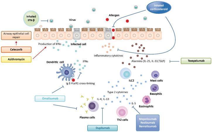
A pathophysiology diagram illustrating the immune response to viral and allergenic stimuli in the airway epithelium and the targets of various asthma therapeutics. The central focus is a ciliated epithelial layer undergoing viral infection and allergen-induced barrier disruption. Pathogen exposure triggers the release of IFNs and 'alarmins' (IL-25, IL-33, TSLP), which activate subepithelial immune cells including dendritic cells, ILC2s, mast cells, basophils, Th2 cells, and eosinophils. Type 2 cytokines (IL-4, IL-13, and IL-5) are shown driving inflammatory pathways. The schematic maps pharmacological interventions to specific nodes: Celecoxib and Azithromycin are linked to airway epithelial cell repair; Inhaled IFN-β targets viral replication; Inhaled Corticosteroids modulate epithelial and cytokine responses; Tezepelumab inhibits alarmins; Omalizumab blocks IgE-FcεRI cross-linking on dendritic cells; Dupilumab targets IL-4/IL-13 signaling; and Mepolizumab, Reslizumab, and Benralizumab inhibit IL-5-mediated eosinophilic inflammation. This diagram provides a comprehensive overview of biological therapies and their mechanisms in managing virally exacerbated asthma.
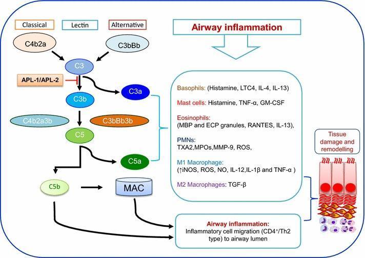
This pathophysiology diagram illustrates the complement cascade's role in asthma-related airway inflammation and tissue remodeling. The flowchart depicts three initiation pathways—Classical (via C4b2a), Lectin, and Alternative (via C3bBb)—converging at C3. A therapeutic intervention point is highlighted where APL-1/APL-2 (Compstatin derivatives) inhibit C3 activation. Downstream, C3 is cleaved into C3a and C3b, leading to C5 convertase formation (C4b2a3b or C3bBb3b) and subsequent cleavage of C5 into C5a and C5b. The anaphylatoxins C3a and C5a are shown activating a variety of inflammatory cells, including Basophils, Mast cells, Eosinophils, PMNs (neutrophils), and M1/M2 Macrophages. Each cell type is listed with its released mediators, such as histamine, cytokines (IL-4, IL-13, TNF-α), and reactive oxygen species (ROS). The terminal pathway shows C5b contributing to the Membrane Attack Complex (MAC). Collectively, these mediators drive inflammatory cell migration (CD4+/Th2 type) to the airway lumen, resulting in the depicted tissue damage and structural remodeling of the bronchial epithelium.
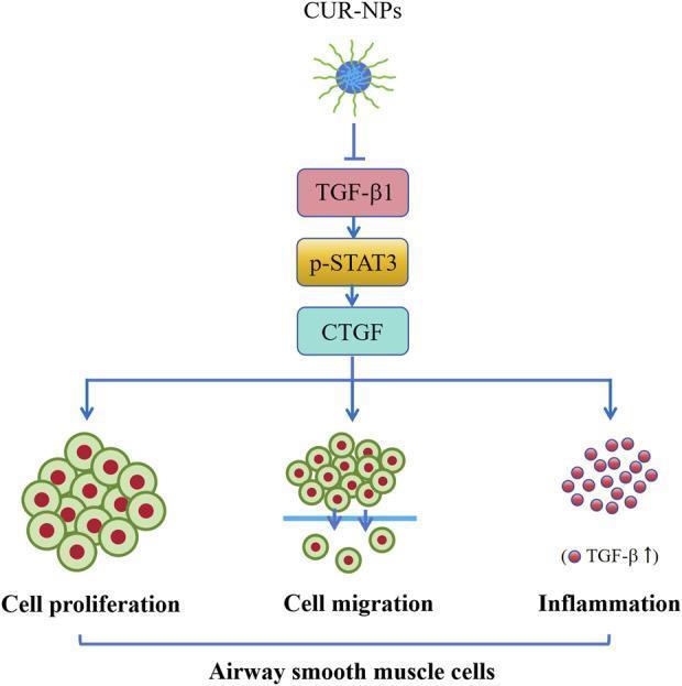
This pathophysiology diagram illustrates the molecular mechanism by which curcumin nanoparticles (CUR-NPs) inhibit asthma-related changes in airway smooth muscle cells (ASMCs). The diagram shows a vertical signaling cascade starting with CUR-NPs exerting an inhibitory effect (indicated by a T-bar arrow) on Transforming Growth Factor-beta 1 (TGF-̢1). This suppresses the downstream signaling pathway consisting of phosphorylated Signal Transducer and Activator of Transcription 3 (p-STAT3) and Connective Tissue Growth Factor (CTGF). The activation of CTGF is linked via branching arrows to three key pathological processes in ASMCs: cell proliferation (depicted as a dense cluster of nucleated cells), cell migration (shown as cells passing through a basement membrane), and inflammation (represented by small red circular markers labeled with 'TGF-̢1 ↑'). The flow indicates that by targeting the TGF-̢1/p-STAT3/CTGF axis, CUR-NPs can mitigate airway remodeling and inflammatory infiltration in conditions like asthma. This pedagogical visual is designed for intermediate to advanced medical education regarding pharmacology and respiratory pathophysiology.
Asthma - Based on Harrison's Principles of Internal Medicine 22e (2025)
Note: The library does not contain Davidson's Principles and Practice of Medicine or the Oxford Handbook of Clinical Medicine as separate titles. The closest equivalent covering the same clinical scope is Harrison's Principles of Internal Medicine 22E (2025), which is used as the primary source below. Content is consistent with the Davidson/Oxford tradition.
Definition
Epidemiology
- Affects ~262 million people globally; prevalence ~7.9% in the USA, ~4.3% worldwide
- More common in children (8.4%) than adults (7.7%); in children males > females (2:1), but in adulthood females > males
- Highest prevalence in the USA among the Puerto Rican population
- Mortality declined globally from 0.44 per 100,000 (1993) to 0.19 (2006), largely due to inhaled corticosteroid (ICS) use; no further improvement since
- Economic burden: ~$82 billion/year in the USA (2013)
Pathophysiology
Airway Hyperresponsiveness
- Direct smooth muscle hyperresponsiveness to agents like histamine or methacholine
- Indirect mechanisms: inflammatory cell activation releasing bronchoconstrictors, sensory nerve stimulation
- Structural changes: airway wall thickening from smooth-muscle hypertrophy/hyperplasia, subepithelial collagen deposition, edema - leads to disproportionate narrowing
Inflammatory Cells
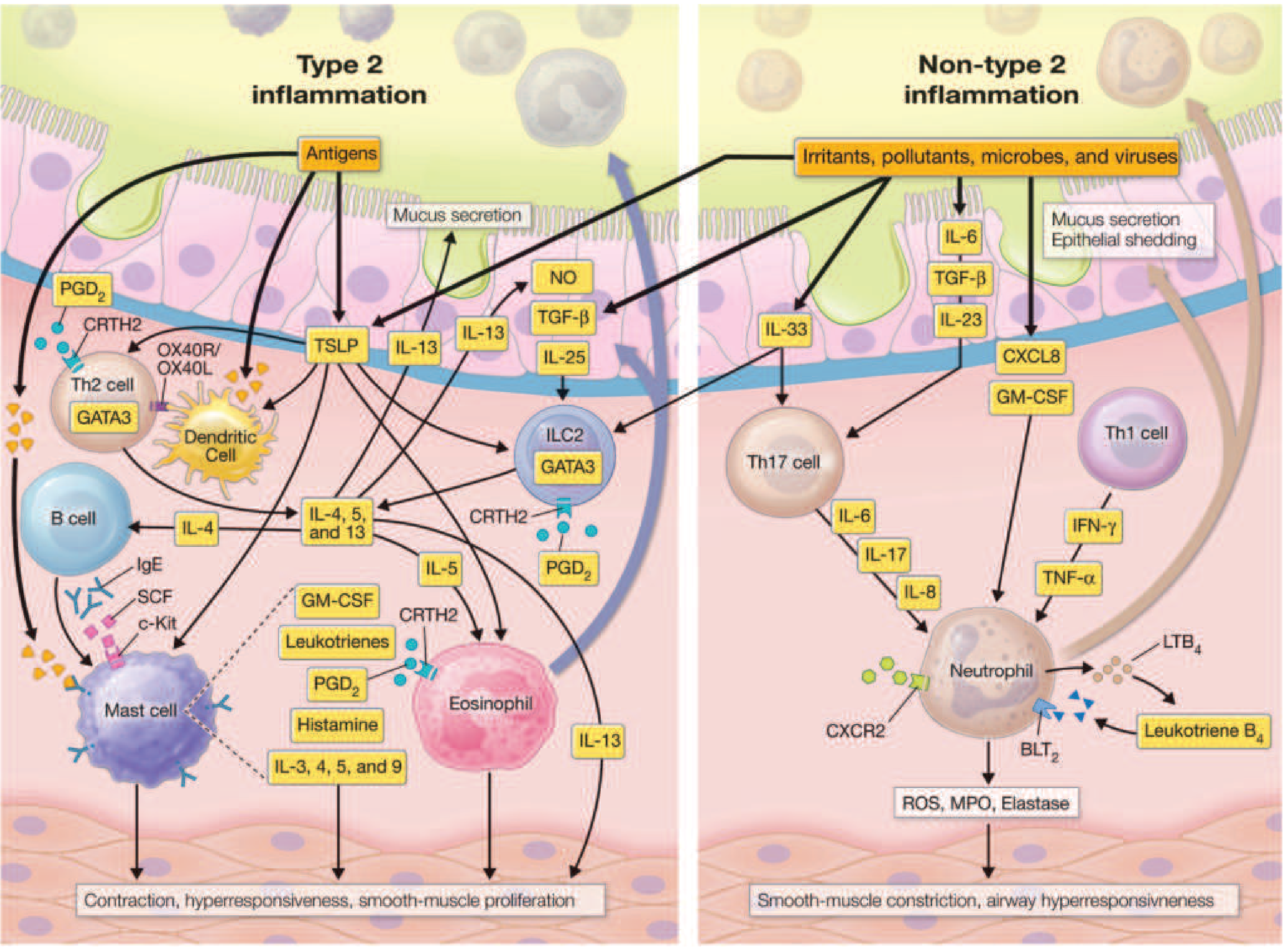
- Mast cells: activated by IgE cross-linking; release histamine, leukotrienes, PGD2, IL-3,4,5,9
- Eosinophils: release major basic protein (disrupts epithelium), cysteinyl leukotrienes; form mucus plugs contributing to severity
- Th2 lymphocytes: orchestrate the T2 response via GATA3
- ILC2s: innate lymphoid cells; also produce T2 cytokines
- Dendritic cells: present antigen; activated by alarmins (TSLP, IL-25, IL-33)
- B cells: stimulated by IL-4 to produce IgE
- Neutrophilic inflammation: driven by IL-6, IL-8, IL-17, TNF-α; may involve Mycoplasma infection; may respond to macrolides
- Pauci-granulocytic asthma: pathologic changes without cellular infiltration (etiology unclear)
Key Mediators
| Category | Mediators | Effects |
|---|---|---|
| T2 cytokines | IL-4, IL-5, IL-13 | Core of T2 inflammation; FDA-approved biologic targets |
| Alarmins | TSLP, IL-25, IL-33 | Initiate T2 cascade from epithelium |
| Cysteinyl leukotrienes (LTC4, LTD4, LTE4) | From eosinophils & mast cells | Potent smooth-muscle constrictors, mucus secretion, microvascular leakage |
| Prostaglandin D2 (PGD2) | From mast cells | Bronchoconstriction, inflammatory cell recruitment |
| Histamine | From mast cells | Bronchoconstriction, edema |
| Non-T2 cytokines | IL-6, IL-8, IL-17, TNF-α | Neutrophilic inflammation, severe asthma |
Airway Remodeling
- Smooth-muscle hypertrophy and hyperplasia
- Subepithelial collagen deposition (fibrosis)
- Goblet cell hyperplasia and mucus hypersecretion
- Microvascular changes and edema
- Results in permanent, partially irreversible airflow limitation in long-standing asthma
Risk Factors and Triggers
Risk Factors for Development (Table 298-1, Harrison's)
- Allergen exposure in atopic individuals
- Occupational exposures
- Air pollution
- Viral infections and Mycoplasma
- Tobacco smoke
- Obesity
- Diet
- Fungi (allergic airway mycoses)
- Reactive airway dysfunction syndrome (RADS) from acute irritants
- High-intensity exercise in elite athletes
Triggers of Airway Narrowing (Table 298-2, Harrison's)
- Allergens
- Irritants (smoke, fumes, chemicals)
- Viral upper respiratory infections
- Exercise and cold/dry air
- Air pollution
- Drugs (NSAIDs/aspirin, beta-blockers)
- Occupational exposures
- Hormonal changes (menstrual cycle, menopause)
- Pregnancy
Clinical Features
- Wheeze (polyphonic, expiratory > inspiratory)
- Dyspnoea (episodic)
- Cough (often nocturnal/early morning; sometimes cough-variant asthma)
- Chest tightness
- Symptoms worsen at night and improve with bronchodilators
- Between attacks, the chest may be entirely normal
- Prolonged expiration, hyperinflation, use of accessory muscles during acute attacks
Diagnosis
Spirometry
- Obstructive pattern: FEV1/FVC < 0.7
- Reversibility: ≥12% AND ≥200 mL improvement in FEV1 after bronchodilator
- Variability: >10% day-to-day variability in PEF (peak expiratory flow)
Bronchial Challenge Tests
- Methacholine or histamine provocation: used when spirometry is normal but asthma is suspected
- Exercise challenge: for exercise-induced symptoms
Additional Evaluation in Severe/Poorly Responsive Asthma
- Allergen testing (skin prick or specific IgE)
- Full blood count (eosinophil count)
- FeNO (fractional exhaled NO - marker of T2 inflammation)
- CT thorax (to exclude structural causes, identify mucus plugs)
- Laryngoscopy (if vocal cord dysfunction suspected)
Comorbidities
- Heart failure
- COPD
- Alpha-1 antitrypsin deficiency
- Airway obstruction (mass or foreign body)
- Inducible laryngeal obstruction (vocal cord dysfunction)
- Bronchiolitis obliterans
- Bronchiectasis
- Tracheobronchomalacia
- Chronic rhinosinusitis +/- nasal polyposis
- Obesity (2-4x increased hospitalization risk)
- GERD (independent predictor of exacerbations)
- Inducible laryngeal obstruction
- COPD (asthma-COPD overlap - see below)
- Anxiety/depression (increase exacerbations)
- Obstructive sleep apnoea
Management
Pharmacological Agents
1. Inhaled Corticosteroids (ICS) - Cornerstone of Treatment
- Beclomethasone, budesonide, fluticasone, ciclesonide
- Reduce airway inflammation, decrease hyperresponsiveness, prevent exacerbations
- Cornerstone of all step-up therapy
- Reduction in mortality attributed primarily to ICS use
2. Beta-2 Agonists (Bronchodilators)
| Class | Agents | Onset | Duration | Use |
|---|---|---|---|---|
| SABA | Salbutamol (albuterol) | 3-5 min | 4-6 h | Acute relief; pre-exercise |
| LABA | Salmeterol, Formoterol | Salmeterol: slow; Formoterol: fast | ~12 h | Combined with ICS only (never monotherapy) |
| Ultra-LABA | Indacaterol, Olodaterol, Vilanterol | Fast | 24 h | Combined with ICS only |
3. Anticholinergics
- Ipratropium (short-acting), tiotropium (long-acting)
- Block cholinergic-mediated bronchoconstriction
- Add-on therapy at Step 4/5; also useful in exercise-induced symptoms in elite athletes
4. Leukotriene Receptor Antagonists (LTRAs)
- Montelukast, zafirlukast
- Alternative/add-on at Step 2
- Note: recent warnings about suicidal ideation with montelukast reduce its appeal
5. Theophylline
- Bronchodilator + mild anti-inflammatory
- Narrow therapeutic index; less favored
- Still used in resource-limited settings
6. Anti-Inflammatory Reliever (AIR) - Major Recent Change in Guidelines
- ICS/formoterol combination used as-needed (instead of SABA alone)
- Rationale: even mild asthma can be fatal; ICS component reduces inflammation at every use
- GINA recommends ICS/formoterol as reliever at ALL steps (including Step 1/intermittent asthma)
- NAEPP recommends ICS/formoterol reliever from Step 3 onward
7. Biologics (Step 5 and beyond)
| Biologic | Target | Indication |
|---|---|---|
| Omalizumab | Anti-IgE | Allergic asthma with elevated IgE |
| Mepolizumab, Reslizumab | Anti-IL-5 | Severe eosinophilic asthma |
| Benralizumab | Anti-IL-5Rα | Severe eosinophilic asthma |
| Dupilumab | Anti-IL-4Rα (blocks IL-4+IL-13) | Moderate-severe T2 asthma |
| Tezepelumab | Anti-TSLP | Severe asthma (broadest indication - T2 and non-T2) |
Stepwise Approach (GINA-adapted, Table 298-5)
| Step | Preferred Controller | Preferred Reliever |
|---|---|---|
| 1 (Intermittent) | As-needed ICS/formoterol | ICS/formoterol |
| 2 (Mild persistent) | Low-dose ICS daily OR as-needed ICS/formoterol | ICS/formoterol or SABA+ICS |
| 3 (Moderate) | Low-dose ICS/LABA | ICS/formoterol |
| 4 (Severe) | Medium-high dose ICS/LABA | ICS/formoterol; add LTRA or LAMA |
| 5 (Very severe) | High-dose ICS/LABA + biologics | ICS/formoterol |
Special Considerations
Asthma Attacks (Acute Exacerbations)
- Precipitated by infections, allergen exposure, poor adherence, or trigger exposure
- Management: oxygen, repeated SABAs (or nebulized salbutamol), systemic corticosteroids, ipratropium, IV magnesium sulfate in severe cases, consider intubation/ICU if failing
Patients at High Risk of Asthma Mortality (Table 298-6)
- History of ICU admission for asthma
- History of intubation for asthma
- Illicit drug use
- Depression
- New diagnosis within the past year
- ≥2 emergency visits in the past 6 months
- Severe psychosocial problems
- Lower socioeconomic status
- Daily oral corticosteroids before admission
Exercise-Induced Bronchoconstriction
- Pre-treatment with SABA raises bronchospasm threshold
- Warm-up exercise and face masks in cold weather help
- Regular SABA use acceptable for frequent exercisers; LTRA can be used regularly
Pregnancy
- Asthma may improve, stay same, or worsen in pregnancy
- Poor control is associated with poor fetal outcomes
- Safe drugs: inhaled salbutamol, beclomethasone, budesonide, fluticasone; formoterol and salmeterol have reassuring data
- Oral corticosteroids: associated with neonatal adrenal insufficiency, preeclampsia, low birth weight, slight risk of cleft palate - but poorly controlled asthma is more dangerous to the fetus
- Avoid PGF2α (bronchoconstriction risk); do not initiate allergen immunotherapy during pregnancy
Aspirin-Exacerbated Respiratory Disease (AERD)
- Triad: asthma + nasal polyposis + aspirin/NSAID sensitivity
- Nasal polyposis in adults with asthma should raise suspicion
- Rare in children
Asthma-COPD Overlap (ACO)
- Features of both conditions coexist
- Management more complex; ICS mandatory (to prevent pneumonia risk as seen in pure COPD with ICS)
Severe Asthma
- Defined as requiring Step 4/5 therapy, or uncontrolled despite maximum therapy
- Requires assessment of: adherence, inhaler technique, comorbidities, triggers, and endotyping before escalating to biologics
Elderly Patients
- Diagnosis may be missed due to underreporting of symptoms
- Comorbidities (COPD, heart failure, GERD) complicate management
Bronchoscopic Therapies (Emerging)
Key Pathophysiology Diagram
Summary Table: Core Concepts
| Feature | Detail |
|---|---|
| Definition | Chronic airway inflammation + reversible obstruction + hyperresponsiveness |
| Hallmark | Airway hyperresponsiveness |
| Most common inflammation | Type 2 (eosinophilic, IgE-mediated) |
| Key cells | Mast cells, eosinophils, Th2 cells, ILC2s |
| Key cytokines | IL-4, IL-5, IL-13; alarmins (TSLP, IL-25, IL-33) |
| Spirometry | Obstructive; ≥12% + ≥200 mL reversibility |
| Cornerstone therapy | ICS |
| Reliever (GINA, all steps) | ICS/formoterol combination |
| Biologics | Anti-IgE, anti-IL-5, anti-IL-4R, anti-TSLP |
| Never use alone | LABA monotherapy |
Management according to Malaysian guidelines
Malaysia Clinical Practice Guidelines Management of Asthma 2024 2023 MOH MTS
"asthma" AND "Malaysia" AND "guideline"
https://www.moh.gov.my/images/04-penerbitan/penerbitan-klini…
Malaysia CPG Management Asthma Adults 2nd edition 2024 full text acute exacerbation assessment severity stepwise
https://mts.org.my/resources/CPG%20Management%20of%20Asthma%…
Asthma Management According to Malaysian Guidelines
- CPG Management of Asthma in Adults, 2nd Edition (2024) - MOH Malaysia / Academy of Medicine Malaysia / MaHTAS
- Quick Reference - Management of Asthma in Adults, 2nd Edition (2024, Updated Oct 2025) - MOH Malaysia
- CPG Management of Childhood Asthma, 4th Edition (2024) - Malaysian Thoracic Society
Key Messages (CPG 2nd Ed, 2024)
- Asthma is a chronic inflammatory airway disease triggered by external stimuli in genetically-predisposed individuals
- Diagnosis is based on typical clinical history, physical examination, and evidence of airway obstruction variability - spirometry is the preferred diagnostic tool
- Assessment of asthma control must be performed before considering stepping up or down
- All asthma patients must be prescribed ICS-containing therapy
- Inhaled SABAs must NOT be used as monotherapy
- All patients must receive self-management education including a written Asthma Action Plan (AAP)
Epidemiology (Malaysia-specific)
- Prevalence in Malaysian adults (≥18 years): 4.5% (National Health and Morbidity Survey 2006)
- Follow-up rate in clinics and hospitals: only 32.6%
- Exacerbation rate: 68.1%, of which 25.8% had >3 exacerbations/year
- Childhood asthma: ~500,000 children aged 6-17; probable asthma 7.1%, doctor-diagnosed 5.3% (NHMS 2023)
- 70% of children had 1-3 exacerbations in the past year; 30% had 4 or more
Diagnosis
- Based on typical symptoms (wheeze, breathlessness, chest tightness, cough) that are variable, episodic, worse at night/early morning
- Spirometry is the preferred tool: demonstrates obstruction (FEV1/FVC < lower limit of normal) with ≥12% and ≥200 mL reversibility post-bronchodilator
- PEF monitoring: >10% diurnal variability supports diagnosis
- Bronchoprovocation testing (methacholine/exercise challenge) when spirometry is normal but asthma is suspected
Assessment of Asthma Control
Asthma Control Test (ACT) - validated in Malay
- Score 25: Fully controlled
- Score 20-24: Partly controlled
- Score <20: Uncontrolled
Level of Control Categories
| Level | Daytime Symptoms | Night Waking | Reliever Use | Activity Limitation | Lung Function |
|---|---|---|---|---|---|
| Well-controlled | ≤2 days/week | None | ≤2 days/week | None | FEV1/PEF ≥80% |
| Partly controlled | >2 days/week | Any | >2 days/week | Any | FEV1/PEF 60-80% |
| Uncontrolled | Most days | ≥1/week | Daily | Yes | FEV1/PEF <60% |
Stable Asthma Management
Assessment Before Initiating Treatment
- Asthma control level
- Risk factors for future exacerbations (prior ICU admission, ≥1 exacerbation/year, low FEV1, poor adherence, heavy SABA use, smoking, comorbidities)
- Modifiable risk factors - address these before stepping up
Initiation of Treatment
| Symptom Burden at Diagnosis | Recommended Starting Therapy |
|---|---|
| Symptoms most days OR night waking ≥1/week OR FEV1 <80% | Maintenance low-dose ICS-LABA OR low-to-medium dose ICS |
| Daily symptoms AND night waking ≥1/week AND FEV1 <80% | Maintenance medium-to-high dose ICS-LABA OR medium-dose ICS-LABA + LAMA |
| Milder/intermittent symptoms | PRN ICS-formoterol OR low-dose ICS + PRN SABA/ICS-SABA |
Stepwise Treatment of Stable Asthma
Reliever Therapy (at all steps)
- ICS-formoterol (preferred, if maintenance already contains ICS-formoterol) - max 12 puffs/day total
- ICS-SABA (alternative reliever)
- SABA (plain salbutamol - acceptable but not preferred; NEVER as sole monotherapy)
Controller Therapy (Maintenance) - Step Up / Step Down
| Step | Maintenance Therapy | Notes |
|---|---|---|
| Step 1 (Intermittent) | PRN ICS-formoterol OR PRN ICS-SABA | No daily controller needed if truly intermittent; still ICS-containing reliever |
| Step 2 (Mild persistent) | Low-dose ICS-LABA OR medium-dose ICS | LTRA may be added if concurrent allergic rhinitis |
| Step 3 (Moderate persistent) | Medium-dose ICS-LABA | Consider phenotypic assessment before stepping up further |
| Step 4 (Moderate-severe) | High-dose ICS-LABA OR medium-dose ICS-LABA + LAMA | Phenotypic assessment mandatory at this stage |
| Step 4 alt | Medium-dose ICS-LABA + LAMA | Consider single inhaler ICS-LABA-LAMA |
| Step 5 (Severe) | High-dose ICS-LABA + LAMA OR single inhaler high-dose ICS-LABA-LAMA | Refer respiratory physician; MDT discussion for biologics; add-on low-dose azithromycin or OCS as alternative |
Note: LTRA (e.g., montelukast) may be added at any step if the patient has concurrent allergic rhinitis.
Step Up and Step Down
- Step up only after confirming: poor adherence is excluded, inhaler technique is correct, triggers are addressed, comorbidities are managed
- Step down when asthma is well controlled for ≥3 months; reduce ICS dose by 25-50%
- Never completely stop ICS in adults
Adherence Assessment
- 10-item version validated in Malay language
- Identifies intentional vs. non-intentional non-adherence
- Poor adherence accounts for up to 50% of poorly controlled asthma referrals
Non-Pharmacological Management
- Written Asthma Action Plan (AAP) for all patients
- Smoking cessation
- Allergen/trigger avoidance
- Breathing exercises (Buteyko, physiotherapy)
- Vaccination (influenza annually; pneumococcal)
- Weight reduction in obese patients
- Treatment of comorbidities (allergic rhinitis, GERD, OSA)
- Regular follow-up (at least 3-6 monthly when stable)
Acute Exacerbation of Asthma
Assessment of Severity (Malaysian CPG)
| Severity | Speech | Posture | Mental Status | RR | PR | SpO2 | PEF |
|---|---|---|---|---|---|---|---|
| Mild-Moderate | Phrases | Sitting up | Not agitated | 20-30/min | 100-120/min | 90-95% | >50% predicted |
| Severe | Words | Sits forward | Agitated; accessory muscles | >30/min | >120/min | <90% | <50% predicted |
| Life-threatening | Silent chest / poor effort | Any | Drowsy / confused / exhausted | Variable | Any | Cyanosis/hypotension | <33% predicted; PaO2 <60 mmHg; normal or raised PaCO2 |
Status asthmaticus = life-threatening asthma that is refractory to initial bronchodilator therapy.
Treatment of Acute Exacerbation
1. Oxygen
- Target SpO2 94-98% (controlled/titrated oxygen, not high-flow unrestricted)
- CO2 retention is NOT usually aggravated by oxygen in asthma
2. Beta-2 Agonists (First-line)
- Salbutamol (albuterol) via oxygen-driven nebulizer: 2.5-5 mg every 15-30 minutes (or continuously in severe cases)
- Alternatively: 4-8 puffs via MDI + spacer in mild-moderate
- IV salbutamol if no improvement with inhaled route
3. Ipratropium Bromide (add-on in severe attacks)
- 0.5 mg nebulized every 4-6 hours (combined with salbutamol)
- Adds bronchodilation via anticholinergic pathway
- Particularly useful in early severe/life-threatening phase
4. Corticosteroids (systemic - mandatory in moderate-severe)
- Prednisolone 40-50 mg orally (preferred if patient can swallow)
- OR Hydrocortisone 100 mg IV 6-hourly if unable to take orally
- Duration: 5-7 days (no taper needed for short course)
- Begin ICS on discharge or continue if already on it
5. Magnesium Sulphate (severe/life-threatening)
- IV MgSO4 2 g over 20 minutes - for severe attacks not responding to initial therapy
- Evidence: reduces hospitalization in severe acute asthma
- Nebulized isotonic MgSO4 as adjunct to salbutamol also has evidence
6. Other treatments
- Heliox (helium-oxygen mixture): may reduce work of breathing; limited evidence
- IV aminophylline: no longer routinely recommended (narrow therapeutic index, no added benefit over beta-agonists alone)
- No sedatives of any kind in acute asthma
- Chest X-ray only if pneumothorax, consolidation, or mechanical ventilation needed
Monitoring During Acute Attack
- Serial PEF measurements
- Continuous SpO2 monitoring
- Conscious level - deterioration = consider intubation
- ABG if not improving or life-threatening features
Criteria for Admission
- PEF <75% after 1 hour of treatment
- SpO2 <94% on air
- No response to initial bronchodilators
- Life-threatening features at any time
- Significant comorbidities or social concerns
- Drowsy, confused, exhausted
- Silent chest
- PaCO2 normal or raised (impending respiratory failure)
- Requiring intubation
Criteria for Discharge (after acute episode)
- PEF >75% predicted or personal best
- SpO2 ≥94% on room air
- Symptoms well controlled on discharge medications for 12-24 hours
- Inhaler technique checked and documented
- Written AAP provided
- Follow-up appointment arranged within 1-2 weeks
Special Groups
Asthma in Pregnancy
- Poorly controlled asthma carries greater risk to fetus than the medications
- Safe drugs: inhaled salbutamol, budesonide, beclomethasone, fluticasone, formoterol, salmeterol
- Oral corticosteroids: use if needed; risk of poorly controlled asthma > drug side effects
- Do NOT initiate allergen immunotherapy during pregnancy
- Avoid PGF2α (bronchoconstriction)
- Monitor closely - asthma may improve, worsen, or be unchanged
Occupational Asthma
- Remove from exposure as early as possible - delay worsens prognosis
- Confirm with serial PEF (at work vs. away from work)
- Treat as per standard asthma stepwise approach
Biologics (Step 5 - Specialist Only)
| Biologic | Target | Indication |
|---|---|---|
| Omalizumab | Anti-IgE | Allergic (atopic) severe asthma, elevated IgE |
| Mepolizumab | Anti-IL-5 | Severe eosinophilic asthma (blood eos ≥300/µL) |
| Benralizumab | Anti-IL-5Rα | Severe eosinophilic asthma |
| Dupilumab | Anti-IL-4Rα | Severe Type 2 asthma (IL-4 + IL-13 pathway) |
| Tezepelumab | Anti-TSLP | Severe asthma, broadest phenotypic coverage |
Referral Criteria to Respiratory Specialist
- Diagnostic uncertainty
- Severe or uncontrolled asthma despite Step 3/4 therapy
- Frequent exacerbations (≥2/year)
- Consideration for biologics
- Occupational asthma
- Complicating comorbidities
- Asthma in pregnancy with poor control
Summary: Malaysian CPG 2024 Management Algorithm
Confirmed Asthma
        ↓
Assess Control + Risk Factors
        ↓
Well-controlled? → Continue / Step Down (after 3 months stable)
Partly controlled? → Step Up
Uncontrolled? → Step Up (after excluding poor adherence/technique)
        ↓
Step 1: PRN ICS-formoterol OR PRN ICS-SABA
Step 2: Low-dose ICS-LABA or Medium-dose ICS
Step 3: Medium-dose ICS-LABA [Phenotypic assessment]
Step 4: High-dose ICS-LABA ± LAMA [Phenotypic assessment]
Step 5: High-dose ICS-LABA-LAMA → Refer specialist → Biologics
        ↓
Reliever at ALL steps: ICS-formoterol (preferred) / ICS-SABA / SABA
        ↓
Non-pharmacological: AAP + education + trigger avoidance