I now have all the key content from Tintinalli's Chapter 152 (the dedicated NF chapter) and Rosen's. I have comprehensive, direct textbook quotes on pathophysiology, clinical features, diagnosis, and treatment. Let me now compile the full answer.
Necrotizing Fasciitis - Comprehensive Answer
(Based on Tintinalli's Emergency Medicine, Chapters 7 & 152; Rosen's Emergency Medicine, Chapter 107)
a) Pathophysiology of Necrotizing Fasciitis, Including the Mechanism of Rapid Tissue Destruction (3 Marks)
Definition
Necrotizing fasciitis (NF) is a rapidly spreading, deep soft tissue infection characterized by fulminant, extensive necrosis of the fascia and subcutaneous tissue, profound systemic toxicity, and high mortality. It is part of a spectrum of necrotizing soft tissue infections (NSTIs).
Portal of Entry
In this patient, the recent cut over the lower leg served as the entry point. The rapid necrotizing process typically begins with direct invasion of subcutaneous tissue from external trauma - IV injection, surgical incision, abscess, insect bite, or ulcer - or from direct spread from a perforated viscus. Predisposing host factors such as diabetes mellitus (as in this case) significantly impair immune response and local tissue defenses, accelerating disease progression.
Microbiology in This Case
- Type I (Polymicrobial) - as indicated by the mixed Gram-positive and Gram-negative blood cultures. This is the most common type, accounting for 55-75% of all NSTIs
- A symbiotic interaction between aerobic and anaerobic organisms drives rapid tissue destruction
- Type II (monomicrobial) - Group A Streptococcus or MRSA; Type III - Vibrio vulnificus
Mechanism of Rapid Tissue Destruction (Step by Step)
Step 1 - Bacterial proliferation and fascial invasion:
Bacteria proliferate in the subcutaneous tissue and invade the deep fascia, releasing exotoxins that initiate the destructive cascade. The infection tracks along fascial planes, which offer minimal anatomical resistance, allowing spread of up to 1 inch per hour.
Step 2 - Polymicrobial synergy (key to Type I NF):
A symbiotic relationship between organisms drives destruction:
- Facultative gram-negative organisms (e.g., E. coli, Klebsiella) consume local oxygen, lowering the oxygen-reduction potential of the tissue
- This anaerobic microenvironment facilitates anaerobic organism growth (e.g., Bacteroides, Clostridium spp.)
- Anaerobic organisms in turn impede phagocyte function, favoring further aerobic bacterial growth
- This mutual amplification produces gas (seen on CT as soft tissue air) and accelerates necrosis
Step 3 - Exotoxin-mediated tissue ischemia and liquefaction:
Bacteria release exotoxins that lead to:
- Tissue ischemia
- Liquefactive necrosis of the fascia and subcutaneous tissue
- Systemic toxicity
- Staphylococcus aureus and streptococci produce exotoxins that trigger TNF-alpha and cytokine release, producing systemic inflammatory response syndrome (SIRS) leading to septic shock, organ failure, and death
Step 4 - Vascular thrombosis causing skin necrosis:
Infection causes vasculitis and thrombosis of perforating blood vessels supplying the overlying skin. This produces ischemic necrosis of the skin and subcutaneous fat secondary to the fascial infection. Because thrombosis of large numbers of capillary beds must occur before skin findings develop, early infection shows little overlying skin change - this explains why the extent of tissue destruction is always far greater than what is visible on the surface.
Step 5 - Progressive spread and systemic collapse:
As the infection progresses, widespread gangrene of the skin, subcutaneous fat, fascia, and even skeletal muscle occurs. Bacteremia (present in 25-30% of cases) drives septic shock and multiorgan failure.
(Tintinalli's Emergency Medicine, Chapter 152 - "Bacteria proliferate, invade subcutaneous tissue and deep fascia, and release exotoxins that lead to tissue ischemia, liquefaction necrosis, and systemic toxicity")
b) Clinical Features, Signs, and Differentiation from Other Soft Tissue Infections (3 Marks)
Clinical Features - Progressive Stages
Early Stage (deceptively benign - mimics cellulitis):
- Severe pain, anxiety, and diaphoresis - classic early symptoms
- Pain out of proportion to physical examination findings, with tenderness extending beyond the area of visible erythema - "perhaps the single most important feature to make the diagnosis early" (Tintinalli, Chapter 152)
- Erythema and edema that can be indistinguishable from cellulitis
- About 10-40% of patients report trauma or a skin break ~48 hours before symptoms begin
Intermediate Stage:
- Brawny (woody, firm) oedema - non-pitting, indurated
- Crepitus from bacterial gas production (present in 13-31% of patients; its absence does NOT exclude the diagnosis)
- Erythema and tenderness extending well beyond the visible margins
- Violaceous (purple-brown) discoloration of overlying skin
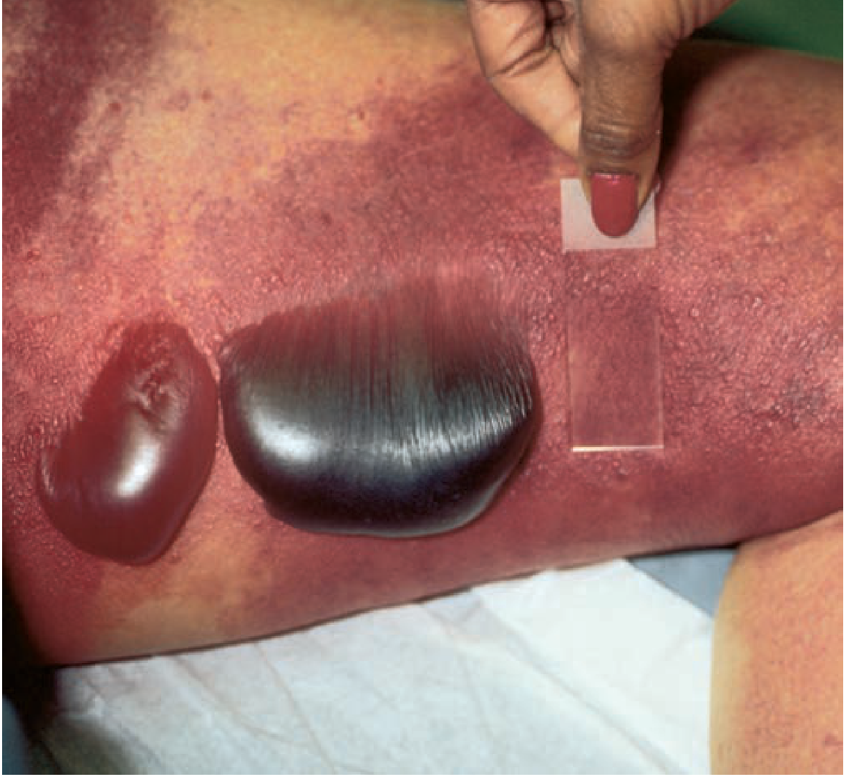
- Development of haemorrhagic/dark bullae with serosanguineous or malodorous discharge - late sign
Advanced Stage:
- Cutaneous anaesthesia/hypesthesia - as nerves are destroyed by ischaemia, the initially exquisitely painful area becomes numb (important differentiating sign)
- Overt skin necrosis and gangrene
- Malodorous "dishwater" discharge
Fig. 152-6: Necrotizing soft tissue infection - large cutaneous haemorrhagic bullae on the leg of a patient with necrotizing fasciitis (Tintinalli's Emergency Medicine)
Systemic Manifestations:
- Low-grade fever with tachycardia out of proportion to the fever
- In fulminant cases: cardiovascular collapse, confusion, rapid deterioration of mental status
- Progression to septic shock and multiorgan failure
(Rosen's Emergency Medicine: "Hallmarks of fasciitis are the presence of marked systemic toxicity and pain out of proportion to local findings. In more advanced cases, there may be deep pain with patchy areas of surface hypesthesia, crepitation, or bullae.")
Differentiation from Other Soft Tissue Infections
| Feature | Cellulitis | Erysipelas | Abscess | Necrotizing Fasciitis |
|---|
| Pain vs. findings | Proportionate | Proportionate | Localised | Pain out of proportion |
| Skin border | Blurred, diffuse | Sharp, raised, demarcated | Fluctuant point | Ill-defined, advancing rapidly |
| Skin discolouration | Red/pink | Bright red | Erythematous rim | Violaceous/bronze/necrotic |
| Bullae | Absent | Rare | Absent | Present (haemorrhagic) |
| Crepitus | Absent | Absent | Absent | May be present (gas) |
| Skin anaesthesia | Absent | Absent | Absent | Present in advanced disease |
| Systemic toxicity | Mild-moderate | Mild-moderate | Mild | Severe and early |
| Response to antibiotics | Responds | Responds | Requires I&D | Does NOT respond - surgery needed |
| Fascial involvement | No | No | No | Yes - spreads along fascia |
| CT findings | Normal fascia | Normal fascia | Collection | Gas along fascial planes, fascial thickening, non-enhancing fascia |
Investigations Supporting Diagnosis
- CT with contrast: Fascial thickening of deep fascial planes; non-enhancing deep fascia (suggesting necrosis); presence of gas (highly specific but found in only ~1/3 of cases) - as in this patient
- MRI: Extensive thickening of intermuscular fasciae with incomplete vascularisation; absence of MRI abnormalities virtually rules out NF
- LRINEC Score (Laboratory Risk Indicator for Necrotizing Fasciitis): Uses CRP >150 mg/L, WBC >15,000/mm³, haemoglobin <13.5 g/dL, sodium <135 mmol/L, creatinine >1.6 mg/dL, glucose >180 mg/dL. Score ≥6 raises suspicion; however, LRINEC alone can miss many cases and should not be used to exclude the diagnosis
- Definitive diagnosis: Surgical exploration - finding grey necrotic fascia, lack of bleeding, no resistance to blunt finger dissection, and "dishwater fluid"
c) Surgical Management of Necrotizing Fasciitis (4 Marks)
Principle: Surgical Emergency - Life-Saving Priority
Necrotizing fasciitis is a surgical emergency. As stated in Tintinalli's: "Successful treatment requires early, aggressive surgical debridement of all necrotic tissue, appropriate broad-spectrum systemic antibiotic therapy, and supportive care."
Delayed definitive debridement remains the single most important risk factor for death.
1. Immediate Surgical Debridement - Role and Technique
Principles of debridement:
- Wide, radical excision of all necrotic tissue - all necrotic skin, subcutaneous fat, and fascia are excised down to and including the infected fascial layer
- The process always extends beyond the area of clinical involvement - the surgeon must probe adjacent tissue with a finger; if it sweeps through without resistance ("finger test" positive), that tissue must also be debrided
- Intraoperative findings confirming NF: grey necrotic fascia, "dishwater" fluid in fascial planes, lack of bleeding from tissues, lack of resistance to blunt dissection, absence of normal tissue attachment to overlying fat
Staged, daily return trips to the operating room:
- A single debridement is almost never adequate
- Patients return to theatre every 24-48 hours for re-exploration and further debridement until no further necrotic tissue is found
- This staged approach is essential - re-inspection is mandatory
Amputation:
- May be required for severe limb disease when infection cannot be controlled with debridement alone, or when limb viability is irretrievably lost
- Diabetic patients (as in this case) have an increased risk of requiring amputation
- Limb salvage should be attempted but must never take priority over life-saving debridement
2. Antibiotic Therapy - Critical Adjunct (Not Replacement for Surgery)
Broad-spectrum empirical antibiotics are started immediately while awaiting surgical source control and culture results.
Empirical regimen (Rosen's/Tintinalli's):
Anti-MRSA coverage (one of):
PLUS broad Gram-negative and anaerobic coverage (one of):
- Piperacillin-tazobactam
- Carbapenem (imipenem/meropenem)
- Ceftriaxone + metronidazole
For confirmed Group A Streptococcal (Type II) NF:
- Penicillin G + Clindamycin - clindamycin is added because it inhibits bacterial protein and exotoxin synthesis (even though bacteria may be resistant to clindamycin alone)
- Tintinalli: "Triple antibiotic therapy with penicillin or a cephalosporin, an aminoglycoside, and clindamycin probably should be used"
Antibiotics are continued until:
- Surgical debridement is complete
- Patient is afebrile for 48-72 hours
- Clinical stabilisation is achieved
- Narrowed based on culture results from intraoperative tissue samples
3. Wound Management After Debridement
- Wounds are left open - primary closure is never appropriate in active NF
- Negative pressure wound therapy (VAC - vacuum-assisted closure): Applied to large open wounds post-debridement; reduces oedema, promotes granulation tissue, bridges the gap to definitive closure
- Skin grafting: Once infection is fully controlled and wound bed is clean and healthy, delayed skin grafting achieves final wound closure
- This reconstruction phase may require input from plastic surgery
4. Hyperbaric Oxygen Therapy (HBO)
Tintinalli's notes HBO as a "valuable adjunct to surgical debridement and antibiotic therapy in necrotizing soft tissue infections, reducing mortality, improving recovery rates, and decreasing both amputation rates and the number of required surgical interventions."
Mechanism of benefit:
- Suppresses growth of anaerobic microorganisms
- Improves bactericidal action of leukocytes that function poorly in hypoxic conditions
- Enhances antibiotic penetration into target bacteria
Protocol: Twice-daily treatments at 2.0-2.5 ATA for 90 minutes with air breaks, until extension of necrosis is halted, then daily for up to 30 sessions.
Note: Not universally available and must never delay surgical debridement.
5. Additional Considerations in Patient Care
Resuscitation and ICU care:
- Aggressive IV fluid resuscitation (massive third-space losses from necrotic tissue)
- Close monitoring of urine output, vitals, and electrolytes
- Vasopressor support if septic shock develops
- Most patients require ICU-level care
Diabetic patient-specific considerations:
- Strict glycaemic control - hyperglycaemia impairs neutrophil function and wound healing
- Assess for peripheral vascular disease - vascular surgery consultation may be needed
- Higher risk of amputation; higher mortality risk
IV Immunoglobulin (IVIG):
- May be considered in Group A Streptococcal NF to neutralise bacterial superantigens
- Limited data - no strong evidence for routine use
Nutritional support:
- Early enteral nutrition supports immune function, wound healing, and recovery through the prolonged post-operative course
Mortality
Adult mortality remains 20-30% despite modern management. Mortality is driven by bacteraemia, septic shock, and multiorgan failure. Factors increasing mortality: age >60 years, delayed surgery, interhospital transfer, positive blood cultures, truncal/perineal involvement, comorbid cancer or renal disease.
Summary
| Component | Key Point (from Tintinalli's/Rosen's) |
|---|
| Pathophysiology | Exotoxins + polymicrobial synergy → fascial necrosis; vascular thrombosis → skin ischaemia; spreads up to 1 inch/hour |
| Hallmark clinical signs | Pain out of proportion; brawny oedema; crepitus; haemorrhagic bullae; skin anaesthesia; marked systemic toxicity |
| Differentiation | Woody induration; gas on CT; skin anaesthesia; non-response to antibiotics; fascial plane gas/thickening on imaging |
| Surgery | Immediate radical debridement; staged daily re-exploration; amputation if needed; wounds left open |
| Antibiotics | Vancomycin + piperacillin-tazobactam (or carbapenem); penicillin + clindamycin for Strep; culture-guided narrowing |
| Adjuncts | HBO therapy; VAC dressing; delayed skin grafting; ICU care; glycaemic control; IVIG (limited evidence) |