
A 60 yrs old male present with intensly itchy lesion all over his body particularly on elbow back inner thighs waist knee joint tibeal surface ear pinna what is probable diagnosis and treatment in detail
Reading File
Finding Sources
Finding Sources
Searching PubMed
scabies treatment permethrin ivermectin
Reading File
Reading File
Reading File
Reading File
Reading File
Reading File
Loading Image
Loading Image
I now have comprehensive information to provide a detailed answer. Let me compile this into a thorough clinical response.
Probable Diagnosis: SCABIES (Sarcoptes scabiei var. hominis)
Why This Diagnosis Fits Perfectly
The clinical triad in your patient - intensely itchy lesions + characteristic distribution + adult male - is textbook scabies. Every site mentioned (elbows, back, inner thighs, waist, knee joints, tibial surface, ear pinna) maps directly to the known predilection sites of the Sarcoptes scabiei mite.
The Causative Agent
Sarcoptes scabiei var. hominis - an obligate human ectoparasite of class Arachnida, family Sarcoptidae. The adult female mite is 0.4 x 0.3 mm - just below naked eye visibility. She burrows into the stratum corneum (0.5-5 mm/day), lays up to 50 eggs over her 30-day lifespan, and the entire life cycle is completed on human skin. A normal host harbors only 3-50 mites, yet the immune hypersensitivity reaction to mite products, eggs, and feces drives intense itching. - Fitzpatrick's Dermatology, Vol. 1-2
Clinical Findings in Detail
Symptoms
- Intense, primarily NOCTURNAL pruritus - the hallmark symptom
- Onset: insidious, typically 4-6 weeks after first infestation (sensitization period); with re-infestation, symptoms appear within 2-3 days
- Some patients remain asymptomatic ("carriers") despite harboring mites
Characteristic Lesion Distribution (Predilection Sites)
| Site | Reason |
|---|---|
| Interdigital web spaces, sides of fingers | Thin stratum corneum, easy burrowing |
| Volar wrists, lateral palms | Same reason |
| Elbows | Present in this patient ✓ |
| Axillae, areolae (women), penis/scrotum (men) | Thin, moist skin |
| Waist/belt line | Present in this patient ✓ |
| Inner thighs/groin | Present in this patient ✓ |
| Knee joints | Present in this patient ✓ |
| Tibial surface | Present in this patient ✓ |
| Ear pinna | Present in this patient ✓ - more common in elderly |
| Head/neck | Usually SPARED in healthy adults (affected in infants, elderly, immunocompromised) |
Skin Lesions
- Burrows - pathognomonic, short (5-15 mm) thread-like, slightly raised, grayish-white or skin-colored tracks in the epidermis
- Papules and vesicles - erythematous, excoriated
- Excoriations - from intense scratching
- Eczematous dermatitis - secondary to hypersensitivity
In a 60-year-old male, ear pinna involvement may suggest early elderly-pattern scabies or prolonged untreated infestation.
Clinical Images
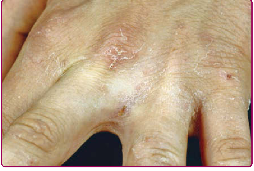
Scabies burrows on the hand (web spaces and knuckles - classic location):
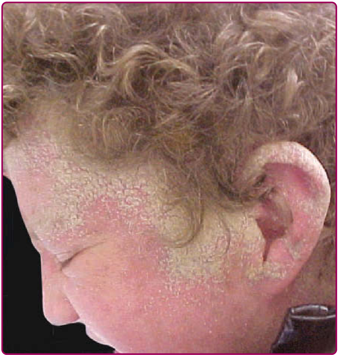
Crusted (Norwegian) scabies affecting the ear region and scalp:
Diagnosis Confirmation
Clinical diagnosis is usually sufficient based on the triad:
- Intense nocturnal pruritus
- Characteristic distribution
- Epidemiologic link (household contact history)
Laboratory confirmation:
-
Skin scraping - scrape a burrow with a scalpel, place in mineral oil on a slide; microscopy reveals mites (330-450 µm female), eggs, or fecal pellets (scybala) - definitive but low sensitivity
-
Dermatoscopy (epiluminescence microscopy) - identifies the "delta-wing jet" sign (mite parts/air bubbles in the stratum corneum); better sensitivity, non-invasive
-
KOH mount or cyanoacrylate adhesive strip preparation
-
PCR of skin swabs - highest sensitivity, but not routine
-
Harrison's Principles of Internal Medicine 22E (2025)
Differential Diagnosis
| Most Likely to Confuse | Distinguishing Features |
|---|---|
| Atopic dermatitis | Flexural, chronic, no burrows, no nocturnal itch predominance |
| Dyshidrotic eczema | Hands/feet only, vesicles, no burrows |
| Contact dermatitis | Exposure history, no burrows |
| Insect bite reactions | No burrows, single or clustered bites |
| Dermatitis herpetiformis | Vesicular, associated with celiac disease |
| Psoriasis | Silvery scales, nail changes, sharper borders |
| Drug eruption | Drug history, different morphology |
- Fitzpatrick's Dermatology, Table 178-1
Complications
- Secondary bacterial infection (impetiginization) - most common; can lead to post-streptococcal glomerulonephritis from Streptococcus pyogenes
- Lymphangitis, septicemia - with crusted scabies
- Bullous pemphigoid - scabies can trigger this autoimmune blistering disorder
Treatment in Detail
Treatment requires two components: (1) scabicidal therapy and (2) environmental/fomite control.
FIRST-LINE TREATMENT
Permethrin 5% Cream (Drug of Choice)
| Parameter | Detail |
|---|---|
| Dose/Application | Apply thinly but thoroughly from jawline to toes (entire body below the head in adults) |
| Special areas | Interdigital spaces, navel, buttock cleft, under fingernails and toenails |
| Duration on skin | Leave on for 8-14 hours overnight, then wash off with soap and water |
| Repeat | Second application 7 days later is essential (not ovicidal against eggs) |
| Pregnancy category | B - safe in pregnancy |
| Mechanism | Pyrethroid - disrupts sodium channel function in mite neurons causing paralysis |
For crusted (Norwegian) scabies: apply daily for 7 days, then twice weekly until cured
Oral Ivermectin (Alternative / Adjunct)
| Parameter | Detail |
|---|---|
| Dose | 200 µg/kg as a single oral dose |
| Repeat | Two doses, 7-14 days apart (not ovicidal, so second dose needed) |
| Indication | Treatment failures, unable to tolerate topicals, crusted scabies, institutional outbreaks |
| Crusted scabies | 3-7 doses over 8-30 days + permethrin + keratolytic |
| Caution | Not FDA-approved for scabies; avoid in children <15 kg and pregnancy |
A 2026 updated systematic review in BMC Infectious Diseases (PMID 42098632) confirms comparable effectiveness of ivermectin vs. permethrin - both are recommended first-line options.
- Red Book 2021 (Committee on Infectious Diseases); Harrison's 22E
SECOND-LINE / ALTERNATIVE AGENTS
| Drug | Dose/Method | Notes |
|---|---|---|
| Crotamiton 10% cream/lotion | Apply to entire body x 2 nights, wash off 24h after last application | Not approved for children; also has antipruritic effect |
| Sulfur 5-10% in petrolatum | Apply nightly x 3 nights, wash off each morning | Oldest treatment; safe in pregnancy and infants <2 months; malodorous |
| Benzyl benzoate 25% (common in developing countries) | Apply x 2-3 consecutive nights | Effective but more irritating; dilute to 12.5% for children |
| Malathion 0.5% lotion/gel | Apply overnight | Used in UK; first-line in some guidelines |
| Lindane 1% lotion | Apply overnight, wash off after 8-12h | Avoid - neurotoxic; reserve for failure of all others; contraindicated in pregnancy, children, epilepsy |
TREATMENT OF CRUSTED (NORWEGIAN) SCABIES
This is a hyperinfestation variant (millions of mites) seen in elderly, immunosuppressed (HIV, transplant), or neurologically impaired patients. Highly contagious.
- Pre-treatment: Apply 6% salicylic acid (keratolytic) to debulk crusts
- Permethrin 5%: Apply to entire body including scalp, face, and ears
- Oral ivermectin: 200 µg/kg - give 3-7 doses over 8-30 days
- Combination of both topical + oral is recommended
- Isolate the patient during treatment
ANTIPRURITIC / SYMPTOMATIC RELIEF
- Oral antihistamines: Chlorpheniramine (sedating - helps nocturnal itch), cetirizine, or hydroxyzine
- Calamine lotion: Topical soothing/antipruritic
- Menthol/methyl salicylate preparations: Topical relief
- Topical corticosteroids: To calm post-treatment persistent itch ("postscabetic itch") - only after scabicidal treatment
- Emollients: For skin barrier support
Important patient counseling: Even after successful treatment, pruritus and rash can persist for up to 4 weeks due to the ongoing hypersensitivity reaction to dead mites and their debris. This is called "postscabetic itch" and does not mean treatment has failed. Do NOT over-retreat with permethrin as it can cause irritant contact dermatitis.
ENVIRONMENTAL / FOMITE CONTROL (Mandatory)
- Wash all clothing and bedding in hot water (>60°C) and dry on high heat, or heat-press
- Items that cannot be washed: seal in a plastic bag for at least 72 hours (mites die off-host within 2-3 days for normal scabies)
- Clean mattresses, furniture, and upholstered surfaces
- Vacuum carpets and floors
CONTACT TRACING AND SIMULTANEOUS TREATMENT
This is a critical point often missed:
All household members and close physical contacts must be treated simultaneously, even if asymptomatic.
Failure to treat contacts is the most common cause of treatment failure and reinfection. Within 1 day of properly applied treatment, the infestation is considered noncommunicable, and the patient may return to normal activities.
- Fitzpatrick's Dermatology; Red Book 2021
Summary Treatment Algorithm for This 60-Year-Old Male
STEP 1: Confirm diagnosis (clinical ± dermoscopy/scraping)
↓
STEP 2: Permethrin 5% cream - apply neck-to-toe overnight
Repeat in 7 days
↓
STEP 3: Treat ALL household contacts simultaneously (Day 0 and Day 7)
↓
STEP 4: Environmental decontamination (laundry, bagging)
↓
STEP 5: Antihistamines for itch relief
↓
STEP 6: Reassure patient - itch may persist 2-4 weeks post-treatment
Topical steroid if postscabetic itch is severe
↓
If treatment failure → Oral ivermectin 200 µg/kg × 2 doses (Day 1 and Day 14)
Sources: Fitzpatrick's Dermatology, Vol. 1-2 | Harrison's Principles of Internal Medicine 22E (2025) | Red Book 2021 (Committee on Infectious Diseases) | Dermatology 2-Volume Set 5e | Innocent et al., 2026, BMC Infect Dis, PMID 42098632


+4 more
A 60 yrs old male present with intensly itchy lesion all over his body particularly on elbow back inner thighs waist knee joint tibeal surface ear pinna what is probable diagnosis and treatment in detail
analyse_media
analyse_media
analyse_media
analyse_media
Now I have analysed all four images in detail. The findings are very important - the images show far more advanced disease than simple scabies. Let me now synthesize everything.
Updated Diagnosis Based on Clinical Images + History
What the Images Actually Show
After careful analysis of all four photographs, this is NOT simple scabies. The findings point to a much more severe, longstanding, and complex dermatological presentation. Here is what each image reveals:
| Image | Site | Key Findings |
|---|---|---|
| Image 1 (waist/flank fold) | Abdominal/flank skin fold | Massive lichenification, dark leathery velvety thickening, severe post-inflammatory hyperpigmentation, exaggerated skin furrows - classic chronic itch-scratch cycle changes |
| Image 2 (thigh/knee) | Extensor thigh + knee + tibial surface | Extremely severe hyperkeratosis, deep bark-like lichenification, grayish-black hyperpigmentation, coarse scaling, pinpoint excoriations - "elephant skin" appearance |
| Image 3 (upper back - wider) | Medial scapular/interscapular area | Background lichenified, hyperpigmented skin + a focal raised indurated plaque with confluent papules/nodules showing vesicles/pustules on surface, with linear red excoriations below |
| Image 4 (upper back - close-up) | Same region, closer | Clustered grouped vesicles (some ruptured) in a herpetiform pattern on dusky erythematous base, hemorrhagic crusting, surrounding hyperpigmentation |
Revised Diagnosis: PRURIGO NODULARIS (with superimposed secondary changes)
Primary diagnosis: Prurigo Nodularis - a chronic, severely pruritic dermatosis caused by the vicious itch-scratch-lichenification cycle, presenting with:
- Intensely pruritic, hyperkeratotic, lichenified nodules and plaques
- Predilection for extensor surfaces (elbows, tibial surfaces), waist, inner thighs, back
- Severe "elephant skin" appearance on the knee/tibial region (Image 2)
- Diffuse lichenification across multiple body surfaces
Secondary/complicating finding: Possible Herpes Zoster (Shingles)
The focal lesion on the upper back (Images 3 and 4) shows a cluster of grouped vesicles on an erythematous/hyperpigmented base in a relatively localized region, which is highly characteristic of Herpes Zoster. This is particularly important in a 60-year-old male who may be immunocompromised from the chronic inflammatory state, or have an underlying condition.
Important note on the ear pinna involvement: In a 60-year-old male, ear pinna + facial/neck involvement by zoster would constitute Ramsay Hunt Syndrome (Herpes Zoster Oticus) if zoster is confirmed. However, if the ear is itchy/pruritic without vesicles/crusting, it may simply be part of the generalized prurigo.
Differential Diagnosis (Ranked by Probability)
| Diagnosis | Supporting Evidence | Against |
|---|---|---|
| Prurigo Nodularis | Bilateral symmetric lichenified plaques, elbows/tibial/back, bark-like skin, extreme chronic changes | |
| Herpes Zoster (on back) | Grouped vesicles, clustered herpetiform pattern, localized area, erythematous base | Non-dermatomal background changes |
| Lichen Simplex Chronicus | Focal lichenified plaques from chronic rubbing | Doesn't explain widespread involvement |
| Crusted (Norwegian) Scabies | Could underlie prurigo in elderly/compromised host | No visible burrows, hyperkeratosis not classical |
| Atopic Dermatitis | Itchy, lichenified | Usually starts in childhood |
| Acanthosis Nigricans | Waist/fold velvety hyperpigmentation | No metabolic workup done |
Workup Required
Given the clinical severity, the following investigations are mandatory:
- Skin scraping with KOH + mineral oil microscopy - rule out crusted scabies (mite/egg identification)
- Tzanck smear from back vesicles - look for multinucleated giant cells (HSV/VZV)
- VZV PCR or DFA from vesicle fluid - confirm zoster
- Skin biopsy (from a nodule/lichenified plaque) - confirm prurigo nodularis histology (eosinophilic infiltrate, neural hyperplasia, pseudoepitheliomatous hyperplasia)
- Full blood count - eosinophilia supports atopic/parasitic etiology
- Fasting blood glucose + HbA1c - acanthosis-like changes at waist suggest insulin resistance/diabetes
- Liver function tests, renal function, thyroid function - systemic causes of pruritus
- HIV serology - consider in severe/atypical presentations
- Serum IgE - elevated in atopic states
Detailed Treatment Plan
STEP 1: Treat the Acute Zoster on the Back (URGENT - IF CONFIRMED)
If the back vesicular lesion is herpes zoster (highly likely given appearance):
| Drug | Dose | Duration |
|---|---|---|
| Acyclovir | 800 mg orally 5 times/day | 7-10 days |
| OR Valacyclovir | 1000 mg 3 times/day | 7 days |
| OR Famciclovir | 500 mg 3 times/day | 7 days |
- Start antivirals within 72 hours of vesicle onset - delays beyond this reduce efficacy
- Add oral analgesic (paracetamol/tramadol) for pain
- If ear pinna involved with vesicles + facial palsy/vertigo → suspect Ramsay Hunt syndrome → needs IV acyclovir + oral prednisolone + ENT referral
STEP 2: Treat Prurigo Nodularis (Long-term Management)
A. Breaking the Itch-Scratch Cycle (First Priority)
| Drug | Dose | Role |
|---|---|---|
| Hydroxyzine | 25-50 mg at night | Sedating antihistamine - breaks nocturnal itch cycle |
| Cetirizine | 10 mg OD | Daytime non-sedating antihistamine |
| Doxepin (low dose) | 10-25 mg at night | Potent H1/H2 blocker + antidepressant effect |
B. Topical Treatment
| Drug | Application |
|---|---|
| Superpotent topical corticosteroid (Clobetasol propionate 0.05%) | Apply to individual nodules under occlusion (polythene wrap) overnight - 2-4 weeks |
| Calcineurin inhibitors (Tacrolimus 0.1% ointment or Pimecrolimus cream) | For face/flexures, or as steroid-sparing alternative |
| Topical capsaicin 0.025-0.1% | Depletes substance P from sensory nerves - reduces neurogenic itch |
| Emollients (generous application) | Restore skin barrier, reduce xerosis-driven itch |
C. Intralesional Treatment (for individual resistant nodules)
- Intralesional triamcinolone acetonide 10-40 mg/mL injected directly into indurated nodules - highly effective
D. Systemic Treatment for Moderate-Severe Prurigo Nodularis
| Drug | Dose | Notes |
|---|---|---|
| Dupilumab (Dupixent) | 300 mg SC every 2 weeks | FDA-approved for prurigo nodularis (2022) - targets IL-4/IL-13 pathway; first-line biologic |
| Naltrexone (low dose) | 2.5-5 mg/day | Opioid antagonist - reduces neurogenic itch |
| Gabapentin/Pregabalin | 300-1800 mg/day | Neuromodulator - excellent for neuropathic/neurogenic itch |
| Mirtazapine | 7.5-30 mg at night | Potent antipruritic + sleep aid |
| Cyclosporine | 3-5 mg/kg/day | Short-term use for severe acute flares; monitor renal function/BP |
| Methotrexate | 10-25 mg/week | Immunosuppressant for refractory cases |
| Thalidomide/Lenalidomide | 50-200 mg/day | Very effective but restricted use due to teratogenicity/neuropathy |
E. Phototherapy
- Narrowband UVB (NB-UVB) - 3x/week for 8-12 weeks - effective for widespread prurigo nodularis
- PUVA - for refractory disease
- Mechanism: immunomodulation and reduction of itch-mediating nerve fibers
STEP 3: Treat the Lichenification (Post-Inflammatory Skin Changes)
- High-potency topical steroids under occlusion to flatten lichenified plaques
- Coal tar preparations (1-5%) - antipruritic, anti-inflammatory, antiproliferative
- Salicylic acid 5-10% in a base - keratolytic for thick hyperkeratotic plaques (like the severe tibial changes)
- Barrier repair emollients applied liberally 2-3 times daily
STEP 4: Investigate and Treat Underlying Cause
Prurigo nodularis is frequently secondary to:
- Diabetes mellitus (waist acanthosis-like changes) - control blood sugar
- Renal failure - treat underlying CKD
- Liver disease - hepatic pruritus
- Hypothyroidism
- Lymphoma/internal malignancy - rule out in a 60-year-old
- Atopic dermatitis - requires specific management
- Psychiatric comorbidity (anxiety/depression) - address with SSRI/referral
STEP 5: Environmental and Lifestyle Measures
- Keep nails short and clean to minimize skin damage from scratching
- Apply ice packs or cold compresses to pruritic areas instead of scratching
- Wear loose, cotton clothing
- Lukewarm (not hot) baths with colloidal oatmeal or oil additives
- Avoid soap directly on affected areas (use soap-free cleansers)
- Low-irritant laundry detergents
- Address psychological stress with counseling/CBT - stress worsens itch
Summary Algorithm
Urgent (Now):
├── Confirm zoster on back → Start valacyclovir 1g TDS x 7 days
│ └── Check ear for Ramsay Hunt syndrome → ENT referral if present
│
Short-term (1-4 weeks):
├── Hydroxyzine at night + cetirizine daytime (break itch cycle)
├── Clobetasol 0.05% under occlusion on nodules
├── Emollients liberally
├── Investigations: skin scraping, Tzanck, bloods, biopsy
│
Medium-term (1-3 months):
├── Treat underlying cause (diabetes/renal/thyroid)
├── Dupilumab (if confirmed prurigo nodularis)
│ or Gabapentin + Mirtazapine
├── NB-UVB phototherapy
│
Long-term:
└── Intralesional triamcinolone for resistant nodules
Psychological support/CBT
Clinical bottom line: This 60-year-old man has chronic severe Prurigo Nodularis as the primary diagnosis, visible across the elbows, waist, thighs, knees, and tibial surfaces as massively lichenified hyperkeratotic plaques. Superimposed on this chronic disease is what appears to be a new episode of Herpes Zoster on the upper back (grouped vesicles), which requires urgent antiviral treatment. Full workup for underlying systemic causes is mandatory at this age.
This is a shared conversation. Sign in to Orris to start your own chat.