
Fracture shaft of femur
"femoral fractures" AND treatment
femoral shaft fracture management guideline adult intramedullary nailing
AP radiograph femoral shaft fracture with intramedullary nail fixation
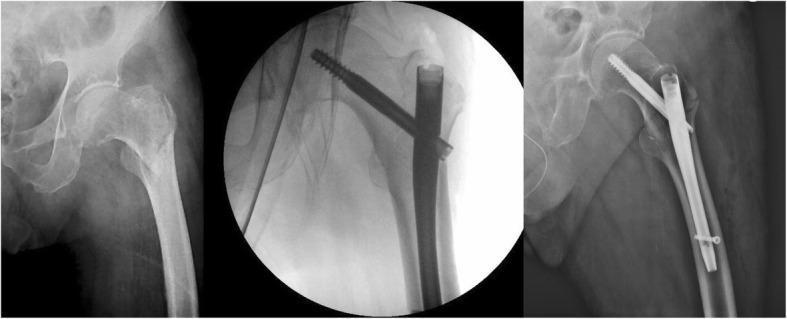
This composite of three X-ray images demonstrates the pre-operative and post-operative management of a proximal femur fracture in an adult patient. The leftmost image is a pre-operative anteroposterior (AP) radiograph of the hip showing a pertrochanteric fracture (AO classification 31.A1). The fracture line involves the intertrochanteric region with visible cortical discontinuity and slight displacement of the proximal femoral shaft relative to the femoral neck. The center image is an intraoperative fluoroscopic view showing the initial placement of an intramedullary nail and a cephalic screw. The rightmost image is a post-operative AP radiograph showing successful internal fixation with a Gamma 3® nail system. Key hardware components include a short intramedullary nail positioned within the femoral canal, a large-diameter cephalic lag screw extending through the nail into the center of the femoral head, and a distal locking screw securing the nail to the femoral shaft. The post-operative view confirms anatomical alignment, fracture reduction, and stable hardware positioning.
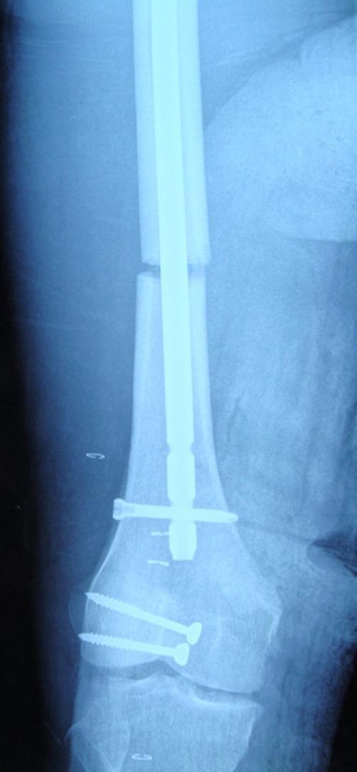
Anteroposterior (AP) radiograph of the distal femur and knee joint showing postoperative orthopedic hardware. The femur exhibits an intramedullary (IM) nail traversing the femoral shaft to stabilize a mid-shaft fracture. A visible transverse radiolucent line indicates the fracture site, which is currently held in alignment by the IM nail. Distal interlocking is achieved with a single transverse cortical screw through the nail. Additionally, two radiopaque malleolar screws are seen in a diagonal orientation across the patella, indicating internal fixation for a patellar fracture. The hardware is properly positioned to maintain reduction of both the femoral and patellar injuries. The radiograph demonstrates a case of ipsilateral femoral shaft and patellar fractures, highlighting the importance of thorough clinical and radiographic evaluation of the knee joint in the presence of high-energy femoral shaft trauma.
Fracture shaft of femur
Causes
- Road traffic collision, pedestrian injury, fall from height, or crush injury
- Gunshot or other penetrating injury may cause open fracture
- Low-energy fracture suggests pathological bone disease, such as tumor, osteoporosis, or metabolic bone disease
- In children, consider non-accidental injury when the history is inconsistent
Types
- Level: proximal, middle, or distal third
- Pattern: transverse, oblique, spiral, wedge, segmental, or comminuted
- Skin status: closed or open
- Displacement: shortening, angulation, rotation, and translation
Clinical features
- Severe thigh pain and inability to bear weight
- Swelling, deformity, shortening, and external rotation of limb
- Tenderness and abnormal mobility
- Large concealed blood loss into the thigh may cause hypovolemic shock
- Examine and document distal pulses, capillary refill, motor and sensory function
- Look for associated injuries: femoral-neck/hip injury, knee ligament injury, patellar injury, pelvic injury, chest and abdominal trauma.
Investigations
- AP and lateral X-rays of the entire femur, including hip and knee
- Pelvic/hip X-ray to exclude ipsilateral neck-of-femur fracture
- CT if fracture extension or associated injury is uncertain
- CBC, blood group and cross-match, coagulation profile, renal function
- Trauma imaging and assessment as indicated by mechanism and examination
Emergency management
- Follow ATLS principles: airway, breathing, circulation, hemorrhage control.
- Give adequate analgesia.
- Immobilize the leg with a traction splint only if appropriate and no suspected pelvic, hip, knee, or lower-leg contraindication.
- Establish IV access, cross-match blood, and treat shock.
- Perform repeated neurovascular checks.
- For open fractures: sterile dressing, urgent IV antibiotics, tetanus prophylaxis, and urgent orthopedic debridement.
Definitive treatment
- Reamed, locked intramedullary nailing is the usual definitive treatment for most adult femoral shaft fractures. It provides stable fixation, permits early mobilization, and has a high union rate. The AAOS overview and AO guidance describe intramedullary nailing as the standard approach where feasible.
- Temporary external fixation may be used in unstable polytrauma, severe open fracture, major soft-tissue injury, or damage-control orthopedics.
- Plate fixation is reserved for selected situations, including certain proximal/distal extensions, inability to nail, or particular fracture configurations.
- Nonoperative traction is uncommon in adults and mainly used where surgery is unavailable or contraindicated.
- Infants and younger children may be treated with traction followed by hip spica casting.
- Children aged about 4-12 years may be managed with elastic stable intramedullary nails, spica/traction in selected cases, external fixation, or plating.
- Older children and adolescents more often need operative fixation. Bailey and Love's Short Practice of Surgery, pediatric femoral shaft fracture section.
Complications
- Hemorrhagic shock
- Fat embolism syndrome, typically 24-72 hours after injury
- Open-fracture infection
- Acute compartment syndrome, though uncommon in the thigh
- DVT and pulmonary embolism
- Associated femoral neck or knee ligament injury
- Delayed union, nonunion, or malunion
- Rotational deformity and limb-length discrepancy
- Knee stiffness and reduced hip/knee function
- Infection or implant failure
- Chronic pain
Prognosis
Explain the ATLS management of a femoral shaft fracture
"femoral fractures" AND trauma
ATLS femoral shaft fracture primary survey hemorrhage traction splint guideline
ATLS management of a femoral shaft fracture
X: Catastrophic external hemorrhage
- Immediately control visible external bleeding with direct pressure and a pressure dressing.
- If the fracture is open, cover the wound with a sterile dressing. Do not repeatedly explore the wound or push protruding bone back.
- Give IV antibiotics and tetanus prophylaxis as early as feasible for an open fracture.
- A femoral shaft fracture can cause major concealed blood loss into the thigh, so treat it as a possible source of hemorrhagic shock even when there is no external bleeding.
A: Airway with cervical-spine protection
- Assess whether the patient can speak and maintain the airway.
- Clear and protect the airway as needed.
- Maintain manual in-line cervical stabilization and use a cervical collar when the mechanism or examination suggests possible spinal injury.
- Secure the airway if there is actual or impending airway compromise, reduced consciousness, or inability to protect the airway.
B: Breathing and ventilation
- Expose and examine the chest: inspect, palpate, percuss, and auscultate.
- Give oxygen and monitor oxygen saturation.
- Identify and immediately treat lethal thoracic injuries, especially tension pneumothorax, open pneumothorax, massive hemothorax, and flail chest.
- In a patient with a femoral fracture and hypoxia or respiratory deterioration, consider associated chest trauma initially. Fat embolism syndrome is usually a later complication rather than a first-minute diagnosis.
C: Circulation with hemorrhage control
- Assess pulse, blood pressure, skin perfusion, mental status, capillary refill, and ongoing bleeding.
- Insert two large-bore IV cannulas, or intraosseous access if needed.
- Send blood for CBC, group and cross-match, coagulation studies, and other trauma blood tests.
- Start warmed blood-product-based resuscitation when hemorrhagic shock is suspected, according to local massive hemorrhage protocol. Avoid excessive crystalloid.
- Look systematically for bleeding in the chest, abdomen, pelvis, retroperitoneum, external wounds, and long-bone fractures.
- Examine and record distal limb perfusion: dorsalis pedis and posterior tibial pulses, capillary refill, temperature, motor function, and sensation.
- Splint the femur early. Gentle alignment and immobilization reduce pain, movement, bleeding, and soft-tissue injury.
D: Disability, neurologic assessment
- Assess GCS, pupils, and limb movement.
- Check sensation and motor function in the affected leg, especially foot and ankle movement.
- Repeat the neurovascular examination after splinting or any manipulation and document findings.
E: Exposure and environmental control
- Fully expose the patient to detect other injuries, including pelvic wounds, open fractures, bruising, and deformity.
- Prevent hypothermia with warm blankets, a warmed environment, and warmed fluids/blood products.
- Log-roll only when appropriate and maintain spinal precautions until spinal injury is excluded.
Adjuncts to the primary survey
- Continuous monitoring: ECG, pulse oximetry, blood pressure, temperature, and urine output when indicated.
- Portable chest and pelvic X-rays in the unstable trauma patient.
- Full-length AP and lateral radiographs of the femur including hip and knee once the patient is stable enough.
- CT trauma imaging only after resuscitation and stabilization, unless it would delay necessary hemorrhage control or surgery.
- Give adequate analgesia, but do not let it delay ABCDE management.
Secondary survey
- Ipsilateral femoral-neck fracture or hip dislocation
- Pelvic fracture
- Knee ligament injury, patellar injury, and distal femoral injury
- Open fracture and soft-tissue damage
- Compartment syndrome
- Fat embolism syndrome during subsequent observation
- DVT and pulmonary embolism risk
Definitive orthopedic management
- Hemodynamically stable patient: early definitive fixation, usually locked intramedullary nailing.
- Unstable polytrauma patient: damage-control orthopedics, commonly temporary external fixation, with later definitive fixation once physiology has normalized.
- Open fracture: urgent orthopedic debridement, antibiotics, tetanus prophylaxis, stabilization, and coordinated soft-tissue management.