
Tinea vesiular
I'll look this up in the medical library for you right away.
Searching sources...
Reading File
Now let me search the library and web in parallel.
Finding Sources
Finding Sources
Searching PubMed
tinea versicolor pityriasis versicolor treatment
Good results. Now let me read the most comprehensive sources - Andrews' Dermatology, Dermatology 2-Volume Set, and Tintinalli's - in parallel.
Reading File
Reading File
Reading File
Let me get the treatment details from Andrews' and Tintinalli's, plus fetch the key clinical image.
Reading File
Reading File
Loading Image
I now have comprehensive information from multiple authoritative textbooks. Here is a full clinical overview:
Tinea Versicolor (Pityriasis Versicolor)
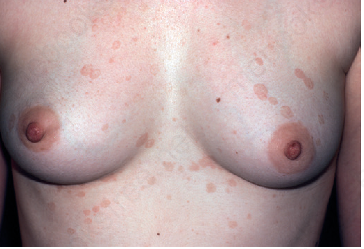
Definition & Etiology
Tinea versicolor is a superficial fungal infection caused by the yeast Malassezia spp. - part of normal cutaneous flora. The major species implicated is M. globosa, with M. restricta, M. sympodialis, M. furfur, M. obtusa, and M. slooffiae also involved.
The organism requires lipids to grow (it is lipophilic), which is why it is more common in young adults when sebaceous gland activity peaks.
Predisposing factors:
- High temperature and humidity (tropical climates)
- Oily skin
- Corticosteroid use (topical or systemic)
- Immunosuppression
Clinical Features
The name "versicolor" = varied colors. Lesions appear as:
| Appearance | Details |
|---|---|
| Hypopigmented macules | Most visible on dark skin; due to abnormally small, poorly melanized melanosomes not transferred to keratinocytes properly; also from dicarboxylic acids (from Malassezia lipid metabolism) inhibiting melanin synthesis |
| Hyperpigmented (brown) macules | More visible on fair skin |
| Pink/salmon macules | On untanned skin |
| Atrophic variant | With effacement of rete ridges, subepidermal fibroplasia, pigment incontinence, elastolysis |
Lesion morphology: Oval-to-round macules/patches/thin plaques with fine, furfuraceous (fluffy) scale - scale may only be apparent on scratching or stretching the skin.
Sites of predilection:
- Sternal region, sides of chest, abdomen, back (seborrheic/oily areas)
- Upper trunk and shoulders (most common)
- Pubis, neck, intertriginous areas (antecubital/popliteal fossae)
- Face - in infants and immunocompromised patients
- Scalp, palms, soles (less common)
- "Inverse" tinea versicolor - flexural involvement
Symptoms: Usually asymptomatic; mild pruritus can occur. Patients most often present for cosmetic concerns. May persist for months to years if untreated.
Diagnosis
KOH preparation (skin scrapings): Short, thick fungal hyphae + large numbers of variously sized spores = classic "spaghetti and meatballs" appearance. Highlighting stains include:
- Parker blue-black ink (mixed 1:1 with 20% KOH)
- 1% Chicago sky blue 6B with 8% KOH
- Gram stain
Wood's lamp: Accentuates pigment changes; may show yellow-green fluorescence of lesions/follicles.
Biopsy: Thick basket-weave stratum corneum with hyphae and spores.
Culture: Rarely needed; requires lipid-enriched media; reserved for cases of suspected malassezia sepsis (e.g., in patients on lipid-rich hyperalimentation).
Differential Diagnosis
| Condition | Distinguishing Feature |
|---|---|
| Seborrheic dermatitis | Erythematous with yellowish, soft/greasy scales; involves glabella, nasolabial folds, scalp |
| Pityriasis rosea | Herald patch; "Christmas tree" distribution; collarette scale |
| Vitiligo | Complete depigmentation, no scale |
| Pityriasis alba | Faint hypopigmented patches, usually children |
| Leprosy | Hypoesthetic patches; peripheral nerve involvement |
| Secondary syphilis | Faint pink <1 cm lesions; peripheral scale; adenopathy; serology positive |
| Confluent and reticulated papillomatosis | Persistent, hyperpigmented, reticulated |
| Nummular eczema / psoriasis | Scale character; distribution |
Treatment
Topical (first-line)
| Agent | Regimen |
|---|---|
| Selenium sulfide 2.5% lotion/shampoo | Daily for 1 week; wash off after 10 min; OR single overnight application (can repeat monthly for prophylaxis) |
| Ketoconazole 2% shampoo | Daily for 1 week, wash off after 3-5 min |
| Zinc pyrithione soap/shampoo | Cost-effective; useful for treatment and prophylaxis |
| Azole creams (clotrimazole, econazole, bifonazole) | Twice-daily applications superior to once-daily |
| Ciclopirox olamine | Topical option |
| Terbinafine (topical) | Effective topically (note: oral terbinafine is not effective for this condition) |
Systemic (for extensive or refractory disease)
| Agent | Regimen |
|---|---|
| Itraconazole | 200 mg once daily x 7 days, OR 400 mg single dose |
| Fluconazole | 400 mg single dose; can repeat monthly |
Note: Oral terbinafine is ineffective for tinea versicolor. This is an important clinical distinction.
Prophylaxis (after initial treatment)
- Weekly selenium sulfide 2.5% or ketoconazole 2% shampoo
- Monthly oral fluconazole
- Monthly overnight ketoconazole, econazole, or bifonazole shampoo application
Key Points to Remember
- Hypopigmentation persists for weeks to months after cure - this is NOT treatment failure. UV exposure may speed pigment normalization.
- Relapse is very common - prophylactic therapy should always be discussed.
- Caused by normal skin flora (Malassezia) undergoing transition from yeast → mycelial (hyphal) phase.
- Oral terbinafine does not work for this condition.
- The scalp can be treated monthly with selenium sulfide shampoo to reduce colonization.
Sources: Andrews' Diseases of the Skin, p. 358 | Dermatology 2-Volume Set 5e | Tintinalli's Emergency Medicine | Textbook of Family Medicine 9e
This is a shared conversation. Sign in to Orris to start your own chat.