Differential diagnosis of 49 yr old male with complains of pain at middle of penis, burning micturition, nocturia
Reading File
Finding Sources
Finding Sources
Finding Sources
Finding Sources
Now I have solid textbook grounding. Let me synthesize a comprehensive differential diagnosis.
Differential Diagnosis — 49-year-old Male: Midpenile Pain, Burning Micturition, Nocturia
This triad points primarily to lower urinary tract pathology with urethral involvement. The key anatomical clue is midpenile/distal urethral pain — burning during micturition in men is typically localized to the distal urethra (pendulous segment).
🔴 Most Likely Diagnoses
1. Urethritis (Infectious)
The most common cause of urethral discomfort + burning micturition in this age group.
- Gonococcal urethritis (Neisseria gonorrhoeae): purulent discharge, dysuria, frequency
- Non-gonococcal urethritis (NGU): Chlamydia trachomatis, Mycoplasma genitalium, Ureaplasma — clear/mucoid discharge, urethral itching, burning
- Urethral discomfort with urination and discharge is the hallmark — Goldman-Cecil Medicine
2. Urethral Calculus (Stone)
A urethral stone lodged in the pendulous (penile) urethra causes:
- Pain localized to the mid/penile shaft
- Burning micturition, obstructive symptoms
- Posterior urethral stones → perineal/rectal pain; pendulous urethral stones → pain at penile tip or shaft
- Campbell-Walsh Urology: "those in the pendulous urethra have pain at the penile tip…patients with primary urethral calculi have more insidious symptoms of persistent pain during voiding, obstructive lower urinary tract symptoms"
3. Benign Prostatic Hyperplasia (BPH)
- BPH is the most common benign prostatic disease in males >50, and symptoms can begin in the late 40s
- Classic LUTS: nocturia, daytime frequency, urgency, dysuria, hesitancy, weak stream
- Differential per Swanson's Family Medicine: "These symptoms include nocturia, daytime frequency, urgency, urge incontinence, and dysuria. Differential diagnosis includes carcinoma of the prostate, neuropathic bladder, chronic prostatitis, and urethral stricture"
4. Chronic Prostatitis / Chronic Pelvic Pain Syndrome (CP/CPPS)
- Perineal, penile, or pelvic pain + irritative voiding symptoms (dysuria, nocturia, frequency)
- Most common prostatitis syndrome in men under 50
- Pain can radiate to shaft/mid-penis; pain during/after ejaculation is characteristic
- Can coexist with urethritis-like symptoms
🟡 Important Considerations
5. Urethral Stricture
- Obstructive + irritative voiding: weak stream, straining, nocturia, dysuria
- Often post-infectious (gonorrhea, chlamydia) or post-traumatic/iatrogenic
- Can cause stasis and secondary UTI/prostatitis
- Smith & Tanagho's General Urology lists urethral stricture in the differential of LUTS + dysuria
6. Cystitis / Urinary Tract Infection (UTI)
- Less common in males of this age, but possible
- Suprapubic pain, frequency, nocturia, burning
- When present, an underlying structural or functional cause (BPH, calculus, stricture) must be sought
7. Interstitial Cystitis / Bladder Pain Syndrome (IC/BPS)
- Chronic pelvic/urethral pain + irritative voiding symptoms in the absence of infection
- Campbell-Walsh Urology: dysfunctional voiding patterns may mimic IC/BPS; nocturia is prominent
- More common in women, but can occur in men — often under-recognized
8. Lichen Sclerosus (BXO — Balanitis Xerotica Obliterans)
- Chronic inflammatory dermatosis affecting glans penis and foreskin
- Presents with pruritus, penile pain, dysuria
- Can cause meatal/urethral stenosis → obstructive + irritative voiding symptoms (nocturia)
- Dermatology 2-Volume Set: "affects glans penis and foreskin; presents with pruritus, pain"
9. Penile Carcinoma / Urethral Carcinoma
- Less common but must be considered with persistent midpenile pain, especially with visible lesion
- Campbell-Walsh Urology differential for penile lesions includes primary tumors
10. Referred Pain — Ureteric/Renal Calculus
- A distal ureteric stone can cause referred pain to the tip or shaft of the penis (via the genitofemoral nerve L1-L2)
- May mimic urethral pain; flank/colicky component may help differentiate
🟢 Rarer / Contextual Causes
| Condition | Clues |
|---|---|
| Reactive arthritis (Reiter's syndrome) | Urethritis + arthritis + conjunctivitis triad |
| Herpes simplex urethritis | Vesicular lesions, recurrent burning |
| Peyronie's disease | Penile plaques, pain during erection |
| Prostate carcinoma | Age-appropriate, obstructive LUTS, weight loss, bone pain |
| Urethral diverticulum | Post-void dribbling, recurrent UTI |
| Penile fracture/trauma | History of injury |
Recommended Workup
| Investigation | Purpose |
|---|---|
| Urinalysis + urine culture | Infection, hematuria |
| Urethral swab (NAAT for gonorrhea/chlamydia) | Urethritis |
| Ultrasound KUB / X-ray pelvis | Calculus |
| PSA + DRE | BPH, prostate carcinoma |
| Uroflowmetry + PVR | Stricture, BPH |
| Cystoscopy / urethrography | Stricture, stone, urethral lesion |
| Serum creatinine | Renal function |
Bottom line: In a 49-year-old male with this symptom triad, urethritis, urethral calculus, BPH, and chronic prostatitis/CPPS are the top four diagnoses to exclude. The specific localization to the middle of the penis makes a urethral stone or urethritis the most anatomically specific first considerations, while BPH explains the nocturia component.
— Smith and Tanagho's General Urology, Campbell-Walsh Wein Urology, Goldman-Cecil Medicine, Swanson's Family Medicine Review
Reasons for convulsion for post channel turp patient in ca prostate pt
Finding Sources
Finding Sources
Reading File
Finding Sources
Reading File
Finding Sources
Loading Image
Excellent — I now have comprehensive, authoritative textbook data. Here is the full answer:
Causes of Convulsion Post-Channel TURP in a Carcinoma Prostate Patient
This is a clinically critical scenario. Convulsions in this context arise from two broad categories: (1) procedural/anesthetic complications and (2) disease-related complications from underlying Ca prostate.
🔴 CATEGORY 1: TURP-Related Causes
1. TURP Syndrome (The Most Important Cause)
The #1 cause to suspect immediately post-TURP.
Definition: Symptomatic hyponatremia + fluid overload from systemic absorption of hypotonic, electrolyte-free irrigating fluid through open prostatic venous sinuses during resection.
Onset: 15 minutes to 24 hours after start of resection. Complicates 10–15% of TURP procedures.
Mechanism of convulsions — three parallel pathways:
| Pathway | Mechanism |
|---|---|
| Hyponatremia | Free water absorption → dilutional hyponatremia → cerebral edema → seizures (typically when Na⁺ < 110–120 mEq/L) |
| Glycine toxicity | Glycine (irrigant solute) allosterically activates NMDA receptors → direct excitatory neurotoxicity → seizures + visual disturbances |
| Ammonia toxicity | Hepatic deamination of glycine → hyperammonemia → encephalopathy, seizures |
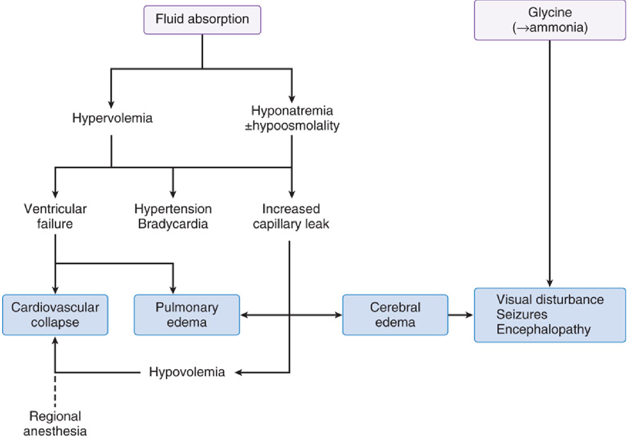
Other TURP syndrome symptoms: nausea/vomiting, confusion, agitation, reduced consciousness, visual disturbance, bradycardia, hypertension → hypotension, pulmonary edema.
Risk factors for TURP syndrome:
- Prolonged resection time (>1 hour)
- High intravesical pressure (>15–25 mmHg)
- Hypotonic irrigants (glycine, sorbitol, mannitol solutions)
- Large gland with open venous sinuses
- Large channel TURP (more venous sinus exposure)
2. Severe Dilutional Hyponatremia (Isolated)
Even without full TURP syndrome, massive fluid absorption causes:
- Na⁺ < 110 mEq/L → seizures + coma
- Na⁺ 120–125 mEq/L → confusion, nausea, headache, agitation
- Cerebral edema drives convulsions
- Miller's Anesthesia: "At Na⁺ concentrations less than 110 mEq/L, symptoms progress to seizures and coma"
3. Hypomagnesemia (Dilutional)
- Fluid absorption dilutes Mg²⁺ → reduced inhibitory control of NMDA receptors → lowers seizure threshold
- Compounds glycine's excitatory effects
- Miller's Anesthesia: "Mg²⁺ can be given for seizures, because its negative control of NMDA receptors counteracts dilutional hypomagnesemia and the excitatory effects of glycine"
4. Anaesthetic-Related Seizures
- Spinal/epidural anaesthesia: local anaesthetic systemic toxicity (LAST) — rare but possible
- General anaesthesia: drug interactions, hypoxia, hypercapnia during intraoperative period
🟡 CATEGORY 2: Carcinoma Prostate-Related Causes
5. Hypercalcemia of Malignancy
- Ca prostate with bone metastases → osteolytic activity → hypercalcemia
- Hypercalcemia causes: confusion, psychosis, seizures (especially acute severe elevations)
- "Bones, stones, groans, and psychic moans"
6. Brain Metastases
- Ca prostate can metastasize to brain (less common than bone, but occurs)
- Cerebral metastases → raised ICP, focal seizures, generalized convulsions
- Leptomeningeal carcinomatosis → seizures
7. Hyponatremia from SIADH (Paraneoplastic)
- Ca prostate can cause paraneoplastic SIADH
- Preoperative or perioperative hyponatremia → seizures independent of TURP syndrome
8. Hormone Therapy-Related
- LHRH agonist flare or anti-androgen withdrawal: can cause metabolic disturbances
- Docetaxel/cabazitaxel (chemotherapy): neurotoxicity in advanced cases
🟢 CATEGORY 3: General Perioperative Causes (apply to any patient)
| Cause | Mechanism |
|---|---|
| Hypoglycemia | Prolonged fasting + diabetic medication → low blood glucose → seizures |
| Hypoxia / Hypercapnia | Airway/respiratory compromise post-anaesthesia |
| Cerebrovascular event | Perioperative stroke — especially in older patients with atherosclerosis |
| Septic encephalopathy | Post-TURP bacteremia/UTI → sepsis → seizures |
| Drug toxicity | Opioid overdose, benzodiazepine paradox, anti-epileptic withdrawal |
| Pre-existing epilepsy | Stress, sleep deprivation, medication non-compliance perioperatively |
Summary Priority Table
| Priority | Cause | Key Clue |
|---|---|---|
| #1 | TURP Syndrome (glycine/hyponatremia) | Onset within 24h, visual disturbance, confusion |
| #2 | Dilutional hyponatremia | Low serum Na⁺ (<120 mEq/L) |
| #3 | Dilutional hypomagnesemia | Low Mg²⁺, compounds NMDA toxicity |
| #4 | Hypercalcemia (bone mets) | Elevated Ca²⁺, bone pain |
| #5 | Brain metastases | Focal neurology, raised ICP signs |
| #6 | Perioperative hypoglycemia | Low blood glucose |
| #7 | Cerebrovascular event | Focal deficits, imaging |
Immediate Workup
- Serum electrolytes: Na⁺, K⁺, Mg²⁺, Ca²⁺, phosphate
- Blood glucose (stat)
- ABG (hypoxia, CO₂)
- Serum osmolality
- Ammonia level
- CT brain (if focal signs or raised ICP suspected)
- Fluid balance review (irrigant absorbed volume)
Treatment of TURP Syndrome Seizures
- Stop irrigation immediately
- Hypertonic 3% saline: 100 mL bolus IV if Na⁺ <120 mEq/L with active seizures; target increase of 2–3 mEq/L acutely; do not correct >10 mEq/L in first 24h (risk of osmotic demyelination)
- IV Magnesium sulphate: for refractory seizures (counters glycine/NMDA excitotoxicity)
- Loop diuretic (furosemide) if hypervolemic
- Benzodiazepines for acute seizure control
— Miller's Anesthesia 10e, Barash Clinical Anesthesia 9e, Campbell-Walsh Wein Urology
Is it possible for turp syndrome with bipolar turp?
Finding Sources
Reading File
Finding Sources
Finding Sources
I have enough authoritative data to give a precise, well-grounded answer.
TURP Syndrome with Bipolar TURP — Is It Possible?
Short Answer: Yes, but in a modified form.
Bipolar TURP eliminates the classic hyponatremia-driven TURP syndrome but does not completely eliminate the risk of a TUR syndrome-like picture. Here's the distinction:
Why Bipolar TURP Was Developed
In monopolar TURP (M-TURP):
- The electrical circuit runs through the patient from resectoscope loop → grounding pad
- Requires a non-ionic, hypotonic irrigant (glycine 1.5%, sorbitol, mannitol) to avoid dispersing the current
- These hypotonic solutions, when absorbed through open prostatic venous sinuses → dilutional hyponatremia + hypoosmolality → cerebral oedema → TURP syndrome
In bipolar TURP (B-TURP):
- The circuit is contained entirely within the resectoscope (current passes between two poles of the instrument itself)
- This allows use of isotonic normal saline (0.9% NaCl) as irrigant
- Result: "The use of iso-osmolar saline in bipolar TURP has reduced the incidence of TUR syndrome" — Campbell-Walsh Wein Urology
What Bipolar TURP Eliminates vs. What Remains
| Feature | Monopolar TURP | Bipolar TURP |
|---|---|---|
| Irrigant used | Hypotonic (glycine/sorbitol) | Isotonic (normal saline) |
| Hypo-osmolar hyponatremia | ✅ High risk | ❌ Eliminated |
| Glycine toxicity | ✅ Present | ❌ Eliminated |
| Ammonia encephalopathy | ✅ Present | ❌ Eliminated |
| Hypervolemia (fluid overload) | ✅ Risk | ✅ Still present |
| Dilutional hyponatremia from massive NS absorption | Low | Still possible (with massive absorption) |
| Visual disturbances (glycine-mediated) | ✅ | ❌ |
The Residual Risk with Bipolar TURP
Per Miller's Anesthesia (10e):
"If saline irrigant is used, surgery should be terminated after 2500 mL has been absorbed. Although the risk for hyposmolar hyponatremia is removed with saline irrigant, the risk for excessive intravascular volume remains."
So with bipolar TURP:
-
Hypervolemia / Fluid Overload — still occurs with large-volume irrigant absorption
- Pulmonary oedema, hypertension, cardiac failure
- This alone can cause cerebral oedema → seizures
-
Dilutional hyponatremia — can still occur with massive saline absorption, though far less severe than with hypotonic irrigants, because isotonic saline does not drop osmolality
-
Dilutional hypomagnesemia — possible with large volume absorption, lowers seizure threshold
-
Hyperchloraemic metabolic acidosis — large-volume normal saline absorption → excess Cl⁻ → acidosis (unique to bipolar)
Practical Implications
| Situation | Risk Level |
|---|---|
| Short procedure, limited absorption | Very low — bipolar is much safer |
| Prolonged resection, large gland, >2500 mL absorbed | Fluid overload risk persists; hypervolemia-related seizures possible |
| Pre-existing cardiac/renal impairment | Higher risk of symptomatic overload even with smaller volumes |
| Ca prostate patient (your scenario) | Compounded by disease-related risks (hypercalcemia, brain mets) |
Bottom Line
Bipolar TURP significantly reduces but does not abolish the risk of TUR syndrome. The classic triad of hyponatremia + glycine toxicity + osmolar disturbance is largely prevented. However, volume overload — which can independently cause cerebral oedema, pulmonary oedema, cardiovascular collapse, and convulsions — remains a real risk, particularly in prolonged resections or in patients with limited cardio-renal reserve.
In a Ca prostate patient undergoing channel TURP, the additional risk factors (large tumour bulk, abnormal venous sinuses from tumour invasion, possible pre-existing metabolic disturbances) mean vigilance is still warranted even with bipolar technique.
— Miller's Anesthesia 10e; Campbell-Walsh Wein Urology; Barash Clinical Anesthesia 9e
Basic blood investigations to be done as a part of regular health update from age of 35 in males and females as per the Indian population guidelines!
Finding Sources
Finding Sources
Finding Sources
Finding Sources
Finding Sources
The library does not contain India-specific preventive health guidelines texts, so I'll draw on established Indian guidelines (ICMR, RSSDI, CSI, IAP, API, NHP) and synthesize a comprehensive, clinically grounded answer.
Recommended Basic Blood Investigations for Regular Health Screening
Indian Population — Starting Age 35 (Males & Females)
Rationale for age 35: India has one of the world's highest burdens of Type 2 diabetes, hypertension, dyslipidaemia, and thyroid disorders, with onset a decade earlier than Western populations. ICMR, RSSDI, and API guidelines recommend initiating metabolic screening by age 35 (or earlier if risk factors present).
🔵 TIER 1 — Core Panel (Everyone, Every Year)
| Investigation | Purpose | Indian Context |
|---|---|---|
| Complete Blood Count (CBC) | Anaemia, infection, haematological disorders | Iron-deficiency anaemia extremely prevalent, especially in women; thalassaemia trait common |
| Fasting Blood Glucose (FBG) | Diabetes/pre-diabetes screening | India has 101 million diabetics; RSSDI recommends annual FBG from age 30–35 |
| HbA1c | 3-month average glucose; diagnoses diabetes | Preferred by RSSDI/ICMR as it doesn't require fasting; more stable |
| Fasting Lipid Profile | Total cholesterol, LDL, HDL, triglycerides, VLDL | Dyslipidaemia is a primary CVD risk driver; Indians have characteristically low HDL + high TG |
| Serum Creatinine + eGFR | Kidney function | CKD prevalence rising; diabetic nephropathy is #1 cause of CKD in India |
| Blood Urea Nitrogen (BUN) | Renal and hepatic function cross-check | |
| Liver Function Tests (LFTs) | ALT, AST, ALP, bilirubin, total protein, albumin | NAFLD affects ~9–32% of urban Indians; alcohol-related liver disease; hepatitis B endemic |
| Thyroid Stimulating Hormone (TSH) | Hypothyroidism/hyperthyroidism screening | India is in an iodine-deficient belt; thyroid disorders affect ~42 million Indians; subclinical hypothyroidism very common in women |
| Urine Routine & Microscopy | Proteinuria, glucosuria, infection | Screens for early diabetic nephropathy, UTI, kidney disease |
🟡 TIER 2 — Strongly Recommended Add-ons (Every 1–2 Years)
| Investigation | Purpose | Notes |
|---|---|---|
| Serum Uric Acid | Gout, metabolic syndrome, CKD risk | High prevalence of hyperuricaemia in Indian males; linked to metabolic syndrome |
| Serum Electrolytes (Na⁺, K⁺) | Electrolyte balance | Especially if on antihypertensives (ACEi, diuretics) |
| Vitamin D3 (25-OH) | Vitamin D deficiency | 70–90% of urban Indians are deficient despite tropical climate; linked to diabetes, CVD, osteoporosis |
| Vitamin B12 | B12 deficiency, neurological risk | Very high prevalence in vegetarians; India has highest vegetarian population globally |
| Fasting Insulin / HOMA-IR | Insulin resistance, metabolic syndrome | Indians have higher insulin resistance at lower BMI ("thin-fat Indian" phenotype) |
| hsCRP (high-sensitivity CRP) | Cardiovascular inflammation marker | Independent CVD risk predictor; useful when lipid profile is borderline |
| Serum Calcium | Bone health, parathyroid, malignancy | Related to Vit D deficiency; osteoporosis screening |
🟠 TIER 3 — Sex-Specific Investigations
Males (Age 35+)
| Investigation | Purpose | Frequency |
|---|---|---|
| PSA (Prostate Specific Antigen) | Prostate cancer screening | Start at 45 (or 40 if family history); annually after 50 |
| Serum Testosterone | Hypogonadism, andropause, metabolic syndrome | If symptoms: fatigue, low libido, weight gain |
| Serum Ferritin | Iron stores, haemochromatosis | Males accumulate iron; ferritin also an inflammatory marker |
| Liver enzymes (GGT) | Alcohol-related liver disease | More relevant in males |
Females (Age 35+)
| Investigation | Purpose | Frequency |
|---|---|---|
| TSH + Free T4 | Hypothyroidism (women affected 8–10× more than men) | Every year; critical in reproductive-age women |
| Serum Iron, TIBC, Ferritin | Iron deficiency anaemia | Very high prevalence in Indian women; menstrual blood loss |
| Blood Glucose (pre-conception / PCOS) | PCOS-related insulin resistance; GDM risk | Especially if PCOS, overweight, family history |
| Anti-TPO antibodies | Autoimmune thyroiditis (Hashimoto's) | If TSH elevated or family history |
| AMH (Anti-Müllerian Hormone) | Ovarian reserve | Age 35–40 if planning pregnancy or PCOS evaluation |
| Prolactin | Menstrual irregularities, galactorrhoea | As indicated |
| CA-125 | Ovarian cancer marker | Only if symptoms or family history — not routine screening |
| Bone mineral density (DEXA) — not blood | Osteoporosis | Age 40+ or post-menopause |
🔴 High-Risk Add-ons (When Risk Factors Present)
| Risk Factor | Additional Tests |
|---|---|
| Hypertension / CVD family history | ECG, echo, homocysteine, Lp(a), ApoB |
| Obesity / Metabolic syndrome | Fasting insulin, HOMA-IR, LFTs, uric acid |
| Family history of diabetes | Post-prandial glucose (PPBG), HbA1c |
| Anaemia | Peripheral blood smear, serum iron, ferritin, B12, folate, reticulocyte count |
| Suspected thyroid disease | Free T3, Free T4, anti-TPO |
| Hepatitis risk | HBsAg, Anti-HCV — India has 40 million HBsAg carriers |
| Family history of cancer | PSA (M), CA-125 (F), CEA, AFP |
| Renal disease risk | Urine microalbumin:creatinine ratio (UACR) |
📅 Suggested Frequency Summary
| Test | Frequency |
|---|---|
| CBC, FBG, HbA1c, lipid profile, LFT, RFT, TSH, urine R/M | Annually |
| Vitamin D, B12, uric acid, electrolytes, ferritin | Every 1–2 years |
| hsCRP, fasting insulin | Every 2 years or when metabolic risk changes |
| PSA (males ≥45), hormone panels | As per age and symptoms |
| HBsAg, Anti-HCV | Once; repeat if high risk |
Key Indian-Specific Considerations
-
"Thin-fat Indian" phenotype — Indians develop metabolic syndrome and insulin resistance at lower BMI (23–25 kg/m²) than Western populations. Metabolic screening should start at lower BMI thresholds.
-
Earlier onset of T2DM — Average age of diabetes onset in India is ~42 years vs. ~52 in the West. HbA1c + FBG from age 30–35 is justified.
-
Low HDL + high triglycerides — characteristic Indian dyslipidaemia pattern; full fasting lipid profile with TG:HDL ratio is essential.
-
Vitamin D & B12 deficiency — paradoxically epidemic despite sunshine and vegetarian diet; both have downstream cardiovascular, neurological, and metabolic consequences.
-
Thyroid disease — iodine deficiency belt, autoimmune thyroiditis, subclinical hypothyroidism; TSH is a non-negotiable annual test in Indian women.
-
Hepatitis B carriage — India is hyperendemic; HBsAg must be checked if not vaccinated or status unknown.
Based on guidelines from ICMR, RSSDI (Research Society for Study of Diabetes in India), Cardiological Society of India (CSI), API (Association of Physicians of India), and Ministry of Health & Family Welfare (NHP India).
How to calculate HOMA iR
Finding Sources
Finding Sources
Finding Sources
The library doesn't have a dedicated HOMA-IR formula section. This is a well-established, straightforward clinical calculation — here's the complete answer:
How to Calculate HOMA-IR
HOMA-IR (Homeostatic Model Assessment of Insulin Resistance) was developed by Matthews et al. in 1985. It is the most widely used clinical surrogate marker of insulin resistance.
The Formula
$$\text{HOMA-IR} = \frac{\text{Fasting Insulin (μIU/mL)} \times \text{Fasting Blood Glucose (mmol/L)}}{22.5}$$
If glucose is reported in mg/dL (as is common in India):
$$\text{HOMA-IR} = \frac{\text{Fasting Insulin (μIU/mL)} \times \text{Fasting Blood Glucose (mg/dL)}}{405}$$
The denominator differs because: 1 mmol/L glucose = 18 mg/dL, and 22.5 × 18 = 405
Requirements
| Parameter | Condition | Unit |
|---|---|---|
| Fasting insulin | 8–10 hours overnight fast | μIU/mL (= mIU/L = μU/mL) |
| Fasting blood glucose | Same sample, same fast | mg/dL or mmol/L |
Both values must be from the same fasting blood draw.
Worked Example
A patient has:
- Fasting insulin = 12 μIU/mL
- Fasting glucose = 95 mg/dL
$$\text{HOMA-IR} = \frac{12 \times 95}{405} = \frac{1140}{405} = \mathbf{2.81}$$
Interpretation
| HOMA-IR Value | Interpretation |
|---|---|
| < 1.0 | Optimal insulin sensitivity |
| 1.0 – 1.9 | Normal (general population) |
| ≥ 2.0 | Early / borderline insulin resistance |
| ≥ 2.5 | Insulin resistance (commonly used clinical cut-off) |
| ≥ 3.0 | Significant insulin resistance |
| > 5.0 | Severe insulin resistance |
Indian-specific note: Some Indian studies use ≥ 2.0 as the cut-off for insulin resistance, as Indians develop metabolic syndrome at lower BMI and lower HOMA-IR values than Western populations.
What HOMA-IR Estimates
The model is based on the feedback loop between the pancreatic β-cell and the liver:
- In a healthy fasted state, the liver produces glucose and the pancreas secretes just enough insulin to maintain euglycaemia
- In insulin resistance: liver requires more insulin to suppress glucose output → fasting insulin rises relative to glucose
- HOMA-IR quantifies this imbalance
Companion: HOMA-β (Beta-Cell Function)
$$\text{HOMA-β} = \frac{20 \times \text{Fasting Insulin (μIU/mL)}}{\text{Fasting Glucose (mmol/L)} - 3.5} \times 100%$$
- Normal ≈ 100%
- Low HOMA-β = impaired β-cell secretory capacity
Limitations of HOMA-IR
| Limitation | Detail |
|---|---|
| Insulin assay variability | Different labs use different assay methods — results not always comparable across labs |
| Not validated in all populations | Cut-offs vary by ethnicity, BMI, age |
| Single time-point | Does not capture dynamic insulin secretion |
| Not for Type 1 DM | No meaningful result when exogenous insulin is used |
| Affected by conditions | Liver disease, renal disease, obesity, medications (steroids, metformin) alter the result |
| Gold standard is different | Euglycaemic hyperinsulinaemic clamp is the true gold standard — HOMA-IR is a surrogate |
Clinical Uses
- Diagnosing metabolic syndrome and pre-diabetes
- Monitoring treatment response in PCOS (HOMA-IR improves with metformin/lifestyle)
- Risk stratification for NAFLD/NASH (high HOMA-IR predicts fibrosis)
- Screening in "thin-fat" Indians — normal BMI but high visceral fat + insulin resistance
- Research and epidemiological studies


Analyse this uroflow report anf
Analyse this urodynamic study and give comments
Finding Sources
Finding Sources
Finding Sources
Finding Sources
This is a shared conversation. Sign in to Orris to start your own chat.