I now have comprehensive data from 5+ authoritative surgical and medical textbooks. Let me compile the full answer.---
Obstructive Jaundice Secondary to Choledocholithiasis (Non-Cholangitis)
A Complete Reference Guide
1. ANATOMY OF THE BILIARY SYSTEM
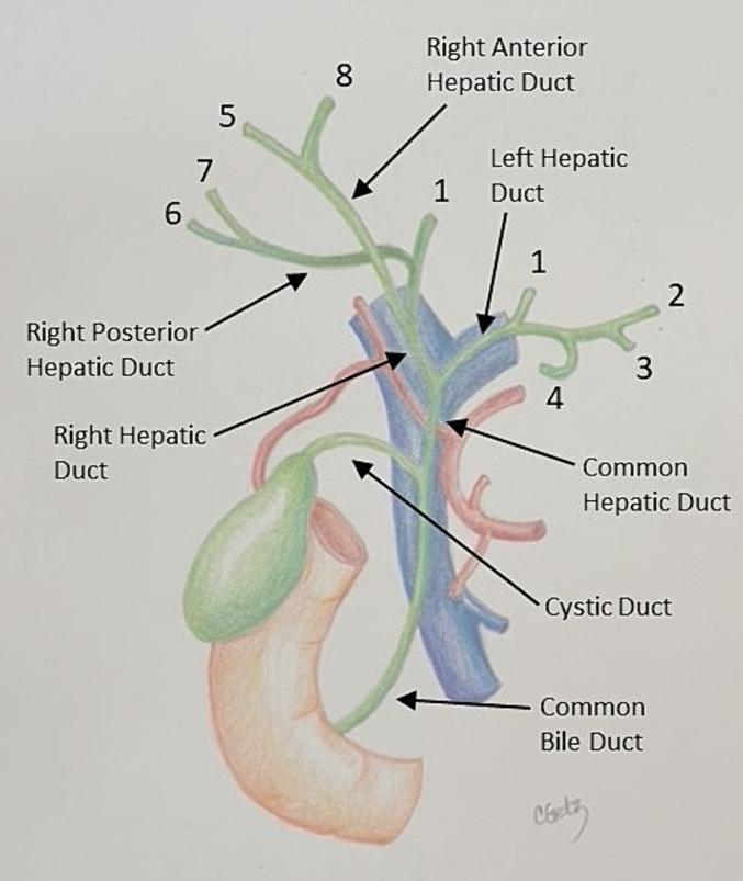
Extrahepatic Biliary Tree
| Structure | Detail |
|---|
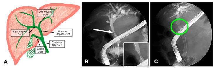
| Right hepatic duct | Drains segments 5, 6, 7, 8 (right anterior + right posterior ducts) |
| Left hepatic duct | Drains segments 1, 2, 3, 4 |
| Common hepatic duct (CHD) | Union of right + left hepatic ducts at porta hepatis |
| Cystic duct | Connects gallbladder to CHD; contains spiral valves of Heister |
| Common bile duct (CBD) | CHD + cystic duct; ~8 cm long, 6 mm normal diameter |
| Ampulla of Vater | CBD + pancreatic duct join, drain through this into 2nd part of duodenum |
| Sphincter of Oddi | Smooth muscle complex controlling bile + pancreatic secretion flow |
CBD Segments (4 parts)
- Supraduodenal - in free edge of lesser omentum (hepatoduodenal ligament), anterior to portal vein, right of hepatic artery
- Retroduodenal - passes posterior to 1st part of duodenum
- Infraduodenal (intrapancreatic) - runs through or behind head of pancreas - most common site for stone impaction
- Intraduodenal (intramural) - pierces wall of duodenum, joins pancreatic duct
Key Relations in Hepatoduodenal Ligament (from left to right):
- Anterior right: CBD
- Anterior left: Hepatic artery proper
- Posterior: Portal vein
(Gray's Anatomy for Students)
2. PHYSIOLOGY OF BILIRUBIN & OBSTRUCTIVE JAUNDICE
Normal Bilirubin Metabolism
Hemoglobin breakdown (RES)
        ↓
Unconjugated bilirubin (fat-soluble, bound to albumin)
        ↓ [Liver - UDP-glucuronosyltransferase]
Conjugated bilirubin (water-soluble)
        ↓ [Secreted into bile canaliculi]
Bile → duodenum → urobilinogen → stercobilin (stool color)
                      ↓ (80% reabsorbed enterohepatic circulation)
                   Urobilinogen → urine (urobilinogen)
What Happens in CBD Obstruction
When a stone obstructs the CBD:
- Conjugated bilirubin cannot be excreted into the duodenum
- Bile canaliculi become congested and rupture
- Conjugated bilirubin regurgitates into hepatic veins and lymphatics
- Result: conjugated (direct) hyperbilirubinemia in the blood
- Bilirubin appears in urine (choluria - dark urine)
- No bilirubin reaches the bowel → no urobilinogen → pale/clay-colored stools
- Secondary: bile salt accumulation → pruritus
- Hepatic backup → hepatomegaly, elevation of ALP and GGT (predominantly cholestatic pattern)
Laboratory Pattern in Obstructive Jaundice
| Parameter | Result |
|---|
| Serum bilirubin | ↑↑ Direct (conjugated) predominantly |
| Urine bilirubin | Present (choluria) |
| Urine urobilinogen | Absent |
| Fecal urobilinogen | Trace to absent (pale stools) |
| Alkaline phosphatase (ALP) | ↑↑↑ (markedly elevated) |
| GGT | ↑↑↑ (most sensitive and specific) |
| AST / ALT | Mildly ↑ (can spike early with acute obstruction) |
| PT / INR | ↑ (decreased vitamin K absorption due to absent bile) |
| Serum albumin | ↓ (prolonged obstruction) |
(Harper's Illustrated Biochemistry 32nd ed; Guyton & Hall Textbook of Medical Physiology)
3. CHOLEDOCHOLITHIASIS - DEFINITION & CLASSIFICATION
Choledocholithiasis = presence of stones in the extrahepatic biliary tree (CBD and hepatic ducts).
Types of CBD Stones
| Primary CBD Stones | Secondary CBD Stones |
|---|
| Origin | Within the bile duct itself | From gallbladder, migrated into CBD |
| Composition | Brown pigment (calcium bilirubinate) | Cholesterol (majority) |
| Associations | Bile stasis, benign biliary strictures, bile duct cysts, sphincter of Oddi dysfunction, post-cholecystectomy | Cholelithiasis (gallstones) - most common type |
| Frequency | Less common | Most common - 10-15% of patients with symptomatic gallstones |
(Mulholland & Greenfield's Surgery 7e)
4. PATHOPHYSIOLOGY OF OBSTRUCTIVE JAUNDICE
Sequence of Events After CBD Stone Impaction
Stone impacts CBD (most often at intrapancreatic / distal segment)
        ↓
Mechanical obstruction of bile flow
        ↓
Biliary back-pressure increases
        ↓
Intrahepatic bile duct dilation → Liver congestion
        ↓
Bile canaliculi rupture → Conjugated bilirubin regurgitates into blood
        ↓
Conjugated hyperbilirubinemia + Choluria + Acholic stools + Pruritus
        ↓
Continued obstruction → Hepatocyte damage
        ↓
Impaired vitamin K absorption (fat-soluble) → Coagulopathy
        ↓
Gut barrier failure + Systemic endotoxemia → Risk of Renal failure (hepatorenal)
        ↓
Bacterial translocation → Risk of Cholangitis (if infection supervenes)
Systemic Effects of Prolonged Biliary Obstruction
| System | Effect |
|---|
| Renal | Hepatorenal syndrome - renal tubular bilirubin deposition, endotoxemia-mediated vasoconstriction |
| Coagulation | PT prolongation due to reduced fat-soluble vitamin K absorption |
| Immune | Endotoxemia (infected bile 95%+), reduced Kupffer cell function |
| Nutritional | Hypoalbuminemia, fat malabsorption |
| Wound healing | Impaired (especially in malignant obstruction) |
| Salt-water | Fluid retention, hyponatremia tendency |
(Pye's Surgical Handicraft 22e)
5. CLINICAL PRESENTATION
Spectrum of Presentation
| Severity | Features |
|---|
| Asymptomatic | Incidental finding on imaging |
| Biliary colic | RUQ pain (most common presentation), intermittent with stone passage |
| Obstructive jaundice | Jaundice + dark urine + pale stools + pruritus ± RUQ pain |
| Gallstone pancreatitis | Epigastric pain radiating to back, raised amylase/lipase |
| Charcot's Triad | Fever + Jaundice + RUQ pain → Cholangitis (not our topic here) |
| Reynolds Pentad | Charcot's triad + Hypotension + Altered mental status → Severe cholangitis |
Key: In non-cholangitis choledocholithiasis, the patient is afebrile with no systemic sepsis signs - just jaundice ± biliary colic.
- 5-20% of patients with symptomatic gallstones have CBD stones
- 10-20% of patients presenting with cholecystitis will also have choledocholithiasis
- CBD stones can pass spontaneously (~30% of low-risk patients)
- Intermittent obstruction causes fluctuating LFTs and bilirubin levels
(Symptom to Diagnosis 4e; Fischer's Mastery of Surgery 8e)
6. DIAGNOSIS
Step 1 - Biochemical Tests
- LFTs have strong negative predictive value (97%) but only 15-50% positive predictive value
- GGT is the most sensitive and specific marker
- Rising ALP disproportionate to transaminases = obstructive pattern
Step 2 - Imaging Modalities
| Modality | Sensitivity for CBD Stones | Notes |
|---|
| Transabdominal USS | 22-60% (poor) | Operator-dependent; identifies dilated CBD (>6mm) and gallstones well |
| CT abdomen | Low | Better for complications; stones may be isodense |
| MRCP | 81-100% sensitivity, 92-100% specificity | Gold standard noninvasive imaging; may miss stones <5mm |
| EUS (Endoscopic USS) | >90% | Best for intermediate-risk patients; if negative, avoids ERCP |
| IOC (Intraoperative cholangiogram) | High | Performed at time of laparoscopic cholecystectomy |
| ERCP | Near 100% | Gold standard diagnostic + therapeutic; reserved for high risk |
(Mulholland & Greenfield's Surgery 7e)
7. RISK STRATIFICATION - ASGE GRADING
ASGE Predictors of Choledocholithiasis (Table 61.5)
| Predictor Category | Specific Predictors |
|---|
| Very Strong | CBD stone seen on transabdominal USS, Ascending cholangitis, Bilirubin >4 mg/dL |
| Strong | Dilated CBD (>6 mm) on USS, Bilirubin elevated (1.8-4 mg/dL) |
| Moderate | Abnormal liver biochemical tests (other than bilirubin), Age >55, Clinical gallstone pancreatitis |
Risk Classification
| Risk Level | Criteria | Probability of CBD Stones | Action |
|---|
| HIGH | Any single Very Strong predictor present | >50% | Proceed directly to ERCP |
| LOW | No predictors at all | <10% | Proceed to Laparoscopic cholecystectomy (± IOC) |
| INTERMEDIATE | All other patients (Strong or Moderate predictors) | 10-50% | EUS or MRCP first, then ERCP if positive |
(American Society for Gastrointestinal Endoscopy guidelines; Mulholland & Greenfield's Surgery 7e)
8. MANAGEMENT ALGORITHM
Suspected Choledocholithiasis (obstructive jaundice, no cholangitis)
                    ↓
         ASGE Risk Stratification
        /           |            \
    HIGH          INTERMEDIATE    LOW
      ↓               ↓            ↓
    ERCP         EUS or MRCP    Lap Chole
  (diagnostic +      ↓          ± IOC
  therapeutic)   Stone found?       ↓
      ↓          Yes → ERCP     No stone
  Stone cleared  No → Lap Chole  confirmed
      ↓
 Lap Cholecystectomy
 (same admission or
  within 2 weeks)
9. TREATMENT OPTIONS
A. Endoscopic Approach (ERCP) - FIRST LINE
ERCP is the primary therapy for choledocholithiasis.
Procedure steps:
- Duodenoscope advanced to 2nd part of duodenum
- Ampulla of Vater cannulated with guide wire
- Cholangiogram performed to map stones and anatomy
- Endoscopic sphincterotomy (EST) - incision of the sphincter of Oddi
- Stone extraction using:
- Balloon catheter (Fogarty-type) - sweeps stones
- Dormia basket - captures and retrieves stones
- For large stones: endoscopic balloon dilation (EBD) of papilla
- For large/impacted stones:
- Mechanical lithotripsy (wire basket crushing)
- Laser lithotripsy (holmium laser)
- Electrohydraulic lithotripsy (EHL)
- Final occlusion cholangiogram to confirm clearance
Success rate: ~90%; retention rate increases with stone size >15 mm
Complications of ERCP:
| Complication | Frequency |
|---|
| Post-ERCP pancreatitis | 3-5% (most common) |
| Cholangitis | 1-3% |
| Bleeding from sphincterotomy | 1-2% |
| Duodenal perforation | <1% |
| Biliary perforation | <1% |
(Mulholland & Greenfield's Surgery 7e; Fischer's Mastery of Surgery 8e)
B. Surgical Approach - Common Bile Duct Exploration (CBDE)
When preferred over ERCP:
- Large, impacted stones
- Multiple failed endoscopic attempts
- Anatomy precluding endoscopy (e.g., previous Roux-en-Y gastric bypass, Billroth II)
- Intermediate-risk patient with IOC confirming stones (single-stage approach)
CBD Exploration Techniques Compared
| Transcystic | Transcholedochal (Choledochotomy) |
|---|
| Indication | Small stones (≤1 cm), CBD <1 cm | Large stones (>1 cm), CBD >1 cm |
| Success | 80-90% | 83-96% |
| Bile leak | 1% | 14% |
| Pancreatitis | 0.5% | 7.3% |
| Reoperation | Low | 7.3% |
| Mortality | Low | 1% |
Choledochotomy closure:
- Primary closure - if all stones removed and CBD clear
- T-tube drainage - if residual stone risk, CBD edema, or concern for retained stones (T-tube cholangiogram performed at day 7-10 post-op)
(Mulholland & Greenfield's Surgery 7e)
C. Percutaneous Approach
Used when ERCP is not feasible:
- Rendezvous procedure: percutaneous transhepatic guide wire passed through bile ducts into duodenum → endoscopist uses this wire for ERCP. Useful in Billroth II anatomy or large duodenal diverticulum.
- Complete percutaneous approach: Transhepatic fistula created, matured for 7-8 days, then used for stone retrieval (multiple sessions required).
D. Laparoscopic-Assisted ERCP
For post-Roux-en-Y gastric bypass patients: laparoscopic gastrostomy in gastric remnant → trocar placed → endoscope passed through trocar to reach ampulla and perform ERCP.
E. Biliary Bypass Surgery (Rare)
For recurrent stones or distal biliary stricture (after excluding malignancy):
- Choledochoduodenostomy (CDD) - simpler, allows future endoscopic access
- Roux-en-Y hepaticojejunostomy - when duodenum cannot be mobilized; limits future endoscopic access
10. ERCP vs CBD EXPLORATION - DECISION TABLE
| Scenario | Preferred Approach |
|---|
| High-risk group - cholangitis, severe gallstone pancreatitis | ERCP before cholecystectomy |
| Intermediate-risk with IOC confirming stones (intraop) | Laparoscopic CBDE (single stage) |
| Failed endoscopy, large impacted stones | Open or laparoscopic CBDE |
| Altered anatomy (gastric bypass, Billroth II) | Percutaneous rendezvous or LA-ERCP |
| Post-cholecystectomy discovery of stones | ERCP (6 weeks after cholecystectomy) |
11. CHOLECYSTECTOMY TIMING
- After ERCP clears the CBD: laparoscopic cholecystectomy same admission or within 2 weeks (to prevent recurrent events, since gallbladder remains the source of secondary stones)
- Single-stage laparoscopic cholecystectomy + CBDE = equivalent duct clearance, equivalent morbidity, but shorter hospital stay compared to two-stage ERCP then cholecystectomy
12. PERIOPERATIVE MANAGEMENT OF JAUNDICED PATIENT (Key Surgical Points)
| Issue | Management |
|---|
| Infection risk (infected bile >95% of cases) | Prophylactic antibiotics |
| Renal failure risk (hepatorenal syndrome) | Aggressive IV fluids perioperatively, 10% Mannitol infusion intraoperatively, urinary catheter with urine output >40 mL/hr target |
| Coagulopathy | Check PT/INR, give IM Vitamin K preoperatively |
| Hypoalbuminemia | Correct nutritional deficits |
| Wound healing | Mass closure with non-absorbable sutures in prolonged jaundice |
| Electrolytes | Avoid excess saline (salt retention tendency); correct fluid deficits |
(Pye's Surgical Handicraft 22e)
13. VISUAL SUMMARY OF ERCP PROCEDURE
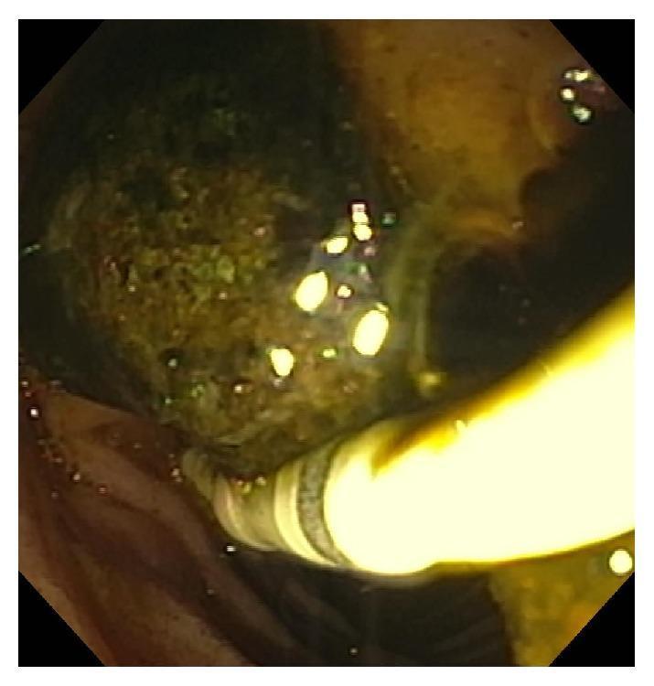
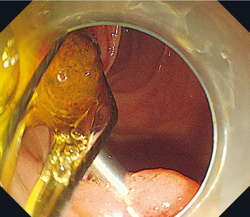
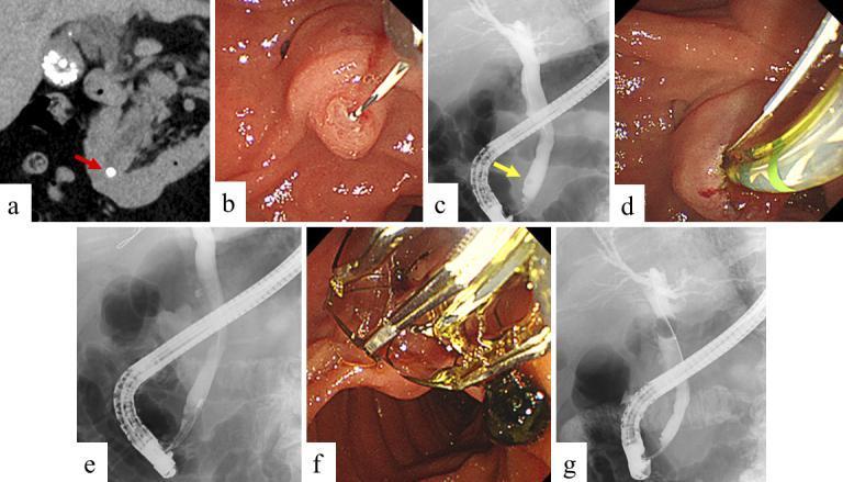
ERCP sequence: (a) CT showing hyperdense stone in distal CBD; (b,d,f) Endoscopic views - cannulation, sphincterotomy, basket extraction; (c,e,g) Fluoroscopy confirming clearance
14. QUICK DIFFERENTIALS - CAUSES OF OBSTRUCTIVE JAUNDICE
| Benign | Malignant |
|---|
| Choledocholithiasis (most common benign) | Cancer of head of pancreas (most common malignant) |
| Mirizzi syndrome | Cholangiocarcinoma (Klatskin tumor - hilar) |
| Benign biliary stricture | Ampullary carcinoma |
| Choledochal cyst | Gallbladder cancer |
| Primary sclerosing cholangitis | Duodenal carcinoma |
| Sphincter of Oddi dysfunction | Metastatic nodes at porta hepatis |
15. KEY NUMBERS TO REMEMBER
| Value | Significance |
|---|
| Jaundice visible at | Bilirubin >2.5-3 mg/dL |
| Normal CBD diameter | ≤6 mm (≤8 mm post-cholecystectomy) |
| CBD dilation on USS | >6 mm = "strong" predictor of stone |
| Bilirubin "Very Strong" predictor | >4 mg/dL |
| Bilirubin "Strong" predictor | 1.8-4 mg/dL |
| CBD stones in symptomatic gallstone disease | 10-15% |
| CBD stones in cholecystitis patients | 10-20% |
| Spontaneous stone passage (low-risk) | ~30% |
| ERCP success rate | ~90% |
| MRCP sensitivity | 81-100% |
| MRCP specificity | 92-100% |
| Transcystic CBDE success | 80-90% |
| Choledochotomy success | 83-96% |
Sources
- Mulholland and Greenfield's Surgery: Scientific Principles and Practice, 7e - Chapter 61
- Fischer's Mastery of Surgery, 8e - Biliary Surgery
- Schwartz's Principles of Surgery, 11e - Jaundice chapter
- Guyton and Hall Textbook of Medical Physiology - Bilirubin physiology
- Harper's Illustrated Biochemistry, 32nd ed - Bilirubin metabolism tables
- Pye's Surgical Handicraft, 22e - Perioperative jaundice management
- Gray's Anatomy for Students - Biliary anatomy
Recent Evidence Note (PubMed 2024-2025): A systematic review (PMID 38555329, Dig Dis Sci 2024) confirmed comparable post-ERCP adverse event rates between symptomatic and asymptomatic choledocholithiasis - supporting active treatment even in asymptomatic cases. A 2025 Bayesian network meta-analysis (PMID 40905968) on balloon dilation duration provides updated guidance on EST + EBD timing for stone extraction.