High yield concepts about Multiple sclerosis and SLE. Add usmle pearls. Condition of these diseases in pregnancy
Reading File
Finding Sources
Finding Sources
Searching PubMed
"multiple sclerosis" AND pregnancy
Searching PubMed
"systemic lupus erythematosus" AND pregnancy
Finding Sources
Finding Sources
Finding Sources
Reading File
Reading File
Finding Sources
Finding Sources
Reading File
Reading File
Finding Sources
Reading File
Reading File
I now have excellent material. Let me compile the comprehensive answer:
Multiple Sclerosis (MS) and Systemic Lupus Erythematosus (SLE)
High-Yield Concepts + USMLE Pearls + Pregnancy
PART 1: MULTIPLE SCLEROSIS
Overview & Epidemiology
- Chronic inflammatory demyelinating disease of the CNS - plaques affect white matter at multiple sites separated in time and space
- Prevalence: ~309/100,000 in the U.S. (~730,000 Americans); ~2.8 million worldwide
- Female:Male = 2-2.5:1 (common autoimmune sex predilection)
- Peak onset: 3rd-4th decade of life (post-pubertal teens to 50s)
- Latitude gradient: more common in temperate zones; decreases near the equator
- Risk factors: cigarette smoking, childhood obesity, low vitamin D, Epstein-Barr virus (EBV)
USMLE Pearl: MS is the most common demyelinating disease. A young woman presenting with relapsing neurologic deficits (optic neuritis + spinal cord symptoms) = think MS first.
Pathology
- Plaques: sharply demarcated patches of demyelination and axonal injury - pink-gray color, primarily in white matter
- Periventricular location is classic (especially around the lateral ventricles)
- Lesions spare the root entry zones of cranial/spinal nerves (peripheral myelin is not affected)
- Pathology = T-cell mediated autoimmune attack on myelin (oligodendrocytes)
USMLE Pearl - Dawson's fingers: Periventricular MS plaques oriented perpendicular to ventricles on sagittal MRI FLAIR = pathognomonic appearance.
Clinical Presentations (High-Yield)
| Symptom | Localization | Pearl |
|---|---|---|
| Optic neuritis | Optic nerve | Painful vision loss; "patient sees nothing, doctor sees nothing" (retrobulbar) - RAPD (Marcus-Gunn pupil) |
| Internuclear ophthalmoplegia (INO) | MLF (medial longitudinal fasciculus) | Impaired adduction ipsilateral eye + nystagmus contralateral eye; bilateral INO in young person = MS until proven otherwise |
| Lhermitte sign | Cervical cord | Electric shock sensation down spine on neck flexion |
| Uhthoff phenomenon | Heat sensitivity | Worsening symptoms with heat (hot bath, fever) |
| Transverse myelitis | Spinal cord | Weakness + sensory level + bladder dysfunction |
| Charcot triad | Cerebellar | Intention tremor + nystagmus + scanning speech |
| Bladder dysfunction | Spinal cord | Urge incontinence most common; neurogenic bladder |
USMLE Pearl: The combination of optic neuritis + bilateral INO in a young woman is nearly pathognomonic for MS. Uhthoff phenomenon is unique to MS (worsening with temperature elevation, not true relapse).
Types of MS
| Type | Frequency | Key Feature |
|---|---|---|
| Relapsing-Remitting (RRMS) | ~85-90% | Discrete attacks with full/partial recovery; most common |
| Secondary Progressive (SPMS) | ~50% of RRMS after 20-40 yrs | Gradual progression after initial RR phase |
| Primary Progressive (PPMS) | ~10-15% | Progressive from onset, no relapses; more common in middle-aged men; treated with ocrelizumab |
| Marburg disease | Rare | Acute fulminant form; fatal within months |
USMLE Pearl: PPMS is the only form treated with ocrelizumab (anti-CD20). Natalizumab (anti-VLA-4/anti-α4-integrin) is used for active RRMS. Both are high-yield treatments.
Diagnosis - McDonald Criteria
- Requires lesions disseminated in time AND space
- MRI brain: >95% abnormal in definite MS - T2/FLAIR hyperintense lesions, periventricular, juxtacortical, infratentorial, spinal cord
- Active lesions: gadolinium-enhancing (T1)
- CSF findings: oligoclonal bands (OCBs) in IgG - present in >95%; elevated IgG index; may see mild pleocytosis
- Visual evoked potentials (VEPs): prolonged latency indicates subclinical optic nerve demyelination
USMLE Pearl: OCBs in CSF are present in MS but NOT in serum (if also in serum, think systemic disease, NOT MS). CSF shows <50 WBCs, mildly elevated protein - markedly elevated protein should suggest an alternative diagnosis.
Treatment (USMLE High-Yield)
| Category | Drug | Pearl |
|---|---|---|
| Acute relapse | High-dose IV methylprednisolone | Shortens duration, does NOT alter long-term outcome |
| 1st-line DMT | Interferon-β, glatiramer acetate | Reduce relapse rate ~30% |
| Oral DMT | Dimethyl fumarate, teriflunomide, fingolimod | Fingolimod: first oral DMT; causes bradycardia (1st dose) + macular edema |
| High-efficacy | Natalizumab, ocrelizumab, alemtuzumab | Natalizumab: risk of PML (JC virus) - check JC antibody titer |
| Symptomatic | Baclofen (spasticity), oxybutynin (bladder), amantadine (fatigue) | |
| Contraindicated | Teriflunomide, mitoxantrone | Teratogenic - contraindicated in pregnancy |
USMLE Pearl: Natalizumab inhibits lymphocyte migration across the blood-brain barrier. Risk of PML is the major complication - requires JC antibody testing. High antibody index = significant PML risk.
MS in Pregnancy
- During pregnancy: disease activity decreases - relapses reduced by ~2/3 in the 3rd trimester (gestational immunosuppression)
- Postpartum: relapses increase significantly in the first 3-6 months postpartum
- Net effect: overall relapse risk across the full pregnancy/postpartum period is not significantly altered
- Breastfeeding may reduce the postpartum relapse rate
- Pregnancy does NOT change the long-term neurologic disability course
- MS does NOT affect fertility, labor, or delivery significantly (slight increase in C-section and SGA infants reported)
Drug management in pregnancy:
- Most DMTs are NOT recommended during pregnancy
- Teratogens (contraindicated): teriflunomide, mitoxantrone - require washout before conception
- Washout period: 0-6 months depending on drug before conception
- Safer options (low placental transfer): glatiramer acetate, interferon-β, natalizumab, ocrelizumab - minimal transfer to breast milk; likely safe for breastfeeding
- Oral DMTs: avoid during lactation
USMLE Pearl: MS does NOT prevent pregnancy. Remind that the relapse rate falls in the 3rd trimester then rebounds postpartum. The baby's risk of developing MS is low (~3-5% if one parent affected). Counsel optimistically.
PART 2: SYSTEMIC LUPUS ERYTHEMATOSUS (SLE)
Overview & Epidemiology
- Chronic multisystem autoimmune disease driven by autoantibodies, complement activation, and immune complex deposition
- Female:Male = 9:1 (classic ratio - highest female predominance of any autoimmune disease)
- Peak onset: women of childbearing age (15-45 years)
- More common and severe in African-American, Hispanic, and Asian women
- Genetic component: concordance 24% in monozygotic twins vs. 2% in dizygotic twins
-
150 susceptibility loci identified (GWAS); strong MHC class II association
USMLE Pearl: ANA is the best screening test (sensitivity ~95%). Specific for SLE: anti-dsDNA and anti-Smith (anti-Sm). Anti-dsDNA correlates with disease activity and lupus nephritis.
Pathogenesis
- Defective clearance of apoptotic debris → nuclear antigens released → ANA production
- Neutrophil NETosis releases nuclear contents → activates plasmacytoid DCs → type I IFN production
- Complement deficiency (C1q, C1r, C1s, C2, C4) → impaired immune complex clearance → tissue deposition
- Immune complexes deposit in kidneys, skin, joints, vessels → inflammation
USMLE Pearl: C1q deficiency is the strongest single-gene risk factor for lupus. Low C3/C4/CH50 during active disease = complement consumption by immune complexes.
Clinical Features - The SOAP BRAIN MD Mnemonic (2019 EULAR/ACR Criteria)
| Domain | Feature | High-Yield Notes |
|---|---|---|
| Skin | Malar (butterfly) rash | Spares nasolabial folds; photosensitive; raised erythema over cheeks/nose bridge |
| Discoid lupus | Scarring, follicular plugging; can occur WITHOUT systemic disease | |
| Photosensitivity | Rash after UV exposure | |
| Oral ulcers | Usually painless | |
| Subacute cutaneous lupus (SCLE) | Anti-Ro/SSA antibodies | |
| Joints | Non-erosive polyarthritis | >75% of patients; hands/wrists most common; Jaccoud arthropathy |
| Serositis | Pleuritis, pericarditis | Pleuritic chest pain; friction rub |
| Renal | Lupus nephritis | Proteinuria, hematuria, RBC casts; worst prognosis |
| Neuropsychiatric | CNS lupus | Psychosis, seizures, stroke, cognitive dysfunction |
| Hematologic | Cytopenias | Hemolytic anemia (Coombs+), leukopenia, lymphopenia, thrombocytopenia |
| Constitutional | Fever, fatigue, weight loss | Common at presentation |
| Cardiac | Libman-Sacks endocarditis | Sterile verrucous vegetations on BOTH sides of mitral valve |
| Pulmonary | Shrinking lung syndrome | Diaphragm weakness; rare but classic |
USMLE Pearl - Libman-Sacks endocarditis: Sterile marantic vegetations on BOTH sides of the valve (usually mitral) - unlike infective endocarditis (irregular, destructive) and rheumatic fever (mitral valve, posterior leaflet only). Associated with antiphospholipid antibodies.
Antibody Panel (Most High-Yield for USMLE)
| Antibody | Association | Specificity/Sensitivity |
|---|---|---|
| ANA | Screening | Sensitive (~95%) but NOT specific for SLE |
| Anti-dsDNA | SLE activity + nephritis | Highly specific (~70%); titer correlates with flares |
| Anti-Smith (Sm) | SLE | Most specific (~99%) for SLE but low sensitivity (~25%) |
| Anti-Ro/SSA | SCLE, neonatal lupus, Sjögren | Causes neonatal heart block in newborns |
| Anti-La/SSB | SLE, Sjögren | Often with anti-Ro |
| Anti-histone | Drug-induced lupus | 95% sensitive for DIL (procainamide, hydralazine, isoniazid) |
| Antiphospholipid | APS (thrombosis, pregnancy loss) | Lupus anticoagulant, anticardiolipin, anti-β2-GPI |
| Anti-ribosomal P | Neuropsychiatric lupus | CNS manifestations |
USMLE Pearl: Anti-Smith is the MOST SPECIFIC antibody for SLE. Anti-dsDNA correlates with DISEASE ACTIVITY (monitor for flares and nephritis). Anti-histone = drug-induced lupus (complement is NORMAL, anti-dsDNA negative).
USMLE Pearl - Drug-induced lupus: Caused by P-HISM - Procainamide (most common), Hydralazine, Isoniazid, Sulfonamides, Minocycline. Spares the kidneys and CNS. Resolves when drug is stopped.
Lupus Nephritis
- Most serious organ manifestation; occurs in 40-60% of SLE patients
- ISN/RPS Classification (Class I-VI):
- Class III/IV: focal/diffuse proliferative nephritis - worst prognosis, most common to cause ESRD
- Class V: membranous - nephrotic syndrome presentation
- Markers of activity: rising anti-dsDNA + falling complement (C3/C4) = flare/nephritis
- Treatment: hydroxychloroquine (all patients) + induction with cyclophosphamide or mycophenolate mofetil (MMF) + steroids; maintenance with MMF or azathioprine
USMLE Pearl: Class IV diffuse proliferative LN = "wire-loop" lesions on EM/LM (subendothelial immune complex deposits). Most likely to progress to renal failure. Treat aggressively.
SLE in Pregnancy
Maternal Risks:
- SLE flares are common during pregnancy and the postpartum period
- Pre-eclampsia occurs at higher rates (especially with lupus nephritis)
- Hypercoagulable state - increased VTE risk
- Disease is best controlled when SLE is in remission for 6 months before conception
Fetal/Neonatal Risks:
- Spontaneous abortion, intrauterine fetal demise, preterm birth, IUGR - all increased
- Neonatal lupus: transplacental passage of anti-Ro/SSA and anti-La/SSB antibodies
- Skin rash, transient thrombocytopenia, liver abnormalities (all transient)
- Congenital complete heart block (CHB) - the feared complication; permanent; may require pacemaker; risk ~2% if mother is anti-Ro+
- Antiphospholipid Antibody Syndrome (APS) co-existing with SLE:
- Recurrent first-trimester pregnancy loss, mid/late pregnancy fetal death
- Thrombosis (arterial and venous)
- Treatment: aspirin + heparin (LMWH) throughout pregnancy - warfarin is contraindicated in pregnancy
Predictors of Poor Pregnancy Outcome (Lancet Rheumatol 2024 - PMID 39153486):
- Active renal disease (lupus nephritis)
- Reduced GFR
- Hypocomplementemia
- Presence of antiphospholipid antibodies
Medications in Pregnancy:
| Drug | Safety | Notes |
|---|---|---|
| Hydroxychloroquine (HCQ) | SAFE - continue | Reduces flares, neonatal CHB risk, preterm birth |
| Prednisone/prednisolone | Relatively safe | Risk of cleft palate (1st trimester high-dose); preferred corticosteroid |
| Azathioprine | Compatible | Preferred immunosuppressant in pregnancy |
| Aspirin (low-dose) | SAFE | Started early to reduce pre-eclampsia and APS complications |
| LMWH (heparin) | SAFE | Anticoagulation for APS - does not cross placenta |
| MMF/cyclophosphamide | CONTRAINDICATED | Teratogenic |
| Methotrexate | CONTRAINDICATED | Teratogenic and abortifacient |
| Warfarin | CONTRAINDICATED | Warfarin embryopathy (nasal hypoplasia, stippled epiphyses) |
| NSAIDs | Avoid in 3rd trimester | Risk of premature closure of ductus arteriosus |
| Belimumab | Avoid | Insufficient data |
USMLE Pearl: The SLE patient who is anti-Ro+ is at risk for neonatal CHB. Continue hydroxychloroquine throughout pregnancy - it reduces CHB risk, prevents flares, and is safe for the fetus. Screen fetus with fetal echocardiogram between 16-26 weeks.
USMLE Pearl: SLE + recurrent pregnancy loss = think antiphospholipid syndrome. Test for lupus anticoagulant, anticardiolipin, and anti-β2-GPI. Treat with aspirin + heparin (NOT warfarin).
QUICK COMPARISON TABLE: MS vs. SLE in Pregnancy
| Feature | MS | SLE |
|---|---|---|
| Effect of pregnancy on disease | Improves (↓ relapses, esp. 3rd trimester) | Can worsen; flares common |
| Postpartum | Increased relapses (1st 3-6 months) | Continued risk of flares |
| Fetal risk | Minimal; slight ↑ IUGR/C-section | High - miscarriage, IUGR, preterm, fetal death |
| Neonatal effect | None specific | Neonatal lupus (anti-Ro → CHB) |
| Key contraindicated drug | Teriflunomide, mitoxantrone | MMF, cyclophosphamide, methotrexate, warfarin |
| Preferred management | Glatiramer acetate, IFN-β (if needed); stop most DMTs | Hydroxychloroquine + low-dose aspirin + azathioprine |
| Breastfeeding | Avoid oral DMTs; large molecules relatively safe | HCQ, prednisolone safe |
Key References
- Adams and Victor's Principles of Neurology, 12th Edition - MS pathology and classification
- Goldman-Cecil Medicine, 2025 - MS diagnosis, treatment, and pregnancy
- Harrison's Principles of Internal Medicine 22E (2025) - SLE pathogenesis and lupus nephritis
- Creasy & Resnik's Maternal-Fetal Medicine - MS and SLE in pregnancy
- Wind M et al., Pregnancy outcome predictors in SLE - Lancet Rheumatol 2024 (PMID 39153486) - active renal disease, hypocomplementemia, and APS predict poor pregnancy outcomes
- Singh M et al. - Autoimmune diseases and adverse pregnancy outcomes - BMC Med 2024 (PMID 38438886) - umbrella review confirming elevated risks across autoimmune conditions
Generate usmle standard high yield questions from multiple sclerosis
Reading File
Finding Sources
Finding Sources
Finding Sources
Finding Sources
Finding Sources
Finding Sources
Finding Sources
Finding Sources
Loading Image
Loading Image
I now have all the content needed - rich textbook data, MRI images, and clinical details. Here are 15 USMLE-standard high-yield questions covering every major tested domain of MS:
USMLE-Standard High-Yield Questions: Multiple Sclerosis
QUESTION 1 - Classic Presentation / Diagnosis
A 28-year-old woman presents with a 3-day history of painful loss of vision in her right eye. She describes difficulty distinguishing colors and notes the pain worsens with eye movement. Examination reveals a right relative afferent pupillary defect. Fundoscopy is normal. Two years ago she had an episode of numbness in her left leg that resolved completely. MRI of the brain and orbits is obtained.
Which of the following MRI findings would MOST strongly support the diagnosis?
- A) Hyperintense lesion in the right optic nerve on T1 with gadolinium enhancement
- B) Multiple periventricular T2/FLAIR hyperintense lesions oriented perpendicular to the lateral ventricles
- C) Diffuse cortical atrophy with sulcal widening
- D) Bilateral basal ganglia T2 hyperintensities
- E) A single enhancing ring lesion with surrounding edema
Correct Answer: B
Explanation: This patient has optic neuritis (painful monocular vision loss, RAPD, normal fundus = retrobulbar inflammation) plus a prior episode of sensory deficits - fulfilling dissemination in time and space. The MRI hallmark of MS is periventricular T2/FLAIR hyperintense ovoid plaques oriented perpendicular to the ventricles (Dawson's fingers on sagittal view). The fundus is normal because the lesion is retrobulbar ("the patient sees nothing, the doctor sees nothing"). A ring-enhancing lesion (E) suggests abscess or primary CNS lymphoma. Cortical atrophy (C) is non-specific and late. T1 gadolinium enhancement in the optic nerve can occur but is not as diagnostically specific for MS as periventricular white matter lesions.
Adams and Victor's Principles of Neurology, 12th Ed.
QUESTION 2 - MRI Imaging (Image-Based)
A 32-year-old man presents with a 6-month history of progressive leg weakness and urinary urgency. MRI of the brain is shown below.
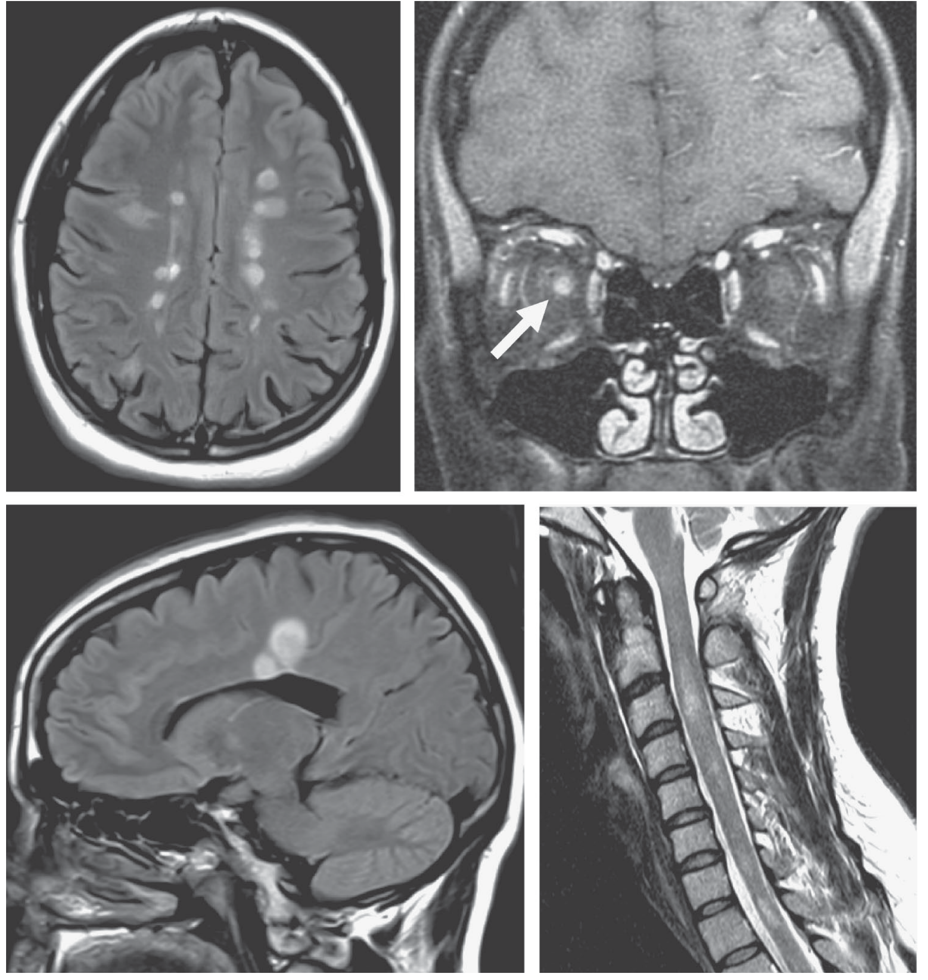
CSF analysis shows: WBC 12 (lymphocytes), protein 55 mg/dL, glucose normal, oligoclonal IgG bands present in CSF but NOT in serum. Which of the following statements about the CSF finding is MOST accurate?
- A) Oligoclonal bands in CSF are 100% specific for MS
- B) Oligoclonal bands present in both CSF and serum support a diagnosis of MS
- C) Oligoclonal bands present in CSF but not in serum indicate intrathecal IgG synthesis
- D) The CSF IgG index is expected to be low in MS
- E) A CSF protein > 100 mg/dL is characteristic of MS
Correct Answer: C
Explanation: Type 2 oligoclonal bands (present in CSF, absent in serum) indicate intrathecal IgG production and are found in >95% of patients with clinically definite MS. If bands are present in BOTH CSF and serum (Type 3 pattern), this suggests a systemic inflammatory process rather than isolated CNS disease. The IgG index is typically elevated (not low) in MS. CSF protein is mildly elevated (rarely >100 mg/dL) - markedly elevated protein should prompt search for an alternative diagnosis. OCBs are supportive but not 100% specific - they also occur in CNS infections, sarcoidosis, and other inflammatory diseases.
Henry's Clinical Diagnosis and Management by Laboratory Methods; Neuroanatomy through Clinical Cases, 3rd Ed.
QUESTION 3 - Eye Movement / Localization
A 26-year-old woman reports intermittent double vision and blurred vision when looking to the right. On examination, when she looks to the right, her left eye fails to adduct fully and her right eye shows horizontal nystagmus. When she looks to the left, both eyes move normally. Convergence is intact.
Where is the lesion responsible for this finding?
- A) Right abducens nucleus (CN VI)
- B) Left oculomotor nucleus (CN III)
- C) Left medial longitudinal fasciculus (MLF)
- D) Right medial longitudinal fasciculus (MLF)
- E) Cerebellar vermis
Correct Answer: C
Explanation: This is right-gaze internuclear ophthalmoplegia (INO) caused by a lesion in the left MLF. On right lateral gaze, the left eye should adduct (mediated by the left CN III, commanded via the right PPRF → left MLF → left CN III nucleus). A left MLF lesion disrupts this pathway, causing failure of left eye adduction with abducting nystagmus of the right eye. Convergence is intact because it uses a different pathway. Bilateral INO in a young woman is pathognomonic for MS. (Choice D would cause a left-gaze INO).
Goldman-Cecil Medicine; K.J. Lee's Essential Otolaryngology
QUESTION 4 - Uhthoff and Lhermitte Phenomena
A 30-year-old woman with known relapsing-remitting MS calls her neurologist reporting that her vision becomes blurry every time she takes a hot bath or exercises. The episode lasts 20-30 minutes and then completely resolves. She also notes an electric shock-like sensation down her spine when she flexes her neck.
Which of the following BEST explains the first symptom?
- A) A new demyelinating plaque in the optic nerve
- B) Thermoregulatory autonomic dysfunction causing vasospasm
- C) Conduction block in previously demyelinated axons due to elevated temperature
- D) Increased intracranial pressure from cerebral edema
- E) Psychogenic exacerbation of symptoms
Correct Answer: C
Explanation: This question tests two classic MS phenomena:
- Uhthoff phenomenon: temporary worsening of symptoms with heat or exercise due to conduction block in demyelinated axons when temperature rises even slightly. This is NOT a new relapse - it reverses on cooling.
- Lhermitte sign: electrical sensation down the spine on neck flexion, caused by a demyelinating lesion in the cervical spinal cord (dorsal columns).
Both are transient and do NOT represent new disease activity or a relapse requiring treatment. Distinguishing Uhthoff phenomenon from a true relapse (lasting >24 hours) is high-yield.
Kanski's Clinical Ophthalmology, 10th Ed.; Goldman-Cecil Medicine
QUESTION 5 - Types of MS
A 52-year-old man presents to his neurologist with a 14-month history of slowly progressive bilateral leg weakness and gait difficulty. He denies any discrete attacks or periods of remission. MRI shows multiple spinal cord lesions but fewer brain lesions compared to typical RRMS. CSF shows oligoclonal bands.
Which of the following disease-modifying therapies has shown efficacy specifically for this patient's type of MS?
- A) Interferon beta-1a
- B) Glatiramer acetate
- C) Natalizumab
- D) Ocrelizumab
- E) Fingolimod
Correct Answer: D
Explanation: This patient has primary progressive MS (PPMS) - progressive neurologic deterioration from disease onset for at least 1 year without relapses. PPMS affects ~10-15% of MS patients and is more common in middle-aged men with predominantly spinal cord involvement. Ocrelizumab (anti-CD20, depletes B cells) is the only FDA-approved therapy for PPMS, reducing disability progression by ~25%. All other options listed are approved for relapsing forms of MS only. PPMS has fewer inflammatory brain lesions and gadolinium-enhancing lesions, reflecting a more neurodegenerative phenotype.
Goldman-Cecil Medicine; Bradley and Daroff's Neurology
QUESTION 6 - Pharmacology / Drug Mechanism
A 29-year-old woman with relapsing-remitting MS is started on a new oral disease-modifying therapy. You counsel her that she must be monitored with cardiac telemetry for 6 hours after the first dose due to the risk of bradycardia and heart block. You also warn her about the risk of macular edema and pulmonary function changes.
Which of the following drugs is being described, and what is its mechanism of action?
- A) Natalizumab - blocks α4-integrin, preventing lymphocyte CNS migration
- B) Fingolimod - sphingosine-1-phosphate receptor modulator, sequesters lymphocytes in lymph nodes
- C) Dimethyl fumarate - activates Nrf2 pathway, reduces oxidative stress
- D) Teriflunomide - inhibits dihydroorotate dehydrogenase, blocks pyrimidine synthesis
- E) Alemtuzumab - anti-CD52, depletes T and B lymphocytes
Correct Answer: B
Explanation: Fingolimod (Gilenya) is the first oral DMT for MS. It is a sphingosine-1-phosphate (S1P) receptor modulator that sequesters lymphocytes in lymph nodes, preventing them from entering the CNS. Key side effects tested on USMLE:
- First-dose bradycardia and AV block → requires 6-hour cardiac monitoring after first dose
- Macular edema → baseline ophthalmologic exam required
- Pulmonary function changes (FEV1 reduction)
- Increased risk of herpes infections
Natalizumab (A) blocks α4-integrin (VLA-4), preventing lymphocyte passage across the blood-brain barrier - its risk is PML (JC virus). Teriflunomide (D) is teratogenic.
Goldman-Cecil Medicine; Katzung's Basic and Clinical Pharmacology, 16th Ed.
QUESTION 7 - Pharmacology / Dangerous Complication
A 34-year-old man with relapsing-remitting MS has been on natalizumab monotherapy for 3 years. His JC virus antibody index has been rising. He now presents with progressive cognitive decline, aphasia, and right-sided weakness over 4 weeks. MRI shows a large non-enhancing, asymmetric white matter lesion in the left hemisphere crossing gyral boundaries.
What is the MOST likely complication and what is the PRIMARY risk factor that should have been monitored?
- A) MS relapse; number of prior relapses
- B) CNS lymphoma; prior use of corticosteroids
- C) Progressive multifocal leukoencephalopathy (PML); JC virus antibody index
- D) Glioblastoma multiforme; duration of natalizumab therapy
- E) Acute disseminated encephalomyelitis; recent vaccination
Correct Answer: C
Explanation: This is natalizumab-associated PML caused by JC virus (JC polyomavirus) reactivation. Risk is stratified by:
- JC antibody seropositivity (and index level - higher index = higher risk)
- Duration of natalizumab (>2 years significantly increases risk)
- Prior immunosuppressive therapy
PML presents with subacute progressive cognitive/motor deficits; MRI shows large, asymmetric, non-enhancing white matter lesions (unlike MS plaques which are small, oval, periventricular). Management: discontinue natalizumab immediately, consider plasma exchange to accelerate drug removal. The JC antibody index should be checked every 6 months in patients on natalizumab.
Goldman-Cecil Medicine; Yamada's Textbook of Gastroenterology, 7th Ed.
QUESTION 8 - CSF Analysis / Diagnosis
A 27-year-old woman presents after an episode of right leg weakness and urinary retention lasting 10 days that spontaneously improved. CSF analysis is performed:
- Opening pressure: normal
- WBC: 18 (80% lymphocytes)
- Protein: 62 mg/dL
- Glucose: normal
- IgG index: 0.78 (elevated)
- Oligoclonal bands: present in CSF, absent in serum
Which of the following is the MOST sensitive test for establishing the diagnosis of MS, and what percentage of confirmed MS patients have abnormal findings?
- A) Oligoclonal bands in CSF; ~60%
- B) MRI brain (T2/FLAIR lesions); >95%
- C) Visual evoked potentials; ~80%
- D) Anti-aquaporin-4 antibody; ~70%
- E) CSF IgG index elevation; ~50%
Correct Answer: B
Explanation: MRI brain is the most sensitive test - abnormal T2/FLAIR findings are present in >95% of patients with clinically definite MS. A normal brain MRI should raise doubt about the diagnosis. Oligoclonal bands are present in >95% of MS patients but require paired CSF/serum analysis. Visual evoked potentials (prolonged P100 latency) are useful for detecting subclinical optic nerve demyelination (~80% sensitive). Anti-aquaporin-4 (NMO-IgG) and anti-MOG antibodies are markers of neuromyelitis optica spectrum disorder (NMOSD) - an important MS mimic. MRI also fulfills dissemination in time and space criteria (McDonald criteria).
Goldman-Cecil Medicine; Grainger & Allison's Diagnostic Radiology
QUESTION 9 - Treatment of Acute Relapse
A 31-year-old woman with known RRMS presents with acute onset left-sided weakness and new urinary incontinence for 5 days. Neurologic exam confirms new left hemiparesis. MRI shows a new gadolinium-enhancing lesion in the right cerebral hemisphere.
What is the MOST appropriate immediate management, and what is the MOST accurate statement about its long-term effect?
- A) Oral prednisone 1 mg/kg/day × 4 weeks; permanently reduces disability
- B) IV methylprednisolone 1 g/day × 3-5 days; shortens duration of relapse but does not alter long-term disability
- C) Plasma exchange; first-line for all MS relapses
- D) Start interferon beta-1a immediately; treats both the relapse and prevents future relapses
- E) Watchful waiting; most relapses resolve spontaneously without treatment
Correct Answer: B
Explanation: High-dose IV methylprednisolone (1 g/day × 3-5 days) is the standard of care for acute MS relapses. Key USMLE fact: it shortens the duration and severity of a relapse but does NOT alter long-term neurologic disability or affect the overall disease course. Oral steroids (choice A) at high equivalent doses may be used but IV dosing is preferred for severe attacks. Plasma exchange (C) is reserved for severe relapses refractory to steroids. Disease-modifying therapies (D) do not treat acute relapses - they prevent future ones. Choice E is incorrect because untreated severe relapses leave permanent residual deficits.
Goldman-Cecil Medicine, 2025
QUESTION 10 - MS and Pregnancy
A 26-year-old woman with relapsing-remitting MS on glatiramer acetate informs you she is 8 weeks pregnant. She is worried about her MS worsening during pregnancy and whether her medication is safe.
Which of the following statements is MOST accurate regarding MS and pregnancy?
- A) Relapse rate increases significantly throughout all trimesters of pregnancy
- B) Relapse rate decreases during pregnancy (especially 3rd trimester) but increases in the first 3-6 months postpartum
- C) Glatiramer acetate should be stopped immediately as it is a known teratogen
- D) Pregnancy increases the long-term risk of disability from MS
- E) Breastfeeding is absolutely contraindicated in all MS patients
Correct Answer: B
Explanation: The PRIMS study established that MS relapses decrease during pregnancy - reduced by ~2/3 in the 3rd trimester due to gestational immunosuppression. However, relapses rebound in the first 3-6 months postpartum. The net effect over the full pregnancy/postpartum period is neutral. Pregnancy does NOT alter long-term disability. Glatiramer acetate has minimal placental transfer and is considered relatively safe (not a known teratogen) - by contrast, teriflunomide and mitoxantrone are teratogens and are contraindicated. Breastfeeding may be protective against postpartum relapses; large-molecule DMTs (glatiramer, interferon, ocrelizumab, natalizumab) have minimal transfer to breast milk and are likely compatible with breastfeeding.
Creasy & Resnik's Maternal-Fetal Medicine; Goldman-Cecil Medicine
QUESTION 11 - MS Mimics / Differential Diagnosis
A 22-year-old woman presents with sudden bilateral vision loss, transverse myelitis with sensory level at T4, and urinary retention. MRI spine shows a longitudinally extensive T2 hyperintensity spanning 4 vertebral segments. Brain MRI shows no periventricular lesions. CSF oligoclonal bands are ABSENT. A specific serum antibody returns positive.
Which antibody is MOST likely positive, and what diagnosis does this suggest?
- A) Anti-Ro/SSA; Sjögren syndrome-associated myelopathy
- B) Anti-AQP4 (aquaporin-4 / NMO-IgG); neuromyelitis optica spectrum disorder (NMOSD)
- C) Anti-MOG (myelin oligodendrocyte glycoprotein); MS
- D) Anti-dsDNA; SLE-associated CNS disease
- E) Anti-GQ1b; Miller Fisher syndrome
Correct Answer: B
Explanation: This is Neuromyelitis Optica Spectrum Disorder (NMOSD), the most important MS mimic. Key distinguishing features:
- Bilateral or rapidly alternating optic neuritis (MS typically unilateral)
- Longitudinally extensive transverse myelitis (LETM) - >3 vertebral segments (MS lesions span <2 segments)
- Area postrema involvement - intractable hiccups/nausea
- Normal brain MRI (or non-MS pattern lesions)
- OCBs absent in ~80% of NMOSD (present in >95% MS)
- Anti-AQP4 antibody positive in ~70-80% of NMOSD
Anti-MOG antibodies (C) suggest MOG-antibody-associated disease (MOGAD), another mimic. This distinction is critical because NMOSD is treated differently from MS (rituximab, eculizumab, inebilizumab - NOT interferon-β, which can worsen NMOSD).
Adams and Victor's Principles of Neurology, 12th Ed.
QUESTION 12 - Pharmacology / Teratogenicity
A 32-year-old woman with RRMS wishes to conceive. She is currently on teriflunomide. Her neurologist decides to switch her therapy before attempting conception.
Which of the following is the MOST accurate statement about teriflunomide in this context?
- A) Teriflunomide is safe in pregnancy; no washout is needed
- B) Teriflunomide requires a washout with cholestyramine or activated charcoal to eliminate the drug rapidly before conception
- C) Teriflunomide can be continued in the first trimester only
- D) Teriflunomide is safe but must be switched after pregnancy is confirmed
- E) Teriflunomide causes cardiac defects specifically in the 3rd trimester
Correct Answer: B
Explanation: Teriflunomide is a known teratogen - it inhibits dihydroorotate dehydrogenase (DHODH), blocking pyrimidine synthesis, and causes fetal harm (embryolethality and teratogenicity in animal studies). Its active metabolite has an extremely long half-life (up to 2 years without intervention). Before conception, an accelerated elimination procedure using cholestyramine 8 g 3x/day × 11 days or activated charcoal 50 g 4x/day × 11 days is required to rapidly clear the drug. Without washout, serum levels may remain detectable for 1-2 years. Mitoxantrone is similarly teratogenic and contraindicated. Compare to glatiramer acetate and interferons which are relatively safer alternatives pre-conception.
Creasy & Resnik's Maternal-Fetal Medicine
QUESTION 13 - Secondary Progressive MS / Disease Course
A 45-year-old woman was diagnosed with relapsing-remitting MS at age 28. Over the last 18 months she has had no distinct relapses but has noticed a steady, progressive increase in leg stiffness, difficulty walking, and urinary frequency. She scores worse on her disability scale than 18 months ago. MRI shows no new gadolinium-enhancing lesions but increased T2 lesion burden.
Which of the following BEST describes her current disease type?
- A) Primary progressive MS
- B) Clinically isolated syndrome
- C) Secondary progressive MS (active)
- D) Acute Marburg MS
- E) Secondary progressive MS (inactive)
Correct Answer: E
Explanation: After initial RRMS, many patients develop secondary progressive MS (SPMS) - characterized by at least 6 months of progressive worsening WITHOUT a distinct relapse. This patient had RRMS for 17 years and now has steady progression without relapses or new gadolinium-enhancing lesions, making this SPMS - inactive (no evidence of current inflammatory activity). If there were superimposed relapses or new MRI activity, it would be active SPMS. PPMS (A) is incorrect because she had an initial relapsing course. The distinction between active and inactive SPMS matters because some DMTs (siponimod) are approved for active SPMS.
Goldman-Cecil Medicine, 2025
QUESTION 14 - Symptom Management
A 35-year-old man with RRMS complains of severe fatigue (the most disabling symptom), significant spasticity in his legs that makes walking difficult, and urinary urgency with incontinence.
Match each symptom with the MOST appropriate pharmacologic treatment:
- A) Fatigue → baclofen; Spasticity → oxybutynin; Bladder → amantadine
- B) Fatigue → amantadine; Spasticity → baclofen; Bladder → oxybutynin
- C) Fatigue → fingolimod; Spasticity → methylprednisolone; Bladder → tamsulosin
- D) Fatigue → modafinil; Spasticity → tizanidine; Bladder → bethanechol
- E) Fatigue → amantadine; Spasticity → diazepam; Bladder → oxybutynin
Correct Answer: B
Explanation: Symptomatic management of MS:
- Fatigue (most common disabling symptom in MS): amantadine (first-line) or modafinil
- Spasticity: baclofen (oral or intrathecal); tizanidine is an alternative
- Urinary urgency/incontinence (detrusor hyperreflexia/overactive bladder): oxybutynin (anticholinergic) or tolterodine; contrast with urinary retention (detrusor areflexia) which would be treated with bethanechol or self-catheterization
- Neuropathic pain: gabapentin, carbamazepine
- Depression: SSRIs
Option D is also partially correct (modafinil for fatigue, tizanidine for spasticity) but the most classic USMLE answer pairing is amantadine/baclofen/oxybutynin.
Goldman-Cecil Medicine
QUESTION 15 - Integrated/Most Challenging
A 24-year-old woman presents with a single episode of painful right eye vision loss 3 months ago that fully resolved. She now has no neurologic complaints and her exam is normal. MRI brain shows 3 periventricular T2-hyperintense lesions and 1 juxtacortical lesion. CSF shows oligoclonal bands. There are no gadolinium-enhancing lesions.
According to McDonald Criteria 2017, which of the following statements is CORRECT?
- A) She cannot be diagnosed with MS because she has had only one clinical episode
- B) She can be diagnosed with MS because CSF oligoclonal bands fulfill dissemination in time criteria
- C) She has a clinically isolated syndrome; MS cannot be diagnosed without a second clinical episode
- D) She requires a repeat MRI in 3-6 months before any diagnosis can be made
- E) She needs a visual evoked potential before any diagnosis can be made
Correct Answer: B
Explanation: The 2017 McDonald Criteria allow MS to be diagnosed after a single clinical attack if MRI demonstrates:
- Dissemination in space (DIS): lesions in ≥2 of the 4 typical MS locations (periventricular, cortical/juxtacortical, infratentorial, spinal cord) - this patient has periventricular + juxtacortical = DIS satisfied
- Dissemination in time (DIT): In the 2017 revision, CSF-specific oligoclonal bands can substitute for DIT when DIS is already demonstrated clinically or on MRI
Therefore, this patient with one clinical attack (optic neuritis) + DIS on MRI + CSF OCBs meets criteria for an MS diagnosis - no need to wait for a second clinical episode. This is high-yield because many students still think a second attack is always required.
Goldman-Cecil Medicine, 2025; Grainger & Allison's Diagnostic Radiology
Summary Answer Key
| Q | Answer | Topic Tested |
|---|---|---|
| 1 | B | Optic neuritis presentation, MRI |
| 2 | C | CSF oligoclonal bands interpretation |
| 3 | C | INO localization to MLF |
| 4 | C | Uhthoff phenomenon mechanism |
| 5 | D | PPMS - ocrelizumab is the only approved therapy |
| 6 | B | Fingolimod - S1P mechanism + first-dose bradycardia |
| 7 | C | Natalizumab → PML (JC virus) |
| 8 | B | MRI is most sensitive test (>95%) |
| 9 | B | IV methylprednisolone shortens relapse, NOT long-term disability |
| 10 | B | MS relapses decrease in pregnancy, rebound postpartum |
| 11 | B | NMOSD - anti-AQP4, LETM, no OCBs |
| 12 | B | Teriflunomide teratogen - accelerated elimination protocol |
| 13 | E | SPMS inactive - progression without new inflammation |
| 14 | B | Symptomatic treatment: amantadine/baclofen/oxybutynin |
| 15 | B | 2017 McDonald Criteria - CSF OCBs satisfy DIT |
Sources: Adams and Victor's Principles of Neurology 12e, Goldman-Cecil Medicine 2025, Harrison's Principles of Internal Medicine 22e, Bradley and Daroff's Neurology in Clinical Practice, Creasy & Resnik's Maternal-Fetal Medicine, Katzung's Basic and Clinical Pharmacology 16e
Generate usmle style high yield questions from SLE. 10 questions
Finding Sources
Finding Sources
Finding Sources
Finding Sources
Finding Sources
Finding Sources
Finding Sources
Finding Sources
Loading Image
Finding Sources
Finding Sources
All the material I need is in hand. Here are the 10 USMLE-standard questions:
USMLE-Style High-Yield Questions: Systemic Lupus Erythematosus (SLE)
10 Questions
QUESTION 1 - Antibody Interpretation / Diagnosis
A 24-year-old African-American woman presents with a 3-month history of fatigue, joint pain in her hands and wrists, a rash across her cheeks, and oral ulcers. Labs show: ANA positive (titer 1:640), anti-dsDNA positive, anti-Smith positive, C3 low, C4 low, CBC showing leukopenia (WBC 2,800) and mild thrombocytopenia (platelets 95,000). Urinalysis shows 2+ protein and RBC casts.
Which combination of antibody findings is MOST specific for SLE, and which antibody level should be monitored to track disease activity over time?
- A) ANA and anti-histone; ANA titer
- B) Anti-dsDNA and anti-Smith; anti-dsDNA
- C) Anti-Ro/SSA and anti-La/SSB; anti-Ro titer
- D) Anti-dsDNA and anti-Ro; complement levels only
- E) ANA and antiphospholipid antibodies; anticardiolipin titer
✅ Correct Answer: B
Explanation: The antibody hierarchy in SLE is high-yield:
- ANA: most sensitive (~95%) but NOT specific - positive in many conditions and normal individuals
- Anti-dsDNA: highly specific (~97%) for SLE; titer correlates with disease activity - rising anti-dsDNA + falling complement = imminent flare (especially lupus nephritis)
- Anti-Smith (anti-Sm): MOST specific (55-100%) for SLE but low sensitivity (~25-30%); does NOT correlate with disease activity
- Anti-histone: drug-induced lupus
- Anti-Ro/SSA: SCLE and neonatal lupus
The RBC casts indicate glomerulonephritis - likely lupus nephritis. The combination of low C3/C4 + rising anti-dsDNA is the classic "storm warning" of a nephritis flare.
Harrison's Principles of Internal Medicine 22E; Harriet Lane Handbook, 23rd Ed.
QUESTION 2 - Renal Pathology (Image-Based)
A 22-year-old woman with known SLE presents with new-onset nephrotic-range proteinuria (4.2 g/day), hematuria, and rising creatinine. Her C3 and C4 are markedly low; anti-dsDNA is highly elevated. A renal biopsy is performed. The image below is from the biopsy (PAS stain, ×400):
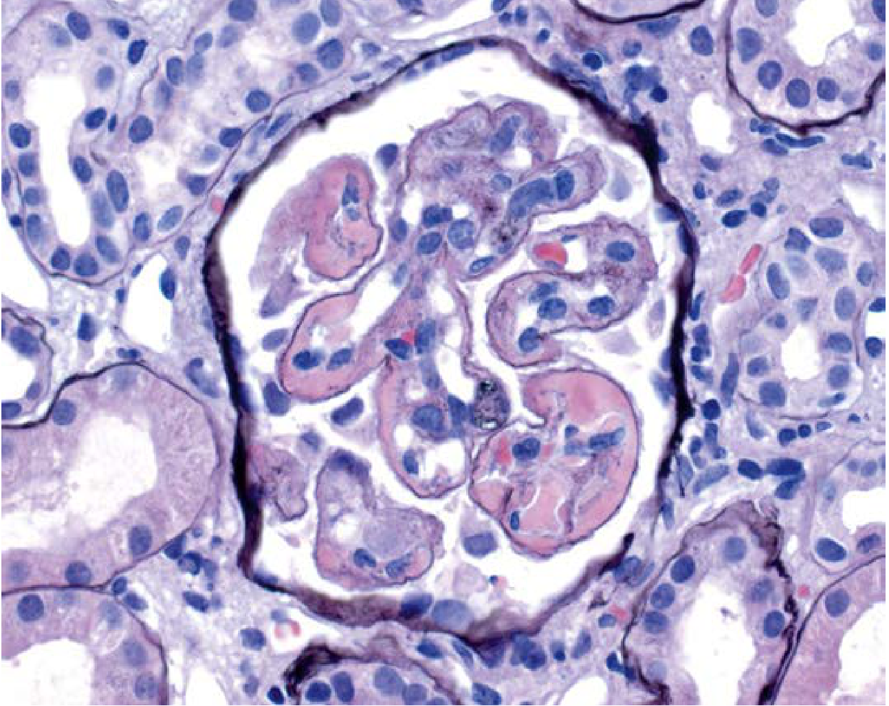
The biopsy shows global endocapillary proliferation with thickening of the glomerular capillary walls. What class of lupus nephritis does this represent, and what is the MOST appropriate induction therapy?
- A) Class II (mesangial proliferative); hydroxychloroquine alone
- B) Class III (focal proliferative); oral prednisone alone
- C) Class IV (diffuse proliferative); mycophenolate mofetil OR cyclophosphamide + high-dose steroids
- D) Class V (membranous); ACE inhibitor monotherapy
- E) Class VI (advanced sclerosis); renal transplant listing
✅ Correct Answer: C
Explanation: The image shows the classic "wire-loop" appearance - intensely eosinophilic/pink thickening of glomerular capillary walls from massive subendothelial immune complex deposits (IgG, IgM, IgA, C3, C1q - the "full house" pattern on immunofluorescence). This is Class IV - Diffuse Proliferative Lupus Nephritis, the most severe and most common form causing ESRD.
ISN/RPS Classification:
| Class | Features | Prognosis |
|---|---|---|
| I | Minimal mesangial | Excellent |
| II | Mesangial proliferative | Good |
| III | Focal (<50% glomeruli) | Moderate |
| IV | Diffuse (≥50% glomeruli) - wire loops | Worst |
| V | Membranous - nephrotic | Variable |
| VI | Advanced sclerosis | Poor |
Induction: mycophenolate mofetil (MMF) OR IV cyclophosphamide + high-dose corticosteroids (both are equivalent; MMF preferred in African-American and Hispanic patients). Maintenance: MMF or azathioprine.
Henry's Clinical Diagnosis; Brenner & Rector's The Kidney; Comprehensive Clinical Nephrology, 7th Ed.
QUESTION 3 - Drug-Induced Lupus
A 68-year-old man with hypertension has been taking hydralazine for 4 years. He now presents with arthralgia, pleuritic chest pain, and a rash. Labs show: ANA positive, anti-histone antibodies positive, anti-dsDNA NEGATIVE, C3 and C4 NORMAL, no renal involvement. CBC is normal.
Which of the following statements is MOST accurate about his condition?
- A) He has true SLE triggered by hydralazine; anti-dsDNA confirms the diagnosis
- B) He has drug-induced lupus (DIL); anti-histone is the hallmark; kidneys and CNS are spared; it resolves on drug discontinuation
- C) He has drug-induced lupus; complement consumption confirms active disease
- D) He requires indefinite hydroxychloroquine treatment
- E) Antinuclear antibodies are negative in drug-induced lupus
✅ Correct Answer: B
Explanation: Classic Drug-Induced Lupus (DIL) - key distinguishing features vs. true SLE:
| Feature | Drug-Induced Lupus | True SLE |
|---|---|---|
| Anti-histone Ab | ✅ Positive (hallmark) | Sometimes positive |
| Anti-dsDNA | ❌ Negative | ✅ Positive |
| Complement | ✅ Normal | Low (consumed) |
| Renal involvement | ❌ Rare/absent | Common |
| CNS involvement | ❌ Rare/absent | Common |
| Course | Resolves when drug stopped | Chronic |
Common causative drugs (P-HISM mnemonic): Procainamide (most common), Hydralazine (2nd most common), Isoniazid, Sulfonamides, Minocycline. Anti-histone antibodies are directed against the H2A-H2B dimer complex. Complement is normal because there is no immune complex deposition in organs.
Quick Compendium of Clinical Pathology, 5th Ed.
QUESTION 4 - Antiphospholipid Syndrome (APS) Paradox
A 30-year-old woman with SLE presents to the ED with acute right leg swelling and pain. Doppler ultrasound confirms DVT. Her labs show: aPTT 62 seconds (prolonged), PT/INR normal, platelet count 88,000. She has a history of two first-trimester miscarriages. Testing for lupus anticoagulant, anticardiolipin IgG, and anti-β2-glycoprotein-I is positive on two occasions 12 weeks apart.
Which of the following BEST explains the apparent paradox in this patient's presentation?
- A) Prolonged aPTT indicates a bleeding disorder, making DVT unlikely
- B) Lupus anticoagulant prolongs aPTT in vitro but is PRO-thrombotic in vivo
- C) Anticardiolipin antibodies are responsible for thrombocytopenia and not thrombosis
- D) The prolonged aPTT is due to heparin contamination of the blood sample
- E) Lupus anticoagulant inhibits the extrinsic coagulation pathway
✅ Correct Answer: B
Explanation: The classic paradox of Antiphospholipid Syndrome (APS):
- In vitro: lupus anticoagulant interferes with phospholipid-dependent coagulation assays → prolonged aPTT (NOT corrected by mixing study with normal plasma)
- In vivo: lupus anticoagulant is strongly PRO-thrombotic → DVT, PE, arterial strokes, recurrent pregnancy loss
This patient meets Sapporo/Sydney criteria for APS: clinical criteria (DVT + ≥2 pregnancy losses) + laboratory criteria (positive on ≥2 occasions, ≥12 weeks apart).
Treatment:
- Acute DVT: heparin bridge → warfarin (target INR 2-3, or 3-4 for arterial events)
- Pregnancy (APS): aspirin + LMWH throughout pregnancy - warfarin is teratogenic
- Note: DOACs (rivaroxaban) are inferior to warfarin in APS - avoid
Tintinalli's Emergency Medicine; Goldman-Cecil Medicine; Adams and Victor's Principles of Neurology, 12th Ed.
QUESTION 5 - Cardiac Manifestation / Libman-Sacks Endocarditis
A 28-year-old woman with SLE and known antiphospholipid antibodies presents for a routine echocardiogram. Echo reveals small, irregular vegetations on BOTH the atrial and ventricular surfaces of the mitral valve leaflets. She is afebrile; blood cultures are negative.
Which of the following BEST characterizes this finding and its most important complication?
- A) Infective endocarditis - treat with IV antibiotics for 6 weeks
- B) Rheumatic heart disease - involves the posterior mitral valve leaflet only with commissural fusion
- C) Libman-Sacks endocarditis - sterile verrucous vegetations on both surfaces of valves; risk of systemic embolism
- D) Nonbacterial thrombotic endocarditis (marantic) - associated with malignancy
- E) Carcinoid heart disease - fibrous plaques on right-sided valves only
✅ Correct Answer: C
Explanation: Libman-Sacks endocarditis is the classic cardiac manifestation of SLE (strongly associated with antiphospholipid antibodies). Key USMLE distinguishing features:
| Type | Valve surface | Features |
|---|---|---|
| Libman-Sacks (SLE/APS) | Both atrial AND ventricular | Sterile, small, flat verrucous; mitral > aortic |
| Rheumatic fever | Atrial surface only | Irregular, along line of closure; mitral most common |
| Infective endocarditis | Irregular, destructive | Culture positive; bulky, mobile |
| NBTE (marantic) | Atrial surface | Associated with DIC/malignancy; bland, small |
Complications: systemic emboli → stroke, mesenteric ischemia. Treat underlying SLE + anticoagulation (especially if APS present). Does NOT require antibiotics.
Robbins Basic Pathology; Dermatology 2-Volume Set 5e
QUESTION 6 - Serositis and Constitutional Manifestations
A 26-year-old woman with SLE presents with fever (38.6°C), pleuritic chest pain worse with breathing, and a friction rub on auscultation. CXR shows small bilateral pleural effusions. ECG shows diffuse ST-segment elevation (saddle-shaped) with PR depression in multiple leads. Anti-dsDNA is elevated; C4 is low.
What is the MOST appropriate initial treatment for her current presentation?
- A) IV antibiotics for presumed bacterial pleuropericarditis
- B) Emergency pericardiocentesis
- C) High-dose NSAIDs (indomethacin) + colchicine
- D) Oral prednisone for lupus serositis
- E) Anticoagulation with heparin for presumed APS
✅ Correct Answer: D
Explanation: This patient has lupus serositis - pleuritis + pericarditis, which are classic SLE manifestations (part of the ACR/EULAR classification criteria). The rising anti-dsDNA + falling complement confirms a lupus flare driving this. Management of lupus serositis:
- Mild: NSAIDs or colchicine (used for idiopathic pericarditis)
- Lupus-related serositis: corticosteroids are preferred, especially when there is laboratory evidence of active SLE flare (rising anti-dsDNA, low complement)
- Colchicine (C) is used for idiopathic pericarditis but corticosteroids are first-line when SLE is the underlying cause
Anticoagulation (E) is incorrect - pericardial effusion in SLE is inflammatory, not thrombotic. Emergency pericardiocentesis (B) is only for cardiac tamponade (absent here). Antibiotics (A) are incorrect - no infection.
Goldman-Cecil Medicine, 2025; Harrison's Principles of Internal Medicine 22E
QUESTION 7 - Neonatal Lupus / SLE in Pregnancy
A 28-year-old woman with SLE (anti-Ro/SSA positive, anti-La/SSB positive) delivers a full-term baby. The infant appears healthy at birth but develops a photosensitive annular rash on the face and scalp at 3 weeks of age. Fetal echocardiogram done at 22 weeks had shown complete AV block (3rd degree heart block).
Which of the following statements is MOST accurate regarding this infant's condition?
- A) The rash and heart block are both permanent and require lifelong treatment
- B) The rash will resolve spontaneously by 6-8 months; the heart block is permanent and may require a pacemaker
- C) Both the rash and heart block will resolve within 6 months as maternal antibodies clear
- D) The rash indicates the infant has developed true SLE; immune suppression is required
- E) Anti-Ro antibodies do not cross the placenta; maternal testing is not relevant
✅ Correct Answer: B
Explanation: Neonatal Lupus Erythematosus (NLE) results from transplacental passage of maternal anti-Ro/SSA (and anti-La/SSB) IgG antibodies. Key facts:
| Feature | Nature | Course |
|---|---|---|
| Skin rash (photosensitive, annular, face/scalp) | Transient - maternal antibodies | Resolves by 6-8 months as antibodies clear |
| Liver abnormalities | Transient | Resolves |
| Thrombocytopenia | Transient | Resolves |
| Congenital CHB (3rd degree) | PERMANENT - fibrosis of AV node | May require pacemaker |
The heart block occurs because maternal anti-Ro antibodies bind to Ro antigens on fetal cardiac conduction tissue → inflammatory damage → fibrosis of the AV node. Risk is ~2% if mother is anti-Ro+; rises to ~15-20% with a previously affected child. Hydroxychloroquine in the mother reduces the risk. Maternal anti-Ro+ pregnancies should have fetal echocardiography every 1-2 weeks between 16-26 weeks.
Goldman-Cecil Medicine; Creasy & Resnik's Maternal-Fetal Medicine; Janeway's Immunobiology, 10th Ed.
QUESTION 8 - Lab Monitoring / Flare Recognition
A 32-year-old woman with SLE on hydroxychloroquine and low-dose prednisone comes for a routine follow-up. She feels well with no current symptoms. Her labs today show: Anti-dsDNA 1:320 (previously 1:40 - markedly elevated), C3 58 mg/dL (low), C4 8 mg/dL (low), creatinine 1.1 mg/dL (previously 0.7), urinalysis: 2+ protein, 10-20 RBCs/hpf.
What is the MOST appropriate next step?
- A) Reassure the patient and repeat labs in 6 months since she feels well
- B) Increase hydroxychloroquine dose
- C) Obtain a renal biopsy to classify lupus nephritis and initiate induction immunosuppression
- D) Start anticoagulation with warfarin
- E) Order antiphospholipid antibodies and defer treatment
✅ Correct Answer: C
Explanation: This patient has subclinical lupus nephritis flare - she is asymptomatic, but the laboratory findings clearly indicate active renal involvement:
- Rising anti-dsDNA (1:40 → 1:320): most reliable marker of flare activity
- Falling C3 and C4: complement consumption by immune complex deposition
- Rising creatinine + hematuria + proteinuria: glomerulonephritis
This is the classic "rising anti-dsDNA + falling complement = lupus nephritis flare" pattern. Feeling well does NOT exclude active organ damage - subclinical nephritis is common. The next step is renal biopsy to classify the lesion (Class III/IV/V) because treatment differs by class. Class IV requires aggressive induction with MMF or IV cyclophosphamide + steroids. Waiting (A) risks irreversible renal scarring.
Tietz Textbook of Laboratory Medicine, 7th Ed.; Comprehensive Clinical Nephrology, 7th Ed.
QUESTION 9 - Treatment / Medications in SLE Pregnancy
A 29-year-old woman with SLE and biopsy-proven Class IV lupus nephritis achieved remission on mycophenolate mofetil (MMF) and hydroxychloroquine. She informs you she is now 6 weeks pregnant.
Which of the following changes to her medication regimen is MOST appropriate?
- A) Continue MMF and hydroxychloroquine throughout pregnancy
- B) Stop hydroxychloroquine immediately due to teratogenicity
- C) Switch MMF to azathioprine; continue hydroxychloroquine
- D) Switch MMF to cyclophosphamide; add low-dose aspirin
- E) Discontinue all immunosuppression during the first trimester only
✅ Correct Answer: C
Explanation: Medication management in SLE pregnancy is heavily tested:
| Drug | Status in Pregnancy | Reason |
|---|---|---|
| Hydroxychloroquine | ✅ CONTINUE - safe | Reduces flares, preeclampsia, neonatal CHB risk |
| Azathioprine | ✅ Safe - preferred IS | Compatible; lacks fetal hepatic enzyme to activate |
| Low-dose prednisone | ✅ Relatively safe | Risk of cleft palate only at high 1st-trimester doses |
| MMF (mycophenolate) | ❌ CONTRAINDICATED | Teratogenic - external ear anomalies, cleft lip/palate |
| Cyclophosphamide | ❌ Contraindicated | Teratogenic; gonadotoxic |
| Methotrexate | ❌ Contraindicated | Abortifacient + teratogen |
| Warfarin | ❌ Contraindicated | Warfarin embryopathy |
| LMWH | ✅ Safe (APS treatment) | Does not cross placenta |
MMF must be stopped before conception or immediately on pregnancy recognition, and switched to azathioprine. Hydroxychloroquine should absolutely be continued - stopping it increases the risk of flares.
Brenner & Rector's The Kidney; Goldman-Cecil Medicine; Comprehensive Clinical Nephrology, 7th Ed.
QUESTION 10 - Integrated / Most Challenging
A 19-year-old woman presents with a 2-month history of fatigue, arthritis in her hands, a raised erythematous rash across her nose and cheeks (sparing nasolabial folds), painless oral ulcers, and pleuritic chest pain. Labs: ANA positive (1:1280 homogeneous), anti-dsDNA positive, anti-Smith positive, anti-Ro positive, CBC: hemoglobin 8.2 g/dL with positive direct Coombs test, WBC 2,600, platelets 78,000. Complement C3 and C4 both low. Urinalysis: 3+ protein, RBC casts.
According to the 2019 EULAR/ACR Classification Criteria, which domain carries the HIGHEST individual point value, and what minimum total score is required for classification as SLE?
- A) Positive ANA (entry criterion); ≥5 points total
- B) Lupus nephritis (renal domain); ≥10 points total
- C) Hemolytic anemia (hematology domain); ≥8 points total
- D) Anti-dsDNA positive (immunology domain); ≥10 points total
- E) Malar rash (mucocutaneous domain); ≥8 points total
✅ Correct Answer: B
Explanation: The 2019 EULAR/ACR SLE Classification Criteria replaced the old ACR 1997 criteria. Key structure:
- Entry criterion: ANA ≥1:80 (must be present; if negative, criteria NOT applicable)
- Minimum score for classification: ≥10 points
- Highest-weighted domains (selected):
| Domain | Criterion | Points |
|---|---|---|
| Renal | Lupus nephritis (Class III/IV/V on biopsy) | 10 |
| Renal | Proteinuria >0.5 g/24h | 4 |
| Neuropsychiatric | Seizures, psychosis | 5 |
| Hematology | Autoimmune hemolytic anemia | 4 |
| Immunology | Anti-dsDNA OR anti-Smith | 6 |
| Immunology | Low C3 AND low C4 | 4 |
| Mucocutaneous | Malar rash | 2 |
| Musculoskeletal | Synovitis ≥2 joints | 6 |
This patient has: ANA (entry) + malar rash (2) + oral ulcers (2) + pleuritis (5) + Class IV nephritis (10) + hemolytic anemia (4) + leukopenia (3) + thrombocytopenia (4) + anti-dsDNA (6) + anti-Smith (6) + low C3+C4 (4) = well above 10. Biopsy-proven lupus nephritis (Class III/IV/V) alone scores 10 points - the single highest-value item.
Tietz Textbook of Laboratory Medicine, 7th Ed.; Harrison's Principles of Internal Medicine 22E
Quick Answer Key
| Q | Answer | Core Concept Tested |
|---|---|---|
| 1 | B | Anti-Sm = most specific; anti-dsDNA = disease activity monitor |
| 2 | C | Class IV lupus nephritis - wire loops; treat with MMF/cyclophosphamide |
| 3 | B | Drug-induced lupus: anti-histone +, complement normal, no renal/CNS |
| 4 | B | APS paradox: lupus anticoagulant = prolonged aPTT but PRO-thrombotic |
| 5 | C | Libman-Sacks: sterile vegetations on BOTH sides of valve |
| 6 | D | Lupus serositis: steroids preferred over NSAIDs when confirmed SLE flare |
| 7 | B | Neonatal lupus: rash transient (6-8 mo); CHB permanent (pacemaker) |
| 8 | C | Rising anti-dsDNA + falling complement = renal biopsy now |
| 9 | C | MMF contraindicated in pregnancy → switch to azathioprine; keep HCQ |
| 10 | B | 2019 EULAR/ACR: lupus nephritis = 10 pts (highest); ≥10 total = SLE |
Sources: Harrison's Principles of Internal Medicine 22E (2025), Goldman-Cecil Medicine 2025, Robbins Pathology, Brenner & Rector's The Kidney, Comprehensive Clinical Nephrology 7th Ed., Creasy & Resnik's Maternal-Fetal Medicine, Tietz Textbook of Laboratory Medicine 7th Ed., Adams & Victor's Principles of Neurology 12th Ed.
Generate list of usmle high yield vignettes from multiple sclerosis and SLE
Finding Sources
Finding Sources
Finding Sources
Finding Sources
Finding Sources
Finding Sources
Finding Sources
Finding Sources
Here is the complete set of 25 USMLE-style vignettes - 13 from MS and 12 from SLE - structured as short clinical scenarios with a targeted answer and a dedicated teaching point for each.
How they're organized:
Each vignette follows the USMLE format:
- A 2-4 sentence clinical scenario (age, sex, symptoms, labs/findings)
- A focused question (diagnosis, mechanism, next step, treatment decision)
- A concise answer
- A "Teaching Point" summarizing exactly what USMLE expects you to know
What's covered across the 25 vignettes:
| MS (13) | SLE (12) |
|---|---|
| Retrobulbar optic neuritis + RAPD | Classic presentation + antibody hierarchy |
| Bilateral INO → MLF localization | Subclinical nephritis (asymptomatic flare) |
| Uhthoff phenomenon + Lhermitte sign | Class IV wire-loop nephritis + treatment |
| CSF oligoclonal bands (Type 2 pattern) | Drug-induced lupus (hydralazine/procainamide) |
| PPMS + ocrelizumab as only therapy | APS paradox (prolonged aPTT + thrombosis) |
| Natalizumab → PML (JC virus) | Libman-Sacks endocarditis |
| Fingolimod first-dose bradycardia | Neonatal lupus (CHB permanent, rash transient) |
| Acute relapse → IV methylprednisolone | Neuropsychiatric SLE vs. steroid psychosis |
| MS in pregnancy (PRIMS pattern) | False-positive VDRL |
| Marburg variant (acute fulminant MS) | MMF contraindicated in pregnancy → azathioprine |
| McDonald 2017 criteria (one attack) | Discoid lupus (mostly no systemic disease) |
| NMOSD mimic (anti-AQP4, LETM) | Shrinking lung syndrome |
| Symptomatic management (amantadine/baclofen/oxybutynin) |
This is a shared conversation. Sign in to Orris to start your own chat.