
Process of ovulation 3d
Reading File
The user wants a 3D visual explanation of the ovulation process - this is a visual/anatomy question. Let me search the medical library for ovulation content and images simultaneously.
Finding Sources
Searching the Web
ovulation process 3D animation steps follicle rupture
Now let me read the key ovulation sections from the medical library - particularly Guyton & Hall (physiology), Langman's (embryology), and Histology atlas for images.
Reading File
Reading File
Reading File
Excellent content and image URLs found. Let me fetch all the relevant images in parallel now.
Loading Image
Loading Image
Loading Image
Generating Image
3D anatomical diagram showing the step-by-step process of ovulation: a cross-section of an ovary showing 1) primordial follicles, 2) growing follicle under FSH stimulation, 3) Graafian follicle with antrum filled with fluid, stigma forming on surface, 4) LH surge triggering follicle rupture and oocyte release, 5) oocyte surrounded by corona radiata being swept into fallopian tube fimbriae, 6) corpus luteum formation. Labels for each stage, scientific medical illustration style, blue and pink color scheme.
Here is a complete, visually-rich breakdown of the ovulation process, drawing from Guyton & Hall, Langman's Embryology, and the Histology Atlas.
Process of Ovulation - 3D
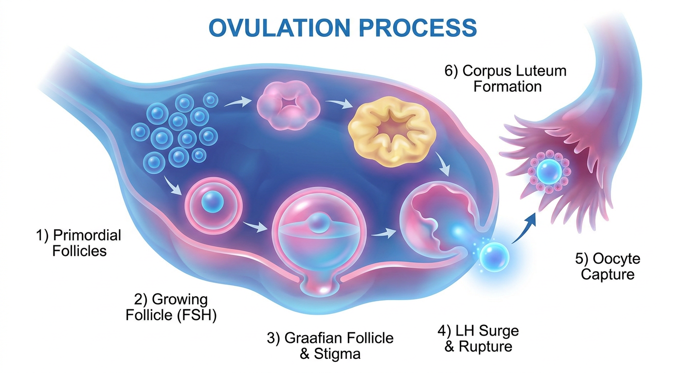
Overview
Ovulation is the release of a secondary oocyte from a mature (Graafian) follicle in the ovary. In a standard 28-day cycle, it occurs on Day 14, triggered by a hormonal surge. The entire rupture event - from first fluid oozing to full expulsion - takes only a few minutes.
Step-by-Step Process
🔵 Phase 1: Follicle Recruitment & Growth (Days 1-7)
Inside each ovary are hundreds of thousands of primordial follicles, each containing an immature egg. At the start of the cycle:
- Rising FSH (follicle-stimulating hormone) from the pituitary recruits a cohort of several primary follicles
- All begin to grow and secrete estrogen, which stimulates the uterine lining
- By Day 7, all but one dominant follicle undergo atresia (degeneration)
The dominant follicle continues developing into a fluid-filled Graafian follicle, with an antrum (cavity) full of follicular fluid, and the oocyte embedded in the cumulus oophorus (granulosa cell mass).
🟡 Phase 2: The LH Surge (Day ~12-13)
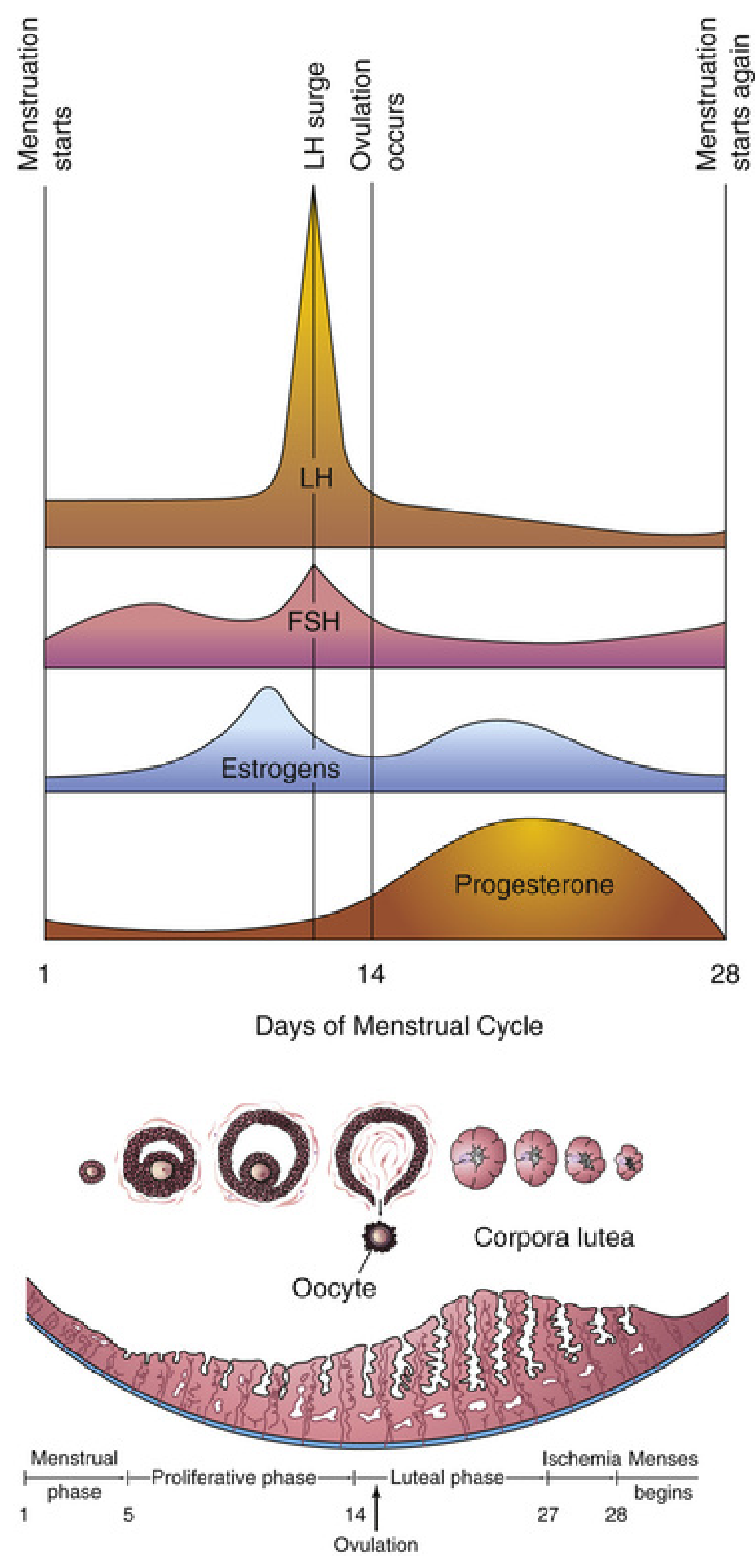
The rising estrogen from the dominant follicle reaches a threshold that triggers a positive feedback at the hypothalamus and anterior pituitary:
- LH secretion surges 6- to 10-fold within ~2 days
- FSH also rises 2- to 3-fold simultaneously
- The LH surge peaks about 16 hours before ovulation
- Ovulation follows the LH peak by 12-24 hours
LH also converts granulosa and theca cells to progesterone-secreting cells, causing estrogen to drop and progesterone to begin rising.
🔴 Phase 3: Follicle Wall Breakdown - The Dual Mechanism
The LH surge triggers progesterone production in the follicle, which then activates two parallel pathways:
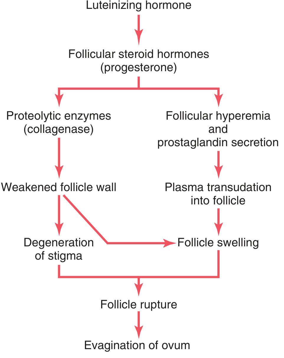
Pathway 1 - Enzymatic Digestion:
- Lysosomes in the theca externa release proteolytic enzymes (collagenase)
- These dissolve the collagen in the follicular capsule wall
- The wall weakens progressively, leading to degeneration of the stigma
Pathway 2 - Vascular & Pressure Changes:
- New blood vessels grow rapidly into the follicle wall (angiogenesis)
- Prostaglandins are secreted into follicular tissues, causing vasodilation and hyperemia
- Plasma transudates into the follicle, dramatically increasing intrafollicular pressure
- Smooth muscle fibers in the theca externa contract (driven by prostaglandins)
Together: follicle swelling + stigma degeneration = follicle rupture.
🟢 Phase 4: Rupture and Oocyte Expulsion
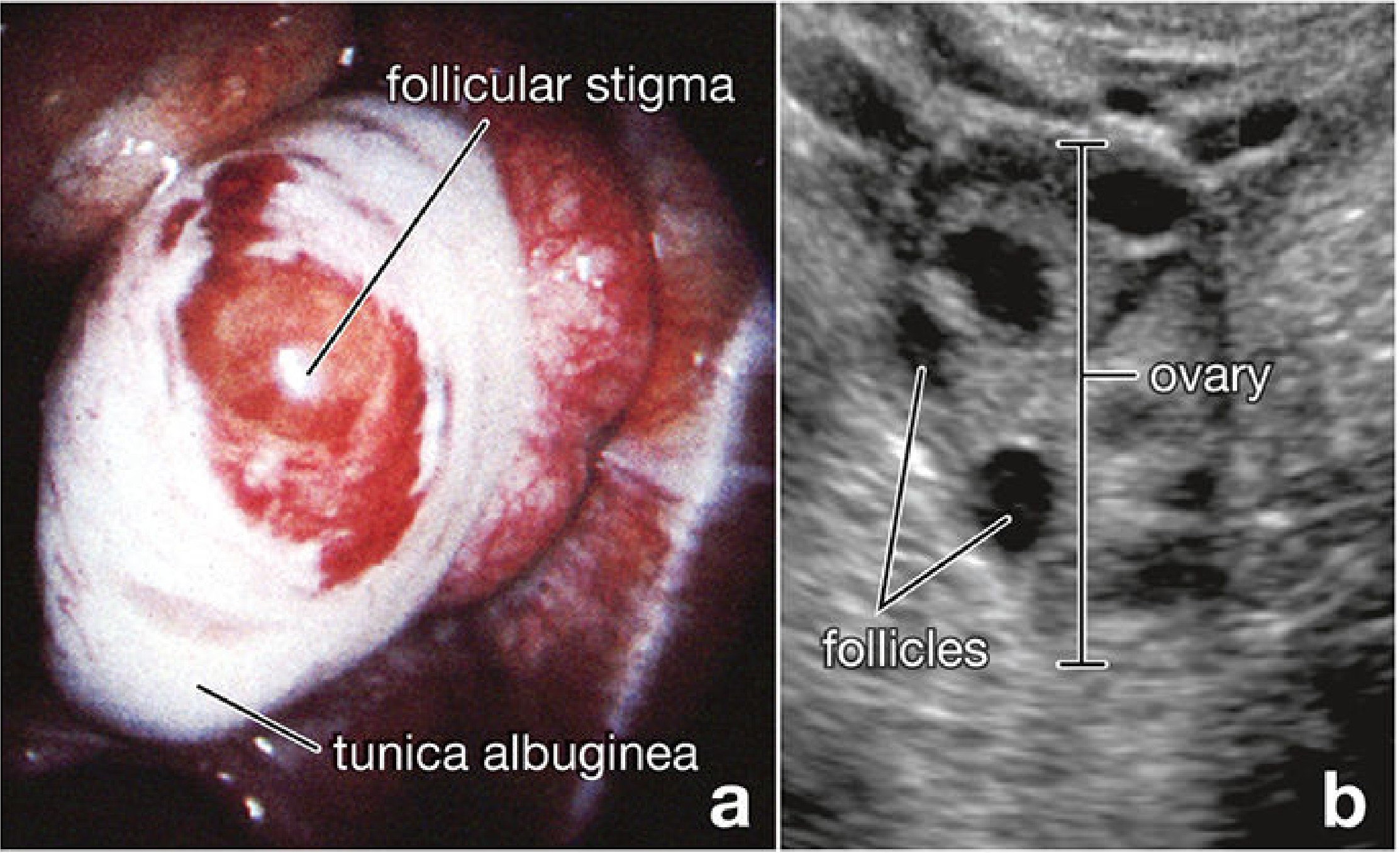
Just before rupture, blood flow stops over a small area of the ovarian surface overlying the bulge - this avascular spot is the stigma (macula pellucida).
The sequence unfolds rapidly:
- The stigma protrudes like a nipple from the follicle surface
- Fluid begins to ooze through the stigma (~30 min after swelling)
- About 2 minutes later, the stigma ruptures widely
- A viscous central fluid is evaginated outward, carrying the oocyte
- The oocyte exits surrounded by the zona pellucida and thousands of granulosa cells arranged radially as the corona radiata - together forming the oocyte-cumulus complex
🟣 Phase 5: Pickup by the Fallopian Tube
At the moment of ovulation, the fimbriae of the fallopian tube sweep closely over the ovarian surface. The cumulus mass (oocyte + corona radiata) adheres firmly to the fimbriae and is actively transported by ciliated epithelial cells into the tube, preventing loss into the peritoneal cavity.
The secondary oocyte remains viable for ~24 hours post-ovulation. If unfertilized, it degenerates as it travels through the tube.
🟤 Phase 6: Corpus Luteum Formation
After expulsion, the remaining granulosa and theca interna cells rapidly transform - they enlarge 2x in diameter and accumulate lipid and lutein pigment, forming the corpus luteum ("yellow body"). This structure secretes progesterone (and some estrogen) to maintain the uterine lining in the luteal phase.
- If fertilization occurs: hCG from the embryo maintains the corpus luteum
- If no fertilization: the corpus luteum degenerates after ~14 days into the corpus albicans (white scar)
3D Animation - Watch It Live
Key Molecules Summary
| Molecule | Role |
|---|---|
| FSH | Recruits and grows follicles |
| Estrogen | Positive feedback triggering LH surge |
| LH | Triggers ovulation; converts cells to progesterone-secreting |
| Progesterone | Activates proteolytic enzymes + smooth muscle contraction |
| Collagenase (proteolytic enzymes) | Digests follicular capsule wall |
| Prostaglandins | Vasodilation + theca muscle contraction |
| Plasminogen activators / MMPs | Enzymatic proteolysis of follicular wall |
| MAPK3/1 (ERK1/2) | Intracellular signaling in follicle cells during rupture |
Sources: Guyton & Hall Textbook of Medical Physiology, p. 1018; Langman's Medical Embryology, pp. 102-103; The Developing Human: Clinically Oriented Embryology, p. 102; Histology: A Text and Atlas, pp. 2209-2210
This is a shared conversation. Sign in to Orris to start your own chat.